. 2024 Sep 20;22(1):852.
doi: 10.1186/s12967-024-05360-y.

STK40 inhibits trophoblast fusion by mediating COP1 ubiquitination to degrade P57Kip2

Affiliations

STK40 inhibits trophoblast fusion by mediating COP1 ubiquitination to degrade P57Kip2

Xia Li et al. J Transl Med. .

Abstract

Background: The syncytiotrophoblast (SCT) layer in the placenta serves as a crucial physical barrier separating maternal-fetal circulation, facilitating essential signal and substance exchange between the mother and fetus. Any abnormalities in its formation or function can result in various maternal syndromes, such as preeclampsia. The transition of proliferative villous cytotrophoblasts (VCT) from the mitotic cell cycle to the G0 phase is a prerequisite for VCT differentiation and their fusion into SCT. The imprinting gene P57Kip2, specifically expressed in intermediate VCT capable of fusion, plays a pivotal role in driving this key event. Moreover, aberrant expression of P57Kip2 has been linked to pathological placental conditions and adverse fetal outcomes.

Methods: Validation of STK40 interaction with P57Kip2 using rigid molecular simulation docking and co-immunoprecipitation. STK40 expression was modulated by lentivirus in BeWo cells, and the effect of STK40 on trophoblast fusion was assessed by real-time quantitative PCR, western blot, immunofluorescence, and cell viability and proliferation assays. Co-immunoprecipitation, transcriptome sequencing, and western blot were used to determine the potential mechanisms by which STK40 regulates P57Kip2.

Results: In this study, STK40 has been identified as a novel interacting protein with P57Kip2, and its expression is down-regulated during the fusion process of trophoblast cells. Overexpressing STK40 inhibited cell fusion in BeWo cells while stimulating mitotic cell cycle activity. Further experiments indicated that this effect is attributed to its specific binding to the CDK-binding and the Cyclin-binding domains of P57Kip2, mediating the E3 ubiquitin ligase COP1-mediated ubiquitination and degradation of P57Kip2. Moreover, abnormally high expression of STK40 might significantly contribute to the occurrence of preeclampsia.

Conclusions: This study offers new insights into the role of STK40 in regulating the protein-level homeostasis of P57Kip2 during placental development.

Keywords: Cell fusion; P57Kip2; Placenta; Preeclampsia; STK40; Trophoblast.


Conflict of interest statement

The authors declare no competing interests.

Figures

Fig. 1
Fig. 1
STK40 interacts with P57. (A) GO cluster analysis of P57-interacting proteins (IPs) identified by protein mass spectrometry analysis. The x-axis denotes the GO term, with the number of IPs indicated on the bars. Enrichment levels are shown on the y-axis. GO terms are color-coded: red bars signify Biological Processes (BP), yellow bars represent Cellular Components (CC), and blue bars indicate Molecular Functions (MF). (B) KEGG signaling pathway analysis of P57 IPs identified by protein mass spectrometry. (C) Venn diagram showcasing the overlap of P57 IPs identified from human villus tissue and Nicolas et al.. (D) Visualization of rigid molecular simulation of docking between P57 and STK40. P57 is illustrated in blue, and STK40 is represented in green. (E) Co-IP validation of protein interaction between P57 and STK40 in human placental villus tissue and HEK-293T cells. IB, Immunoblotting; Input, protein lysis buffer; IP, immunoprecipitation; IgG, negative control
Fig. 2
Fig. 2
Expression pattern of STK40 during trophoblast fusion. (A) Cell immunofluorescence detection of PHT purity. CK7 labels trophoblast, Vimentin labels stromal cells, DAPI stains nuclear; Scale: 50 μm. (B) Cell immunofluorescence detection of STK40 expression pattern in the spontaneous fusion model of PHT. E-CAD labels cell membranes, DAPI stains nuclear; Scale: 50 μm. (C) Western blot analysis of STK40, E-CAD, and CGβ protein expression patterns in PHT and BeWo cells. 3 h, primary VCT; 48 h, primary SCT; DMSO, solvent control; FSK, cell fusion inducer. (D) Immunofluorescence staining of STK40 expression localization in human villi from 6–8 weeks of gestation age. CK7 labels cytotrophoblasts, DAPI stains nuclear. Scale: 50 μm. (E) Immunofluorescence staining of sequential sections showing STK40 and CGβ expression in human villi from 6–8 weeks of gestation age. CGβ labels the syncytiotrophoblast, DAPI stains nuclear. Scale: 50 μm
Fig. 3
Fig. 3
Overexpression of STK40 inhibited VCT fusion. (A) The cell viability of BeWo cells for days 1, 2, 3, 4, and 5 in Vector and STK40-OE groups detected by CCK8. (B) EdU assay assesses the number of EdU-positive cells in BeWo cells with Vector and STK40-OE groups. (C) Cell immunofluorescence detection of BeWo cells fusion with or without FSK induction in Vector and STK40-OE groups. E-CAD labels cell membranes. Scale: 50 μm. (D) Western blot analysis of STK40, E-CAD, and CGβ expression in BeWo cells with or without 48 h FSK induction in Vector and STK40-OE groups. GAPDH was used as a loading control. Vector, empty vector; STK40-OE, STK40 overexpression. ** P < 0.01, *** P < 0.001
Fig. 4
Fig. 4
Knockdown of STK40 promoted VCT fusion. (A) The cell viability of BeWo cells for days 1, 2, 3, 4, and 5 in Vector and sh-STK40 groups detected by CCK8. (B) EdU assay assesses the number of EdU-positive cells in BeWo cells with Vector and sh-STK40 groups. (C) Cell immunofluorescence detection of BeWo cells fusion with or without FSK induction in Vector and sh-STK40 groups. E-CAD labels cell membranes. Scale: 50 μm. (D) Western blot analysis of STK40, E-CAD, and CGβ expression in BeWo cells with or without 48 h FSK induction in Vector and sh-STK40 groups. GAPDH was used as a loading control. Vector, empty vector; sh-STK40, knockdown of STK40. ** P < 0.01, *** P < 0.001
Fig. 5
Fig. 5
Search for STK40 interaction site with by P57 truncation mutation strategy. (A) A schematic diagram of P57 domain, depicting the CDK-binding domain, Cyclin-binding domain, Proline-rich region (PAPA), proline-alanine repeats (QT), PCNA-binding region, and Skp2-dependent degradation domain. (B) Schematic diagram illustrating the construction of Myc-P57 truncation mutants. (C) Line chart displaying the GC content within the coding sequence of P57. (D) Co-IP validation of the interaction between STK40 and truncated mutants of P57 in HEK-293T cells. IB, Immunoblotting; Lystates: protein lysis buffer; IP: immunoprecipitation; IgG: negative control. (E) Flow cytometry analysis of cell cycle progression in BeWo cells with Vector and STK40-OE groups. (F) Flow cytometry analysis of cell cycle progression in BeWo cells with Vector and sh-STK40 groups. * P < 0.05, ** P < 0.01
Fig. 6
Fig. 6
STK40 promoted ubiquitination degradation of P57 through COP1. (A) Western blot analysis of STK40, E-CAD, CGβ, and P57 in BeWo cells with or without 72 h FSK induction in Vector and STK40-OE groups. GAPDH was used as a loading control. (B) Western blot analysis of STK40, E-CAD, CGβ, and P57 in BeWo cells with or without 72 h FSK induction in Vector and sh-STK40 groups. GAPDH was used as a loading control. (C) RT-qPCR was utilized to measure the mRNA expression of STK40 and P57 in BeWo cells in Vector and STK40-OE groups. (D) RT-qPCR was utilized to measure the mRNA expression of STK40 and P57 in BeWo cells in Vector and sh-STK40 groups. (E) Co-IP was performed to validate the protein-protein interactions between P57, COP1, and HA-STK40 in HEK-293T cells. (F) Western blot analysis was conducted to detect the ubiquitination levels of P57. ns, no statistical significance; *** P < 0.001
Fig. 7
Fig. 7
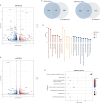
Transcriptome shows potential downstream targets of STK40. (A) Volcano maps illustrating DEGs in Vector and STK40-OE groups. Red points indicate up-regulation DEGs, blue indicates down-regulation DEGs, and the gray point represents non-significant DEGs. (B) Volcano maps showcasing DEGs in Vector and sh-STK40 groups. (C) Venn diagram displaying the overlap between up-regulated DEGs after STK40 overexpression and down-regulated DEGs after knockdown of STK40. And the Venn diagram of the down-regulated DEGs after STK40 overexpression and the up-regulated DEGs after knockdown of STK40. (D) GO analysis of the potential downstream targets of STK40. (E) KEGG pathway enrichment analysis of potential downstream targets of STK40
Fig. 8
Fig. 8
Abnormally high expression of STK40 in PE placenta. (A) Expression levels of STK40 in normal placenta and PE placenta from the GSE149812 dataset. (B) Immunofluorescence detection of STK40 in normal placenta and PE placenta. CK7 labels cytotrophoblasts, DAPI stains nuclear; Scale: 50 μm. (C) Western blot analysis of STK40 in normal placenta and PE placenta

Similar articles

References

    1. Io S, Kondoh E, Chigusa Y, Kawasaki K, Mandai M, Yamada AS. New era of trophoblast research: integrating morphological and molecular approaches. Hum Reprod Update. 2020;26:611–33. - PubMed
    1. Redman CWG, Staff AC, Roberts JM. Syncytiotrophoblast stress in preeclampsia: the convergence point for multiple pathways. Am J Obstet Gynecol. 2022;226:S907–27. - PubMed
    1. Burton GJ, Redman CW, Roberts JM, Moffett A. Pre-eclampsia: pathophysiology and clinical implications. BMJ. 2019;366:l2381. - PubMed
    1. Renaud SJ, Jeyarajah MJ. How trophoblasts fuse: an in-depth look into placental syncytiotrophoblast formation. Cell Mol Life Sci CMLS. 2022;79:433. - PMC - PubMed
    1. Liu Y, Fan X, Wang R, Lu X, Dang Y-L, Wang H, et al. Single-cell RNA-seq reveals the diversity of trophoblast subtypes and patterns of differentiation in the human placenta. Cell Res. 2018;28:819–32. - PMC - PubMed

Substances

LinkOut - more resources