. 2024 Sep 17:15:614-633.
doi: 10.18632/oncotarget.28637.

Integrated stress response (ISR) activation and apoptosis through HRI kinase by PG3 and other p53 pathway-restoring cancer therapeutics

Affiliations

Integrated stress response (ISR) activation and apoptosis through HRI kinase by PG3 and other p53 pathway-restoring cancer therapeutics

Xiaobing Tian et al. Oncotarget. .

Abstract

Restoration of the p53 pathway has been a long-term goal in the field of cancer research to treat tumors with mutated p53 and aggressive clinical behavior. p53 pathway restoration in p53-deficient cancers can be achieved by small molecules via p53-dependent or p53-independent processes. Hereafter p53-independent restoration of p53-pathway-signaling in p53-deficient/mutated tumors is referred to as 'restoration of the p53 pathway'. We compare activation of p53 target genes by novel compounds PG3 and PG3-Oc, that activate p53-target genes in a p53-independent manner, and four mutant p53-activating compounds while Nutlin-3a is used as negative control. PG3 and PG3-Oc upregulate p21, PUMA, and DR5 in five cancer cell lines with various p53 mutational statuses through ATF4 (Activating Transcriptional Factor 4) and integrated stress response (ISR) independent of p53. Mutant p53-targeting compounds induce expression of the 3 major downstream p53 target genes and ATF4 in a highly variable and cell-type-dependent manner. PG3 treatment activates ATF4 through ISR via kinase HRI (Heme-Regulated Inhibitor). ATF4 mediates upregulation of PUMA, p21, and NAG-1/GDF15 (Nonsteroidal anti-inflammatory drug-activated gene 1). We note that PUMA mediates apoptosis through activation of caspase-8 in HT29 cells and potentially caspase-10 in SW480 cells. We provide a novel mechanism engaged by PG3 to induce cell death via the HRI/ATF4/PUMA axis. Our results provide unique insights into the mechanism of action of PG3 as a novel cancer therapeutic targeting p53 pathway-like tumor suppression.

Keywords: ATF4; ClpP; HRI; integrated stress response (ISR); mutant p53.


Conflict of interest statement

CONFLICTS OF INTEREST

W.S.E-D. is a co-founder of Oncoceutics, Inc., a subsidiary of Chimerix, p53-Therapeutics, Inc., and SMURF-Therapeutics, Inc. Dr. El-Deiry has disclosed his relationships with Oncoceutics/Chimerix, p53-Therapeutics, Inc., and SMURF-Therapeutics and potential conflicts of interest to his academic institution/employer and is fully compliant with NIH and institutional policy that is managing this potential conflict of interest.

Figures

Figure 1
Figure 1. PG3 inhibits cell proliferation and growth in a p53-indepent manner.
(AE) Cell viability assay, dose response curves and IC50 value measurements in a panel of cancer cell lines and normal cells. Cells were treated with different concentrations of PG3, PG3-Oc (Oc), obatoclax (Ob), prodigiosin (P) or DMSO for 72 h. Luciferase activity was imaged by the IVIS Imaging System after treatment. Cell viability data were normalized to those of DMSO treatment control in each cell line and data analyses were performed using PRISM4 software. IC50 data are expressed as the mean ±SD (normal; n = 3).
Figure 2
Figure 2. p53-dependent inhibition of cell proliferation is only observed at specific concentrations.
(AJ) Cell viability assay, dose response curves and IC50 value measurements in a panel of cancer cell lines and normal cells. Cells were treated with different concentrations of ZMC1, CP-313908, APR-246, Ellipticine, Nutalin-3a or DMSO for 72 h. Luciferase activity was imaged by the IVIS Imaging System after treatment. Cell viability data were normalized to those of DMSO treatment control in each cell line and data analyses were performed using PRISM4 software. IC50 data are expressed as the mean ±SD (normal; n = 3).
Figure 3
Figure 3. PG3 does not activate p53-responsive reporter activity.
SW480 cells carrying a p53-responsive luciferase reporter were used for assay of functional restoration of mutant p53. Cells were seeded in 96-well plates (5 × 10⁴ cells/well) and were treated with compounds indicated for 6 and 15 h, respectively. Then, D-luciferin was added to each well with final concentration 100 μg/mL, and cells were imaged by using an IVIS Imaging System to detect luciferase activity.
Figure 4
Figure 4. Activation of ATF4 and up-regulation of typical p53 target genes.
(AE) Western blot analysis of p53 protein levels and upregulation of ATF4, PUMA, DR5 and p21. Cells were treated for 24 h with indicated compounds, concentrations and cell lines. Abbreviations: S.E.: Short exposure; L.E.: Long exposure.
Figure 5
Figure 5. PG3 induces apoptosis in p53-null and p53-mutant cancer cell lines.
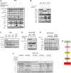
(AE) Dose-response and time-course analysis of cleavage of caspase-8, -3, cleaved PARP and PUMA in PG3-treated HT29, SW480 and HCT116 p53−/− cells by western blot using the indicated antibodies. (FH) Flowcytometry analysis of cell death. (I) HT29 and HT29-PUMA−/− and (J) SW480 cells were co-treated with PG3 and pan-caspase inhibitor Z-VAD-fmk for 48 h. (K) HCT116 p53−/− cell were co-treated with PG3 and Z-VAD-fmk for 24 hours.
Figure 6
Figure 6. ATF4 is a key regulator and mediates PUMA expression that is required for PG3-induced apoptosis.
(A) HT29 and (B) HCT116 p53−/− cells were transfected with Control and ATF4 siRNAs, and at 32 hours after transfection, the cells were treated with 1 μM PG3 for 16 h. Then cell lysates were prepared, and Western blotting was performed using the indicated antibodies. (C) SW480 cells were transfected with siControl, siPUMA, siNAG-1, siCaspase-9 for 24 hours, and then treated with PG3 for 24 hours. Western blots were performed using the indicated antibodies. (D) HT29 and (E) SW480 cells were transfected with siControl, siCaspase-8 for 24 hours and then treated with PG3 for 48 and 24 hours, respectively. (F) SW480 cells were transfected with siControl, siCaspase-8 and siCaspase-9 for 24 hours and then treated with PG3 for 24 hours. Western blots were performed using the indicated antibodies. (G) SW480 cells were co-treated with PG3 and caspase-10 inhibitor for 24 hours, and then Western blots were performed using the indicated antibodies.
Figure 7
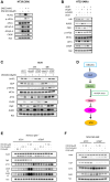
Figure 7. PG3 induces upregulation of ATF4 through ISR via HRI.
(A) HT29 cells were treated with PG3, GSK2606414, TG and ONC201 respectively, or co-treatment with PG3/GSK2606414, PG3/TG, and PG3/ONC201 for 15 h. (B) MEF, MEFPERK−/− and MEFGCN2−/− cells were treated with PG3 and ONC201 for 16 hours. (C) HT29 cells were transfected with siControl, siPKR, siHRI and siPKR/siHRI for 24 hours and then treated with PG3 for 16 hours. (D) HCT116 p53−/− cells were transfected with siControl and siHRI for 24 hours and then treated with PG3 for 16 hours. (E) HCT116 p53−/− cells were treated with PG3 and ONC201 for indicated times. (F) PANC1-sgCtrl, -sgHRI and -sgPKR cell lines were treated with PG3 and imipridones (ONC201, ONC206 and ONC212) for 16 hours. Western blot was performed using indicated antibodies. (G) Proposed model of PG3-induced cell apoptosis through HRI/ATF4/PUMA axis. Abbreviation: L.E.: Long exposure.
Figure 8
Figure 8. Inhibition of heme biosynthesis leads to upregulation of ATF4.
(A) HT29 cells were treated with SA and ONC201 for 20 hours. Western blotting was performed using the indicated antibodies. (B) HT29 cells were transfected with siCtrl, siHRI, siClpX, siALAS1, siClpX/siALAS1 and siClpX/siALAS1/siHRI for 48 hours. (C) HT29 cells were transfected with siCtrl, siLonP1, siClpP for 24 or 48 hours respectively, and then treated with ONC201 for 24 hours. (D) Proposed model of ONC201-induced cell apoptosis through ClpP/ALAS1/HRI/ATF4 axis. (E, F) Silencing of ClpP in HCT116 p53−/− and MDA-MB-468 cells and then treated with PG3, PG3-Oc and ONC201. Western blot was performed using indicated antibodies.

Similar articles

References

    1. Freed-Pastor WA, Prives C. Mutant p53: one name, many proteins. Genes Dev. 2012; 26:1268–86. 10.1101/gad.190678.112. - DOI - PMC - PubMed
    1. Muller PA, Vousden KH. Mutant p53 in cancer: new functions and therapeutic opportunities. Cancer Cell. 2014; 25:304–17. 10.1016/j.ccr.2014.01.021. - DOI - PMC - PubMed
    1. Mantovani F, Collavin L, Del Sal G. Mutant p53 as a guardian of the cancer cell. Cell Death Differ. 2019; 26:199–212. 10.1038/s41418-018-0246-9. - DOI - PMC - PubMed
    1. Bykov VJN, Eriksson SE, Bianchi J, Wiman KG. Targeting mutant p53 for efficient cancer therapy. Nat Rev Cancer. 2018; 18:89–102. 10.1038/nrc.2017.109. - DOI - PubMed
    1. Olivier M, Hollstein M, Hainaut P. TP53 mutations in human cancers: origins, consequences, and clinical use. Cold Spring Harb Perspect Biol. 2010; 2:a001008. 10.1101/cshperspect.a001008. - DOI - PMC - PubMed

MeSH terms

LinkOut - more resources