. 2024 Sep 17;134(18):e164535.
doi: 10.1172/JCI164535.

HLA A*24:02-restricted T cell receptors cross-recognize bacterial and preproinsulin peptides in type 1 diabetes

Affiliations

HLA A*24:02-restricted T cell receptors cross-recognize bacterial and preproinsulin peptides in type 1 diabetes

Garry Dolton et al. J Clin Invest. .

Abstract

CD8+ T cells destroy insulin-producing pancreatic β cells in type 1 diabetes through HLA class I-restricted presentation of self-antigens. Combinatorial peptide library screening was used to produce a preferred peptide recognition landscape for a patient-derived T cell receptor (TCR) that recognized the preproinsulin-derived (PPI-derived) peptide sequence LWMRLLPLL in the context of disease risk allele HLA A*24:02. Data were used to generate a strong superagonist peptide, enabling production of an autoimmune HLA A*24:02-peptide-TCR structure by crystal seeding. TCR binding to the PPI epitope was strongly focused on peptide residues Arg4 and Leu5, with more flexibility at other positions, allowing the TCR to strongly engage many peptides derived from pathogenic bacteria. We confirmed an epitope from Klebsiella that was recognized by PPI-reactive T cells from 3 of 3 HLA A*24:02+ patients. Remarkably, the same epitope selected T cells from 7 of 8 HLA A*24+ healthy donors that cross-reacted with PPI, leading to recognition and killing of HLA A*24:02+ cells expressing PPI. These data provide a mechanism by which molecular mimicry between pathogen and self-antigens could have resulted in the breaking of self-tolerance to initiate disease.

Keywords: Autoimmunity; Diabetes; Immunology; Structural biology; T cell receptor.


Figures
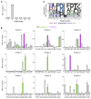
Figure 1
Figure 1. Sizing scan and positional scanning CPL screening of 4C6 T cells.
(A) The 4C6 T cell clone was incubated overnight with sizing scan mixtures of defined amino acid length (x axis) using C1R-HLA A*24:02 as antigen-presenting cells. Assay supernatants used for MIP-1β ELISA. Data points shown for duplicate conditions. (B) Based on the results of the sizing scan, a 9-mer positional scanning CPL (PS-CPL) screen was performed, using 4C6 T cells and antigen-presenting cells and ELISA as in A. Key: Green bars indicate amino acid present in the natural PPI epitope; and magenta bars and arrows show amino acid present in the superagonist peptide (shared amino acid residues are underlined). Data points shown for duplicate conditions. A replicate assay gave similar results. (C) Motif logo plot summarizing the amino acid preference of 4C6 at each position of the PS-CPL.
Figure 2
Figure 2. Cellular and biophysical analyses of the PPI and superagonist peptides.
(A) Sensitivity of 4C6 T cell clone to PPI (LWMRLLPLL) and superagonists (NMPRLFPIV and QLPRLFPLL) peptides in a titration assay. Incubation overnight with C1R–HLA A*24:02 as antigen-presenting cells. Assay supernatants used for MIP-1β ELISA. Data points shown for duplicate conditions. Replicate assay including all candidate superagonist peptides tested for 4C6 as shown in Supplemental Figure 2. Underlined amino acid residues are the same as for the PPI peptide. Highlighted residues were present in both superagonist peptides. The most potent superagonist, QLPRLFPLL, was used for downstream experiments. (B) Staining of the 4C6 T cell clone with irrelevant (AYAQKIFKIL from CMV), PPI, and superagonist PE-conjugated tetramers. Tetramer used alone or following pretreatment with the protein kinase inhibitor (PKI) dasatinib. Mean fluorescence intensity of staining is displayed. Stained for CD8 APC-Vio770 and the viability stain VIVID. (C) SPR analysis of 4C6 TCR recognition of LWMRLLPLL (green) and QLPRLFPLL (magenta). SPR response to 10 serial dilutions of 4C6 was measured (left and center). KD values were calculated using nonlinear fit curve (y = [P1 × x]/[P2 + x]) (right).
Figure 3
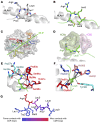
Figure 3. Structural analysis of the 4C6 TCR with superagonist and PPI peptides.
(A) Structure of HLA A*24:02–QLPRLFPLL. Peptide shown as gray sticks, with MHC α-helix (gray) shown for orientation. Dotted lines represent VdW interactions. (B) Structure of 4C6-HLA A*24:02–LWMRLLPLL. Peptide shown as green sticks, with MHC α-helix (gray) shown for orientation. Dotted lines represent VdW interactions. (C) Top-down view of 4C6 TCR “footprint” on HLA A*24:02–LWMRLLPLL. 4C6 CDR loops shown as colored cartoon, with the peptide shown as green sticks. Green line and number indicate crossing angle. (D) Close-up of 4C6:HLA A24:02–LWMRLLPLL structure focusing on residues Arg4 and Leu5 (green sticks), which form a peg-in-hole formation inside the 4C6 TCR (lines and surface). (E and F) LWMRLLPLL peptide residues Arg4 (E) and Leu5 (F) shown as green sticks. Important 4C6 TCR residues are labeled. Black dotted lines indicate VdW interactions. Red dotted lines indicate hydrogen bonds. (G) Heatmap of 4C6 TCR contacts with the LWMRLLPLL peptide.
Figure 4
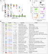
Figure 4. 4C6 T cells cross-react with peptides derived from bacterial proteomes.
PS-CPL data for 4C6 (Figure 1) was used to screen a database of infectious bacteria and the top 20 peptides selected for testing. (A) Peptide titrations using 4C6 with the top 20 bacteria peptides (listed in B). Incubation overnight with C1R–HLA A*24:02 as antigen-presenting cells. Assay supernatants used for MIP-1β ELISA. Data points shown for duplicate conditions. (B) Peptide sequence and origin. Scoring indicates prediction of how likely the peptide is to be recognized by 4C6 T cells, with the best-scoring peptide at the top. EC50 of activation in bold indicates peptides that acted as superagonists of the 4C6 T cell (ligand more potent than natural insulin sequence). One of the peptides gave no response (NR), and 2 of them gave ambiguous EC50 values (~) according to GraphPad Prism. (C) Staining of 4C6 T cell clone with pMHC tetramers bearing CMV irrelevant epitope (AYAQKIFKIL), PPI (LWMRLLPLL), superagonist (QLPRLFPLL), and K. oxytoca (RYPRLFGIV and SLPRLFPLL) PE tetramers. Staining performed without PKI. Mean fluorescence intensity of staining is shown. Stained for CD8 APC-Vio770 and the viability stain VIVID.
Figure 5
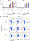
Figure 5. K. oxytoca peptides are real epitopes and prime PPI-reactive T cells from a healthy donor.
(A) Genes encoding enterochelin esterase (also known as DUF3327 containing protein) (gene: fes) or glutathione ABC transporter ATP-binding protein (gene: gsiA) from K. oxytoca were expressed in C1R cells (with or without HLA A*24:02) using lentivirus, then used in overnight activation assays with PPI LWMRLLPLL peptide–reactive CD8+ T cell clone 4C6. Irrelevant protein IMP2 (gene: IGF2BP2) was used as a negative control. Peptides (10–5 M) RYPRLFGIV from enterochelin esterase and SLPRLFPLL from glutathione ABC transporter ATP-binding protein were used as positive controls. Supernatants used for MIP-1β and TNF ELISA. Error bars represent standard deviation of triplicate conditions. (B) Purified CD8+ T cells from an HLA A*24+ healthy donor (BB51) were primed with K. oxytoca (Kleb) SLPRLFPLL or PPI LWMRLLPLL peptides at 10–5 M. Unprimed T cells were cultured without peptide. The lines were stained with HLA A*24:02–irrelevant (AYAAAAAAL), Kleb SLPRLFPLL, and PPI tetramers. Percentages are for viable CD3+ tetramer+ cells.
Figure 6
Figure 6. Klebsiella tetramers co-select PPI-specific T cells in nondiabetic donors.
(A) PBMCs from 8 HLA A*24+ healthy donors were enriched in parallel with HLA A*24:02 Klebsiella SLPRLFPLL or CMV-AYAQKIFKIL PE-conjugated tetramers and anti-PE magnetic beads. After 2 weeks of expansion with allogeneic PBMCs and PHA, the T cell lines were stained with irrelevant (HLA A24:02 AYAAAAAAL; not shown in the schematic), PPI (LWMRLLPLL), and Klebsiella or CMV (depending on the enrichment) tetramers. (B) Tetramer staining of enriched T cell lines from donor BB64. Percentage tetramer+ is shown. (C) Percentage of Kleb, PPI, and irrelevant tetramer staining for Kleb tetramer–enriched lines from 7 of 8 donors. Donor BB72 did not have Kleb T cells, so those data are not shown. Donor BB64 staining is shown in B. Performed as single-staining conditions. (D) Percentage of CMV, PPI, and irrelevant tetramer staining for CMV tetramer–enriched lines from 7 of 8 donors. Donor BB64 staining shown in B. Performed as single-staining conditions.
Figure 7
Figure 7. T cell lines enriched with Klebsiella peptide recognize and kill HLA A*24:02+ target cells expressing PPI.
T cell lines created from healthy HLA A*24+ donors by enrichment with HLA A*24:02 Klebsiella–SLPRLFPLL tetramers were found to be cross-reactive with LWMRLLPLL from PPI. CMV (AYAQKIFKIL) T cell lines from the same donors were used in parallel. Left and middle: T cells lines were incubated with K562s expressing HLA A*24:02 (K562 A24) with or without PPI (K562 A24 PPI). Reactivity toward 10–6 M PPI peptide was assessed using C1R HLA A*24:02 (C1R A24) as antigen-presenting cells. CD3/CD28 beads used as a positive control for T cell activation. Data points shown for duplicate conditions. Right: T cell lines from BB57 incubated (6 hours) with chromium 51–labeled K562 A24 and K562 A24 PPI cells. Background killing with K562 A24 cells was subtracted from killing with K562 A24 PPI cells. Performed in triplicate with error bars showing SEM.

Comment in

  • Microbial mimics supersize the pathogenic self-response

Similar articles

References

    1. Daar AS, et al. The detailed distribution of HLA-A, B, C antigens in normal human organs. Transplantation. 1984;38(3):287–292. doi: 10.1097/00007890-198409000-00018. - DOI - PubMed
    1. Bottazzo GF, et al. In situ characterization of autoimmune phenomena and expression of HLA molecules in the pancreas in diabetic insulitis. N Engl J Med. 1985;313(6):353–360. doi: 10.1056/NEJM198508083130604. - DOI - PubMed
    1. Foulis AK, et al. Aberrant expression of class II major histocompatibility complex molecules by B cells and hyperexpression of class I major histocompatibility complex molecules by insulin containing islets in type 1 (insulin-dependent) diabetes mellitus. Diabetologia. 1987;30(5):333–343. doi: 10.1007/BF00299027. - DOI - PMC - PubMed
    1. Itoh N, et al. Mononuclear cell infiltration and its relation to the expression of major histocompatibility complex antigens and adhesion molecules in pancreas biopsy specimens from newly diagnosed insulin-dependent diabetes mellitus patients. J Clin Invest. 1993;92(5):2313–2322. doi: 10.1172/JCI116835. - DOI - PMC - PubMed
    1. Serreze DV, et al. Major histocompatibility complex class I-deficient NOD-B2mnull mice are diabetes and insulitis resistant. Diabetes. 1994;43(3):505–509. doi: 10.2337/diab.43.3.505. - DOI - PubMed

MeSH terms