. 2024 Sep 2:14:1417946.
doi: 10.3389/fcimb.2024.1417946. eCollection 2024.

Fusobacterium nucleatum induces invasive growth and angiogenic responses in malignant oral keratinocytes that are cell line- and bacterial strain-specific

Affiliations

Fusobacterium nucleatum induces invasive growth and angiogenic responses in malignant oral keratinocytes that are cell line- and bacterial strain-specific

Ajith Selvaraj et al. Front Cell Infect Microbiol. .

Abstract

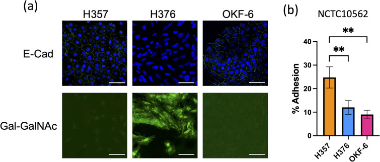
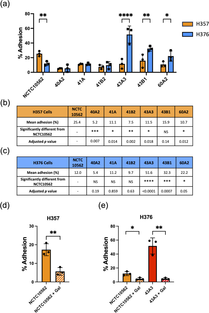
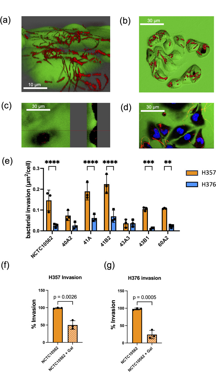
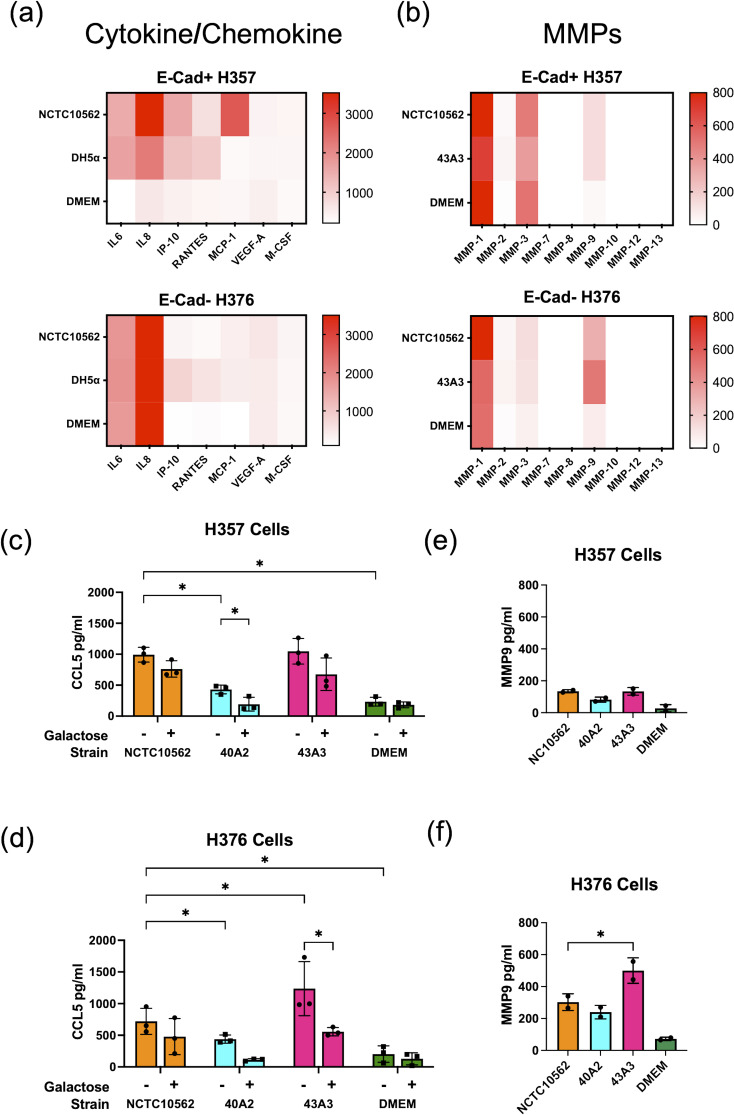
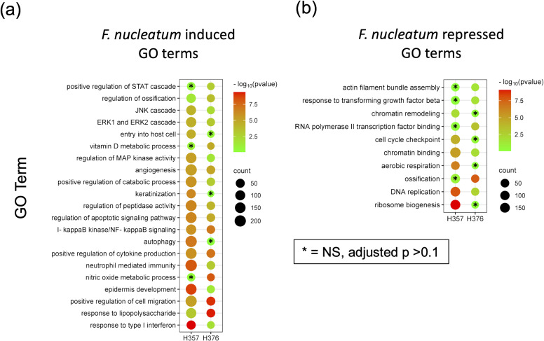
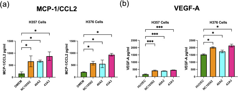
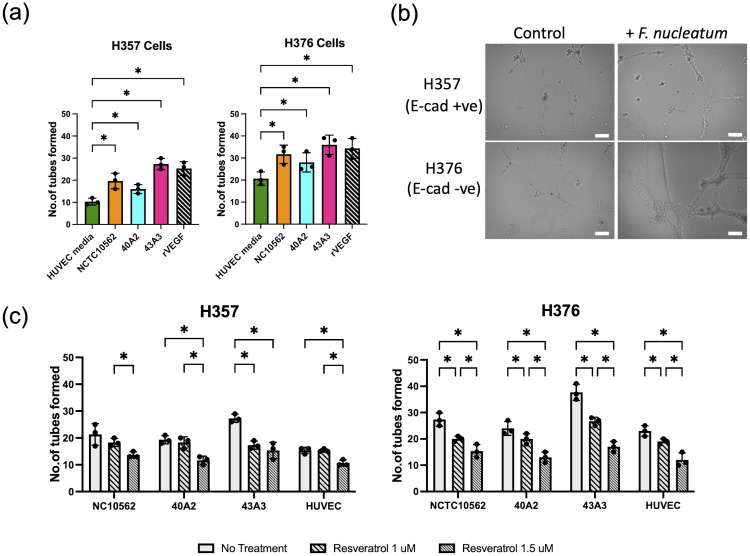
Fusobacterium nucleatum is an anaerobic commensal of the oral cavity recently reported to be associated with cancers of the gastrointestinal tract and oral squamous cell carcinoma (OSCC). In this study, we investigate the impact on oral keratinocytes of infection with a genetically diverse set of strains of F. nucleatum subsp. polymorphum recovered from patients with oral dysplasia (n=6). We employed H357 oral keratinocytes derived from a stage 1 OSCC and H376 cells derived from a stage 3 OSCC. Adhesion phenotypes were strain specific, with 3/6 clinical isolates examined exhibiting higher adherence to the stage 3 H376 cell line. Conversely, intracellular invasion was greatest in the H357 cells and was associated with specific transcriptional responses including autophagy and keratinization. Infection of both H357 and H376 cell lines induced transcriptional and cytokine responses linked to cancer cell migration and angiogenesis. F. nucleatum infection induced greater levels of MMP9 secretion in the H376 cell line which was associated with enhanced motility and invasion phenotypes. Additionally, the degree of F. nucleatum induced invasive growth by H376 cells varied between different clinical isolates of F. nucleatum subsp. polymorphum. Blockage of CCL5 signalling using the inhibitor metCCL5 resulted in reduced keratinocyte invasion. F. nucleatum infection also induced expression of the pro-angiogenic chemokine MCP-1 and the angiogenic growth factor VEGF-A resulting in increased capillary-like tube formation in HUVEC cells, most significantly in H376 cells. Treatment of HUVEC cells with resveratrol, a VEGF-A signalling inhibitor, significantly attenuated F. nucleatum induced tube formation. Our data indicate that the outcomes of F. nucleatum-oral cell interactions can vary greatly depending on the bacterial genotype and the malignant phenotype of the host cell.

Keywords: Fusobacterium nucleatum; angiogenesis; inflammation; invasion; oral cancer.


Conflict of interest statement

The authors declare that the research was conducted in the absence of any commercial or financial relationships that could be construed as a potential conflict of interest.

Figures

Figure 1
Figure 1
Characterisation of oral keratinocyte cell lines. (A) Top row shows immuno-staining of H357, H376 and TERT-1/OKF6 cells for E-cadherin expression using anti-E-cadherin antibody (staining green fluorescent junctions) and DAPI stained nuclei in blue. Bottom row shows staining of H357, H376 and TERT-1/OKF6 cells with FITC labelled peanut agglutinin. The scale bar represents 75 µm in length. (B) Adhesion of F. nucleatum subsp polymorphum strain NCTC10562 to H357, H376 and TERT-1/OKF6 cells (** = ANOVA p <0.001).
Figure 2
Figure 2
Adhesion of F. nucleatum subsp. polymorphum clinical strains to oral keratinocytes. (A) Adhesion of F. nucleatum subsp. polymorphum strains to E-cadherin positive H357 and E-cadherin negative H376 cells. Significant differences between cell lines indicate by asterisks (adjusted p= *<0.05, **<0.01, *** < 0.001, **** < 0.0001). (B, C) Comparisons of adhesion of NCTC10562 and the indicated clinical isolates of F. nucleatum subsp. polymorphum to (B) H357 and (C) H376 cells using ANOVA with Dunnett’s multiple comparison test. (D) Impact of galactose (60 mM) on the adhesion of NCTC10562 to H357 cells (t-test p= **<0.01). (E) Impact of galactose (60 mM) on the adhesion of strains NCTC10562 and 43A3 to H376 cells (t-test p= *<0.05, **<0.01).
Figure 3
Figure 3
Invasion of oral keratinocytes by F. nucleatum subsp. polymorphum. (A) Propidium iodide (PI) stained cells of F. nucleatum subsp. polymorphum NCTC10562 interacting with H357 oral keratinocytes. (B) 3D confocal image of H376 oral keratinocytes with internalised cells of F. nucleatum subsp. polymorphum NCTC10562 stained with PI. (C) Saggital-section of H357 epithelial cells showing internalised bacteria of strain NCTC10562. (D) Confocal Z stack image of invasion assay of H357 cells (nuclei stained using Hoescht) being invaded by strain NCTC10562. (E) Bacterial invasion of H357 and H376 oral keratinocytes quantified using Imaris software as µm2 of bacteria per cell. Significant differences in the invasion of H357 and H376 cells indicated by asterisks (ANOVA adjusted p = ** < 0.01, *** <0.001, **** <0.0001). (F) Impact of galactose (60 mM) on the invasion of H357 cells by strain NCTC10562 with t-test p value. (G) Impact of galactose (60 mM) the invasion of H376 cells by strain NCTC10562 with t-test p value.
Figure 4
Figure 4
Response of H357 and H376 oral keratinocytes to F. nucleatum infection. (A) Expression of selected cytokines and chemokines (pg/ml) detected in a multiplex assay (Eve Technologies, Canada) in cell culture media (DMEM) recovered from H357 and H376 cells infected with F. nucleatum subsp. polymorphum NCTC10562 (MOI 10:1) or E. coli DH5α (MOI 50:1). (B) Expression of MMPs (pg/ml) detected in a multiplex assay (Eve Technologies, Canada) in cell culture media (DMEM) recovered from H357 and H376 cells infected with F. nucleatum subsp. polymorphum NCTC10562 or 43A3. (C) ELISA of CCL5 expression in H357 cells and (D) H376 cells uninfected (DMEM only) and infected with the indicated strains of F. nucleatum subsp. polymorphum in the presence or absence of galactose. Asterisk indicates significant differences identified in 2-way ANOVA corrected for multiple comparisons (adjusted p= *<0.05). (E) ELISA results of MMP9 expression in H357 and (F) H376 cells uninfected (DMEM only) and infected with the indicated strains of F. nucleatum subsp. polymorphum. Asterisk indicates significant difference from NCTC10562 in ANOVA with Dunnett’s test for multiple (adjusted p= *<0.05).
Figure 5
Figure 5
RNA-Seq analysis of the response of H357 and H376 oral keratinocytes to infection with F. nucleatum 23726. Oral keratinocytes were infected with F. nucleatum for 3 h at an MOI of 10:1. Plot shows the results of Gene Ontology (GO) term enrichment analysis in the (A) upregulated and (B) down regulated gene sets in both cell lines. Adjusted p values were generated using the Benjamini-Hochberg FDR method. The most significant non-redundant GO terms were selected for display. Cell lines with adjusted p values >0.1 are indicated by asterisks. A full list can be seen in Supplementary Tables S3 , S4 .
Figure 6
Figure 6
Scratch wound assay. (A) Representative images of scratch wounds in H357 cells (top row) and H376 cells (bottom row). Left panel shows cell at time 0 and right panel after 24 h infections with strain NCTC10562. Arrows indicate scratch wound width (µM). Scale bar=100 μm. (B) Wound closure measurements in H357 cells and (C) H376 cells following incubation with DMEM (control) or infections with the indicated strains. Cell migration was assessed by calculating the amount of wound closure after 24 h infection. Treatments significantly different from the control (DMEM) are indicated by asterisks (ANOVA adjusted p = * <0.05, ** < 0.01, **** <0.0001).
Figure 7
Figure 7
Analysis of invasive phenotypes in H357 and H376 cells in response to CM from cells infected with F. nucleatum subsp. polymorphum. (A) Representative image showing stained oral keratinocytes following migration across the matrix coated filter. The white lines represent scale bar=100 μm (B) Comparison of migration of H357 and H376 keratinocytes following treatment with CM from the indicated strains. Asterisks indicate results of 2-way ANOVA (adjusted p= *<0.05). (C) Analysis of H357 and (D) H376 cell invasion following treatment with CM from the indicated strains with and without pre-treatment with the inhibitor metCCL5. Asterisks indicates CM treatments significantly different from DMEM and significant reductions in invasion following metCCL5 treatment (2-way ANOVA adjusted p= *<0.05).
Figure 8
Figure 8
Analysis of secretion of (A) MCP-1/CCL2 and (B) VEGF-A by H357 and H376 oral keratinocytes following F. nucleatum infection. ELISA quantification was carried out on culture medium (DMEM or HUVEC) following 24 h infection with the indicated strains of F. nucleatum subsp, polymorphum (MOI 10:1). Significant differences from uninfected cells was calculated using ANOVA with Dunnett’s test for multiple comparisons (adjusted p = *<0.05, ***<0.001).
Figure 9
Figure 9
Analysis tube formation by HUVEC cells. (A) Quantification of tube formation in HUVEC cells exposed to conditioned HUVEC medium from H357 and H376 oral keratinocytes following F. nucleatum infection (24 h infection, MOI 10:1). Recombinant VEGF-A (rVEGF; 50 ng/ml) is included as a positive control. Significant differences from uninfected control (HUVEC medium) was calculated using ANOVA with Dunnett’s test for multiple comparisons (adjusted p = *<0.05). (B) Representative images of HUVEC cells after 12 h exposure to conditioned HUVEC medium obtained from H357 cells (top row) or H376 cells (bottom row) that were uninfected (control) or infected with F. nucleatum strain 43A3 (+ F. nucleatum). The white lines represent scale bar=100 μm. (C) Inhibition of HUVEC tube formation by resveratrol (1 μM and 1.5 μM). Significant differences between treatments were identified using 2-way ANOVA (q = *<0.05).

Similar articles

References

    1. Abdalla Z., Walsh T., Thakker N., Ward C. M. (2017). Loss of epithelial markers is an early event in oral dysplasia and is observed within the safety margin of dysplastic and T1 OSCC biopsies. PloS One 12, 17. doi: 10.1371/journal.pone.0187449 - DOI - PMC - PubMed
    1. Abed J., Emgård J. E. M., Zamir G., Faroja M., Almogy G., Grenov A., et al. . (2016). Fap2 mediates fusobacterium nucleatum colorectal adenocarcinoma enrichment by binding to tumor- expressed gal-galNAc. Cell Host Microbe 20, 215–225. doi: 10.1016/j.chom.2016.07.006 - DOI - PMC - PubMed
    1. Brazier J. S., Citron D. M., Goldstein E. J. C. (1991). A selective medium for Fusobacterium spp. J. Appl. Bacteriol. 71, 343–346. doi: 10.1111/j.1365-2672.1991.tb03798.x - DOI - PubMed
    1. Brennan C. A., Garrett W. S. (2019). Fusobacterium nucleatum — symbiont, opportunist and oncobacterium. Nat. Rev. Microbiol. 17, 156–166. doi: 10.1038/s41579-018-0129-6 - DOI - PMC - PubMed
    1. Bullman S., Pedamallu C. S., Sicinska E., Clancy T. E., Zhang X., Cai D., et al. . (2017). Analysis of Fusobacterium persistence and antibiotic response in colorectal cancer. Science 358, 1443–1448. doi: 10.1126/science.aal5240 - DOI - PMC - PubMed

MeSH terms

Substances

Grants and funding

The author(s) declare financial support was received for the research, authorship, and/or publication of this article. GM and CH are funded to carry out this work by the Irish Health Research Board (Grant no. ILP-POR-2019-030). AS was funded by a Provost’s Award PhD Scholarship from Trinity College Dublin.

LinkOut - more resources