Review
. 2024 Aug 28:28:101210.
doi: 10.1016/j.mtbio.2024.101210. eCollection 2024 Oct.

Tailoring biomaterials for skin anti-aging

Affiliations
Review

Tailoring biomaterials for skin anti-aging

Xin Dan et al. Mater Today Bio. .

Abstract

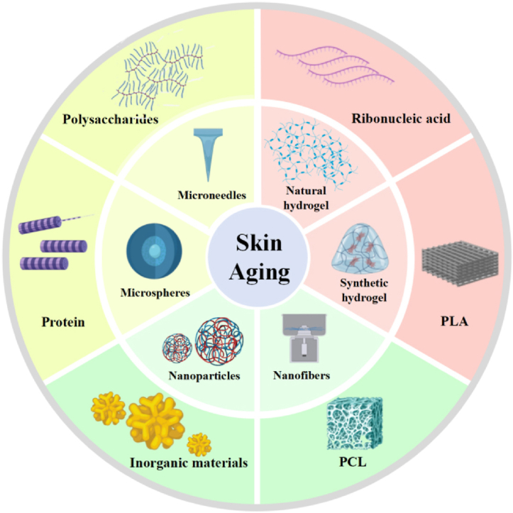
Skin aging is the phenomenon of degenerative changes in the structure and function of skin tissues over time and is manifested by a gradual loss of skin elasticity and firmness, an increased number of wrinkles, and hyperpigmentation. Skin anti-aging refers to a reduction in the skin aging phenomenon through medical cosmetic technologies. In recent years, new biomaterials have been continuously developed for improving the appearance of the skin through mechanical tissue filling, regulating collagen synthesis and degradation, inhibiting pigmentation, and repairing the skin barrier. This review summarizes the mechanisms associated with skin aging, describes the biomaterials that are commonly used in medical aesthetics and their possible modes of action, and discusses the application strategies of biomaterials in this area. Moreover, the synergistic effects of such biomaterials and other active ingredients, such as stem cells, exosomes, growth factors, and antioxidants, on tissue regeneration and anti-aging are evaluated. Finally, the possible challenges and development prospects of biomaterials in the field of anti-aging are discussed, and novel ideas for future innovations in this area are summarized.

Keywords: Biomaterials; Medical aesthetics; Plastic surgery; Skin anti-aging; Tissue regeneration.


Conflict of interest statement

The authors declare that they have no known competing financial interests or personalrelationships that could have appeared to influence the work reported in this paper.

Figures

Image 1
Graphical abstract
Fig. 1
Fig. 1
Mechanisms of skin aging and progress in anti-aging research. (A) Changes in collagen, elastic fibers, and glycosaminoglycans in normal skin, endogenous aging skin, and photoaging skin. Reprinted from Ref. [33] with permission from Elsevier. (B) One of the latest advances in skin anti-aging: cytomegalovirus immune response actively removes senescent cells. Reprinted from Ref. [34] with permission from Cell Press. (C) One of the latest advances in skin anti-aging: an innovative bioswitchable miRNA inhibitor delivery system based on tetrahedral framework DNA. Reprinted from Ref. [35] with permission from Wiley. Abbreviations: CD4 CTL, cytotoxic CD4+ T cells; HCMV, human cytomegalovirus; miR, microRNA/miRNA.
Fig. 2
Fig. 2
Biocompatibility and biodegradability requirements for biomaterials. (A) Electrospun membranes with three different surface topologies (random, aligned, and latticed). (i) Surface morphology and workflow on the mouse surface. (ii) Immunofluorescence staining for foreign body reactions. (iii) Histological staining of foreign body reactions. (iv) Cellular composition of foreign body reactions. v) Observations on the surface of mice. Reprinted from Ref. [58] with permission from the American Association for the Advancement of Science. (B) Hyaluronic acid and collagen injected into mice. (i) Schematic diagram of workflow. (ii) Schematic diagram of hyaluronic acid and collagen under electron microscopy. (iii) Three-dimensional simulation image of degradation rate. (iv) Magnetic resonance imaging performance during degradation. Reprinted from Ref. [61] with permission from Springer.
Fig. 3
Fig. 3
Surface physicochemical properties of biomaterials. (A) Cells exhibit different proliferation and morphologies on surfaces with different roughness levels. Reprinted from Ref. [68] with permission from Cell Press. (B) Morphology and adhesion behavior of cells on positive and negatively variable charges. Reprinted from Ref. [71] with permission from Mary Ann Liebert, Inc. (C) Biomaterials modified by different chemical functional groups have different functions for cells. Reprinted from Ref. [73] with permission from the American Chemical Society. Abbreviations: CM-, Carboxymethylated; CNF, cellulose nanofibril; nPCL, negatively charged PCL; pPCL, positively charged PCL; TO-, TEMPO oxidized.
Fig. 4
Fig. 4
Silk protein. (A) Hierarchical structure of silk fibroin and its processing conditions. (i) Schematic diagram of the hierarchical structure of silk fibroin in different extraction steps. (ii) Extraction of silk fibroin from silkworm cocoons to produce regenerated silk fibroin solution. Reprinted from Ref. [81] with permission from Nature Portfolio. (B) Different forms of filamentous proteins. Reprinted from Ref. [83] with permission from Elsevier. (C) Penetration of silk protein nanoparticles in the skin. (i) Transmission electron microscopy images of silk fibroin nanoparticles and fluorescent fibroin nanoparticles. (ii) Fluorescence micrographs of a cross-section of mouse skin at 2 and 6 h after administration of rhodamine B solution and illumination of the same cross-section in bright and dark fields. Reprinted from Ref. [84] with permission from Elsevier.
Fig. 5
Fig. 5
Hyaluronic acid. (A) Schematic of the chemical structure of hyaluronic acid. Reprinted from Ref. [95] with permission from Elsevier. (B) Collagen regeneration in skin tissue after injection of hyaluronic acid fillers. Reprinted from Ref. [97] with permission from Lippincott Williams & Wilkins. (C) Schematic and in vivo experimental performance of hyaluronic acid hydrogels as tissue fillers. Reprinted from Ref. [98] with permission from Elsevier. (D) Changes in skin wrinkles at different times after hyaluronic acid injection and after stopping injection. Reprinted from Ref. [99] with permission from Wiley. Abbreviations: CL-, cross-linked; COL, collagen; HA, hyaluronic acid.
Fig. 6
Fig. 6
Microspheres. (A) Schematic diagram of the preparation process of microspheres. Reprinted from Ref. [199] with permission from Keai Publishing ltd. (B) Comparison of the morphology between porous microspheres and non-porous microspheres. Reprinted from Ref. [180] with permission from MDPI. (C) Scanning electron microscopy images of PLLA and PCL microspheres, changes occurring in the dorsal skin of hairless mice after implantation of microspheres, and histological manifestations of type I collagen deposition observed after implantation. Reprinted from Ref. [202] with permission from Wiley. (D) Electron microscopy photos of PLLA microspheres of different sizes/hematoxylin and eosin staining and immunofluorescence staining of microspheres prepared from different biomaterials implanted in the dermis or subdermal tissue. Reprinted from Ref. [181] with permission from Oxford University Press. Abbreviations: PCL, polycaprolactone; PLLA, poly (L-lactic acid).
Fig. 7
Fig. 7
Microneedles. (A) Microneedle patches loaded with exosomes. () Schematic diagram of implantation in mice. () Extracellular matrix image of a microneedle patch. () Fluorescent staining image of skin after implantation. Reprinted from Ref. [205] with permission from BMC. (B) A novel anti-aging microneedle. () Schematic diagram of microneedle for anti-aging therapies. () Microneedle penetration and degradation ability. Reprinted from Ref. [207] with permission from Wiley.
Fig. 8
Fig. 8
Hydrogels. (A) A stem cell-derived extracellular matrix injectable hydrogel for collagen production in the dermis. (i) Sol-gel transition images. (ii) Distribution of extracellular vesicles in untreated and hydrogel-treated mice. (iii) Effect of collagen production in mice in vivo. Reprinted from Ref. [211] with permission from ACS Publications. (B) Highly bioactive THPCcross-linked recombinant collagen hydrogel implant significantly improves skin quality after photoaging. (i) Degree of cross-linking. (ii) Skin microscopy and ultrasonic skin imaging after implantation in mice. Reprinted from Ref. [218] with permission from Elsevier. Abbreviations: Sol, solute; Gel, gelation; EV, extracellular vesicle; TS, thermosensitive; BDDE, 1,4-butanediol diglycidyl ether; EDC, 1-Ethyl-3-(3-dimethylaminopropyl)carbodiimide; GA, glutaraldehyde NHS,N-Hydroxysuccinimide; PEG, Polyethylene glycol; TC-THPC,THPC-crosslinked triplehelical recombinant collagen.
Fig. 9
Fig. 9
Nanoparticles. (A) Dopamine nanoparticles for anti-aging treatment. (i) Schematic representation of a nanoparticle antioxidant and anti-aging cells. (ii) Transmission electron microscopy images of PDA NPs and UPDA NPs. Reprinted from Ref. [190] with permission from Elsevier. (B) A manganese dioxide nanoparticle promotes skin anti-aging. (i) Schematic diagram of the reaction process of MnO2 NPs with H2O2. (ii) MnO2 NPs scavenge ROS in vivo in senescent mice and Masson's trichrome staining. Reprinted from Ref. [220] with permission from Wiley. Abbreviations: D-gal, D-galactose.

Similar articles

References

    1. Haydont V., et al. Age-related evolutions of the dermis: clinical signs, fibroblast and extracellular matrix dynamics. Mech. Ageing Dev. 2019;177:150–156. doi: 10.1016/j.mad.2018.03.006. - DOI - PubMed
    1. Rorteau J., et al. [Functional integrity of aging skin, from cutaneous biology to anti-aging strategies] Med. Sci. 2020;36(12):1155–1162. doi: 10.1051/medsci/2020223. - DOI - PubMed
    1. He X., et al. Research progress on skin aging and active ingredients. Molecules. 2023;28(14):5556. doi: 10.3390/molecules28145556. - DOI - PMC - PubMed
    1. Jomova K., et al. Reactive oxygen species, toxicity, oxidative stress, and antioxidants: chronic diseases and aging. Arch. Toxicol. 2023;97(10):2499–2574. doi: 10.1007/s00204-023-03562-9. - DOI - PMC - PubMed
    1. Nishikori S., et al. Resistance training rejuvenates aging skin by reducing circulating inflammatory factors and enhancing dermal extracellular matrices. Sci. Rep. 2023;13(1) doi: 10.1038/s41598-023-37207-9. - DOI - PMC - PubMed

LinkOut - more resources