. 2024 Sep 16;57(1):66.
doi: 10.1186/s40659-024-00546-6.

PTN from Leydig cells activates SDC2 and modulates human spermatogonial stem cell proliferation and survival via GFRA1

Affiliations

PTN from Leydig cells activates SDC2 and modulates human spermatogonial stem cell proliferation and survival via GFRA1

Xueheng Zhao et al. Biol Res. .

Abstract

Background: Spermatogonial stem cells (SSCs) are essential for the maintenance and initiation of male spermatogenesis. Despite the advances in understanding SSC biology in mouse models, the mechanisms underlying human SSC development remain elusive.

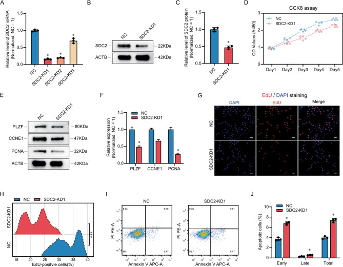
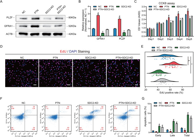
Results: Here, we analyzed the signaling pathways involved in SSC regulation by testicular somatic cells using single-cell sequencing data (GEO datasets: GSE149512 and GSE112013) and identified that Leydig cells communicate with SSCs through pleiotrophin (PTN) and its receptor syndecan-2 (SDC2). Immunofluorescence, STRING prediction, and protein immunoprecipitation assays confirmed the interaction between PTN and SDC2 in spermatogonia, but their co-localization was observed only in approximately 50% of the cells. The knockdown of SDC2 in human SSC lines impaired cell proliferation, DNA synthesis, and the expression of PLZF, a key marker for SSC self-renewal. Transcriptome analysis revealed that SDC2 knockdown downregulated the expression of GFRA1, a crucial factor for SSC proliferation and self-renewal, and inhibited the HIF-1 signaling pathway. Exogenous PTN rescued the proliferation and GFRA1 expression in SDC2 knockdown SSC lines. In addition, we found downregulation of PTN and SDC2 as well as altered localization in non-obstructive azoospermia (NOA) patients, suggesting that downregulation of PTN and SDC2 may be associated with impaired spermatogenesis.

Conclusions: Our results uncover a novel mechanism of human SSC regulation by the testicular microenvironment and suggest a potential therapeutic target for male infertility.

Keywords: Apoptosis; Pleiotrophin; Proliferation; Self-renewal; Spermatogonial stem cells; Syndecan-2.


Conflict of interest statement

No authors declare competing interests.

Figures

Fig. 1
Fig. 1
Single-cell sequencing reveals cellular communication among testicular cells. (A) UMAP cluster analysis of human testicular scRNA-seq data showing the distribution of different testicular cell types in two dimensions. (B) Total number and weight of cellular communication between testicular cells, with the direction and thickness of the arrows reflecting the direction and strength of intercellular signaling, respectively. (C) Dot plot showing the cellular signals emitted by Leydig cells towards SSCs and differentiating spermatogonia, with redder colors indicating a higher probability of that signaling pattern, and the dot size indicating the p-value. (D) Ligand and receptor circle plot showing the PTN signaling pathway between testicular cells, the thickness of the line represents the strength of the signaling. (E) Violin diagram showing the distribution of conventional PTN signaling pathway molecules in the testis. (F) Ligand and receptor interactions of the PTN signaling pathway in testicular cells. SSCs: spermatogonial stem cells, Diffing. Spg: differentiating spermatogonia, L: leptotene spermatocytes, Z: zygotene spermatocytes, P: pachytene spermatocytes, D: diplotene spermatocytes, RS: round spermatids, ES: elongated spermatids, LCs: Leydig cells, SCs: Sertoli cells, ECs: endothelial cells, PTM: peritubular myoid cells, Mø: macrophages
Fig. 2
Fig. 2
Expression patterns of PTN and SDC2 in normal human testicular tissues. (A) Double immunofluorescence localization of SDC2 with GFRA1, KIT, γH2AX and PCNA. Most SDC2-positive cells co-expressed GFRA1 and PCNA, and SDC2 rarely co-localized with KIT and occasionally co-localized with γH2AX. (B) Box plots showing the co-expression of SDC2 with multiple marker molecules in A. (C) Double immunofluorescence localization of PTN with CYP11A1 in testicular tissues. (D) Box plots showing the proportion of double-positive cells in C. (E) Double immunofluorescence localization of PTN with SDC2 in testicular tissues. (F) Box plots showing the proportion of double-positive cells in E. (G) The STRING database predicts reciprocal proteins for PTN, where SDC2 may have interactions with PTN. (H) Protein immunoprecipitation to detect the interaction of PTN and SDC2. The white dotted line in the fluorescent image indicates the edge of the seminiferous tubule. Scale bar, 50 μm
Fig. 3
Fig. 3
SDC2 knockdown impairs proliferation and self-renewal of human SSC lines in vitro. (A) qPCR analysis of SDC2 mRNA levels after small interfering RNA (siRNA) transfection. (B) Western blot analysis of SDC2 protein levels after knockdown. (C) Bar graphs showing the relative levels of SDC2 protein in the NC and knockdown groups, normalized to GAPDH. (D) CCK8 assay measuring cell proliferation, showing that SDC2 knockdown significantly inhibited SSC proliferation. (E) Western blot analysis of SSC self-renewal and proliferation-related proteins, including PLZF, CCNE1, and PCNA, showing that SDC2 knockdown significantly reduced the expression of these proteins. (F) Bar graphs showing the relative protein levels of PLZF, CCNE1, and PCNA in the NC and knockdown groups, normalized to GAPDH. (G) EdU assays assessing cellular DNA synthesis, showing that SDC2 knockdown significantly decreased the number of EdU-positive cells. (H) Ridgeline plots showing the distribution of EdU-positive cells in the NC and knockdown groups. (I) Flow cytometry combined with Annexin V to detect cell apoptosis, showing that SDC2 downregulation significantly increased cell apoptosis. (J) Bar graphs showing the apoptotic percentage of SSCs in the NC and knockdown groups. * indicates P < 0.05
Fig. 4
Fig. 4
RNA sequencing identifies downstream genes of SDC2. (A) Heatmap of the top 50 DEGs showing the concordance of gene expression after SDC2 knockdown. (B) Volcano plot displaying the expression of all genes after SDC2 knockdown. 90 genes were significantly down-regulated and 96 genes were significantly up-regulated by SDC2 knockdown. Red color indicates up-regulated genes and green color indicates down-regulated genes. (C) KEGG enrichment analysis of all significantly down-regulated genes. (D) qPCR validation of randomly selected DEGs confirming the reliability of RNA sequencing. (E) Violin plot showing the distribution of the top 20 DEGs in human testis. GFRA1 and NUP88 are highly expressed in SSCs. (F) Western blot analysis verifying the differential expression of GFRA1 in the NC and knockdown groups
Fig. 5
Fig. 5
Exogenous PTN rescues the phenotypic defects induced by SDC2 deficiency. (A) Western blot analysis of PLZF and GFRA1 expression levels in the NC, PTN, SDC2-KD and PTN + SDC2-KD groups. The results showed that PLZF and GFRA1 expression was significantly increased in the PTN group compared with the NC group, while PLZF and GFRA1 expression was significantly decreased in the SDC2-KD group, and PLZF and GFRA1 expression in the PTN + SDC2-KD group was comparable to that of the NC group, suggesting that PTN could restore the expression of PLZF and GFRA1. (B) Bar graphs showing the relative levels of PLZF and GFRA1 in (A), normalized to the NC group. (C) CCK8 assay measuring cell proliferation in the four groups, indicating that exogenous PTN alleviated the proliferation defect caused by SDC2 knockdown. (D) EdU assays assessing cellular DNA synthesis in the four groups, showing that PTN supplementation rescued the DNA synthesis impairment induced by SDC2 deficiency. (E) Violin plots showing the EdU positivity rates of the four groups in (D). (F) Flow cytometry combined with Annexin V to detect cell apoptosis in the four groups, showing that PTN addition reduced the apoptosis induced by SDC2 deficiency. (G) Bar graphs showing the apoptotic percentage of SSCs in the four groups. * indicates significant down-regulation compared to the NC group, and # indicates significant up-regulation compared to the NC group, P < 0.05. Scale bar in (D) is 50 μm
Fig. 6
Fig. 6
Expression of PTN and SDC2 in testicular tissues of patients with OA and NOA. (A) Double immunofluorescence localization of PTN and SDC2 in testicular tissues with varying spermatogenic potential. (B) Box plots showing the proportion of PTN- and SDC2-positive cells in different testicular tissues. (C) Western blot analysis of PTN and SDC2 protein levels in different testicular tissues. (D) Bar graphs comparing the relative expression of PTN and SDC2 in different testicular tissues. Normal denotes testicular tissues with normal spermatogenesis, HS denotes testicular tissues with hypospermatogenesis, Spc MA denotes testicular tissues with spermatocyte maturation arrest, and Spg MA denotes testicular tissues with spermatogonia maturation arrest. The scale bar in A is 50 μm, * indicates significant difference from the normal group, P < 0.05
Fig. 7
Fig. 7
Graphic abstracts of this study. Using single-cell sequencing analysis and in vitro experiments, we found that Leydig cell-derived PTN promotes proliferation and self-renewal of human spermatogonial stem cells through binding to its ligand SDC2, and that their dysregulation may be associated with non-obstructive azoospermia

Similar articles

References

    1. Qiao J, Wang Y, Li X, Jiang F, Zhang Y, Ma J, et al. A Lancet Commission on 70 years of women’s reproductive, maternal, newborn, child, and adolescent health in China. Lancet. 2021;397(10293):2497–536. - PubMed
    1. Agarwal A, Baskaran S, Parekh N, Cho CL, Henkel R, Vij S, et al. Male infertility. Lancet. 2021;397(10271):319–33. - PubMed
    1. Tharakan T, Luo R, Jayasena CN, Minhas S. Non-obstructive azoospermia: current and future perspectives. Fac Rev. 2021;10:7. - PMC - PubMed
    1. Saitou M, Hayashi K. Mammalian in vitro gametogenesis. Sci (New York NY). 2021;374(6563):eaaz6830. - PubMed
    1. Brinster RL, Zimmermann JW. Spermatogenesis following male germ-cell transplantation. Proc Natl Acad Sci U S A. 1994;91(24):11298–302. - PMC - PubMed

MeSH terms

LinkOut - more resources