[Preprint]. 2024 Sep 6:2024.09.05.611512.
doi: 10.1101/2024.09.05.611512.

Calpains Orchestrate Secretion of Annexin-containing Microvesicles during Membrane Repair

Affiliations

Calpains Orchestrate Secretion of Annexin-containing Microvesicles during Membrane Repair

Justin Krish Williams et al. bioRxiv. .

Abstract

Microvesicles (MVs) are membrane-enclosed, plasma membrane-derived particles released by cells from all branches of life. MVs have utility as disease biomarkers and may participate in intercellular communication; however, physiological processes that induce their secretion are not known. Here, we isolate and characterize annexin-containing MVs and show that these vesicles are secreted in response to the calcium influx caused by membrane damage. The annexins in these vesicles are cleaved by calpains. After plasma membrane injury, cytoplasmic calcium-bound annexins are rapidly recruited to the plasma membrane and form a scab-like structure at the lesion. In a second phase, recruited annexins are cleaved by calpains-1/2, disabling membrane scabbing. Cleavage promotes annexin secretion within MVs. Our data supports a new model of plasma membrane repair, where calpains relax annexin-membrane aggregates in the lesion repair scab, allowing secretion of damaged membrane and annexins as MVs. We anticipate that cells experiencing plasma membrane damage, including muscle and metastatic cancer cells, secrete these MVs at elevated levels.


Figures

1.
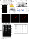
1.. Annexin-containing extracellular vesicles are distinct from exosomes.
(A) Immunoblots show distribution of EV markers across an iodixanol gradient of the conditioned medium 100k × g pellet fraction. Samples were taken from low (F2-Fraction #2) to high density (F14-Fraction #14). (B) Immunoblots show depletion of annexins from exosomes after immunoprecipitation with anti-CD63 beads from the conditioned medium 100k × g pellet fraction (W1-wash #1, W2-wash #2). (C) Immunoblots show degradation of EV markers in the conditioned medium 100k × g pellet fraction after treatment with indicated combinations of proteinase K (ProK) and 0.1% Triton X-100 (TX-100). For EGTA treatments, 5 mM EGTA was introduced into the medium prior to centrifuging the conditioned medium at 100k × g. (D) Schematic illustrating the separation of EVs for quantitative proteomics using sucrose step gradients is shown. (E) Volcano plot shows enriched proteins in the high buoyant density fractions (red) relative to the low buoyant density fractions (green). P-values were calculated using a t-test.
2.
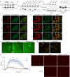
2.. Annexin-containing extracellular vesicles are shed from the repair scab after plasma membrane damage.
(A) Representative confocal micrographs of FM1–43 infiltration are shown. Image times are relative to the first image taken post ablation. Large arrows in pane I and II indicate the site of ablation. Small arrows in pane II and III indicate the staining of intracellular compartments in an ablated (lower arrows) and a non-ablated cell (upper arrows). Large arrows in pane III and IV indicate extracellular vesicles. Scale Bars: 5 μm. (B) Representative confocal micrographs of ANXA2-mScarlet recruitment after ablation are shown. Image times are relative to the first image taken post ablation. White arrows in pane I and II indicate the site of ablation. Arrow in pane III indicates an extracellular vesicle. Scale Bars: 5 μm. (C) Sytox Green staining over time in the absence of extracellular Ca2+ after cells pretreated with the indicated concentration of SLO were rapidly heated from 4°C to 37°C. (D) Membrane-protected luminescence in medium fractions at indicated time points after ANXA2-Nluc or CD63-Nluc cells pretreated with 200 ng/μl SLO were rapidly heated from 4°C to 37°C is shown. (E) EV production index from ANXA2-Nluc or CD63-Nluc cells treated with increasing concentrations of SLO is shown.
3.
3.. Annexins within extracellular vesicles are shifted in apparent molecular weight.
(A) Immunoblots of gradient fractions show enrichment of indicated EV markers after 200 ng/mL SLO treatment, with or without 1 mM Ca2+ in the media. F2, F3, and F4 refer to buoyant fractions of a sucrose step gradient of the conditioned medium 100k × g pellet fraction. (B) Immunoblot analysis of cell lysates after treatment with indicated combinations of 1 mM Ca2+ and 200 ng/mL SLO is shown. (C) Immunoblots show separation of indicated EV markers after 200 ng/mL SLO treatment. F2-F15 refer to fractions #2–15 of an iodixanol gradient of the conditioned medium 100k × g pellet fraction, moving from low to high density.
4.
4.. Calpain-1/2 cleave annexins which are then shed in microvesicles.
(A) Immunoblot analysis of cytosol fractions after incubation with or without 1 mM Ca2+. (B) Immunoblot analysis of cytosol fractions after incubation with or without 1 mM Ca2+, with a range of concentrations of calpastatin domain I inhibitor (Calpain Inh.). (C) In gel fluorescence of JF646-labeled, recombinant annexin A2-Halo incubated with indicated concentration of purified, porcine calpain-1. Arrows indicate uncleaved and cleaved product. (D) Mapping of tryptic peptides to the first 50 amino acids of annexin A2. Mass spectrometry analysis of recombinant annexin A2 (red text) is compared to tryptic digest-mass spectrometry analysis of recombinant annexin A2 treated with purified, porcine calpain-1 (yellow highlight). (E) Immunoblot analysis of cell lysate and conditioned medium 100k × g pellet fraction after treating cells with 200 ng/ml SLO in the presence or absence of 10 μM calpain inhibitor, ALLN. (F) Schematic illustrating the recombinant annexin A2-Halo reporter, labeled with a 5WS maleimide quencher on the N-terminus and a JF646 halo ligand on the C-terminus. (G) JF646 fluorescence of an in vitro reaction containing 5 μM self-quenched annexin A2 with or without 0.5 μM porcine calpain-1. (H) Representative confocal micrographs of dequenched annexin A2-Halo-JF646 fluorescence in cells. Times are relative to the first image taken post addition of 1 mM Ca2+. White arrows in pane II indicate puncta on the cell periphery. Arrow in pane III indicates an extracellular vesicle. Scale Bars: 10 μm. (I) Total protein (Sypro Ruby staining) and immunoblot analysis of substate trapping experiment, using 3xFlag (–), 3x-Flag C115S calpain-1 (dCAPN-I), or 3x-Flag C105S calpain-2 (dCAPN-II) as bait is shown. Arrows indicate calpain proteins. (J) Table listing proteins identified in EGTA elutions from 3xFlag, 3x-Flag C115S calpain-1 (dCAPN-I), or 3x-Flag C105S calpain-2 (dCAPN-II) capture experiments. Proteins are listed by Exponentially Modified Protein Abundance Index (emPAI) in the dCAPN-I elution. Keratin proteins were not included in the list.
5.
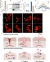
5.. Calpain cleavage attenuates the membrane binding and scabbing activity of annexin A2.
(A) Immunoblot analysis of lysates from cells expressing wildtype ANXA2-HA or ANXA2[P27D, P28D]-HA. Ca2+ (1 mM) was added to lysis buffer where indicated. (B) Immunoblot analysis of lysates from cells expressing wildtype ANXA2-HA or ANXA2-HA with the indicated N-terminal truncations. Ca2+ (1 mM) was added to lysis buffer where indicated. (C) Representative confocal micrographs of ANXA2-mScarlet (wt-mScarlet), ANXA2(18–339)-mNeonGreen (Tr-mNeonGreen)-expressing cells. Image times are relative to the addition of 400 ng/ml SLO. Scale Bars: 5 μm. (D) Immunoblot analysis of lysates and anti-HA immunoprecipitation elutions from cells expressing wildtype ANXA2-HA (wt-HA), ANXA2(18–339)-HA (Tr-HA), ANXA2[P27D, P28D]-HA (Mt-HA), or no HA construct. (E) Representative confocal micrographs of ANXA2-mScarlet (wt-mScarlet) or S100A10-mNeonGreen-expressing cells. Image times are relative to the addition of 400 ng/ml SLO. Arrows indicate cells with annexin A2 and S100A10 translocating to the membrane. Scale Bars: 5 μm. (F) Representative widefield micrographs of wildtype or annexin A2 knockout cells, with 2.5 μM Sytox Green added for 6 min. SLO (200 ng/ml) was added with Sytox Green in the indicated panels. Scale Bars: 100 μm. (G) Representative confocal micrographs of FM1–43 stained (2.5 μM) wildtype or annexin A2 knockout cells, 100 sec after laser ablation. Scale Bars: 5 μm. (H) Quantification over time of the repair scab intensity from FM1–43 stained (2.5 μM) wildtype or annexin A2 knockout cells. 6 cells were ablated for each condition. (I) Representative widefield micrographs of Texas Red-labeled 200 nm liposomes mixed with the indicated combinations of 300 nM annexin A2, annexin A2-S100A10, or Calpain-1.
6.
6.. Inhibition of annexin A2 cleavage decreases annexin A2+ microvesicle secretion during membrane repair.
(A) EV production index from ANXA2-Nluc cells treated with the indicated combinations of 200ng/ml SLO and 20 μM calpain Inhibitor, ALLN. (B) Immunoblot analysis of lysates from wildtype cells (wt), annexin A2 knockout cells (KO), and annexin A2 knockout cells expressing wildtype annexin A2-Nluc (KO + wt) or ANXA2[P27D, P28D]-Nluc (KO + Mt). (C) EV production index from wildtype ANXA2-Nluc (wt) or ANXA2[P27D, P28D]-Nluc (Mt) cells treated with or without SLO. (D) Quantification over time of the repair scab intensity from FM1–43 stained (2.5 μM) annexin A2 knockout cells rescued with ANXA2-mScarlet (wt) or ANXA2[P27D, P28D]-mScarlet (Mt). Cells (6) were ablated for each condition. (E) Confocal micrographs of 8 laser ablated annexin A2 knockout cells rescued with either wild-type ANXA2-mScarlet (wt) or ANXA2[P27D, P28D]-mScarlet (Mt). Images are 2 min, 30 sec after ablation. Scale Bars: 5 μm. (F) Schematic depicting the current model of plasma membrane repair and annexin+ MV secretion.

Similar articles

References

    1. Kalluri R., LeBleu V. S., The biology, function, and biomedical applications of exosomes. Science 367, eaau6977 (2020). - PMC - PubMed
    1. Jeppesen D. K., et al., Reassessment of Exosome Composition. Cell 177, 428–445.e18 (2019). - PMC - PubMed
    1. Koerdt S. N., Gerke V., Annexin A2 is involved in Ca2+-dependent plasma membrane repair in primary human endothelial cells. Biochim. Biophys. Acta Mol. Cell Res. 1864, 1046–1053 (2017). - PubMed
    1. Sønder S. L., et al., Annexin A7 is required for ESCRT III-mediated plasma membrane repair. Sci. Rep. 9, 6726 (2019). - PMC - PubMed
    1. McNeil P. L., Khakee R., Disruptions of muscle fiber plasma membranes. Role in exercise-induced damage. Am. J. Pathol. 140, 1097–1109 (1992). - PMC - PubMed

Publication types

LinkOut - more resources