. 2024 Sep 13:12:RP87935.
doi: 10.7554/eLife.87935.

Vangl2 suppresses NF-κB signaling and ameliorates sepsis by targeting p65 for NDP52-mediated autophagic degradation

Affiliations

Vangl2 suppresses NF-κB signaling and ameliorates sepsis by targeting p65 for NDP52-mediated autophagic degradation

Jiansen Lu et al. Elife. .

Abstract

Van Gogh-like 2 (Vangl2), a core planar cell polarity component, plays an important role in polarized cellular and tissue morphology induction, growth development, and cancer. However, its role in regulating inflammatory responses remains elusive. Here, we report that Vangl2 is upregulated in patients with sepsis and identify Vangl2 as a negative regulator of The nuclear factor-kappaB (NF-κB) signaling by regulating the protein stability and activation of the core transcription component p65. Mice with myeloid-specific deletion of Vangl2 (Vangl2ΔM) are hypersusceptible to lipopolysaccharide (LPS)-induced septic shock. Vangl2-deficient myeloid cells exhibit enhanced phosphorylation and expression of p65, therefore, promoting the secretion of proinflammatory cytokines after LPS stimulation. Mechanistically, NF-κB signaling-induced-Vangl2 recruits E3 ubiquitin ligase PDLIM2 to catalyze K63-linked ubiquitination on p65, which serves as a recognition signal for cargo receptor NDP52-mediated selective autophagic degradation. Taken together, these findings demonstrate Vangl2 as a suppressor of NF-κB-mediated inflammation and provide insights into the crosstalk between autophagy and inflammatory diseases.

Keywords: NF-κB signaling; PDLIM2; Vangl2; human; immunology; inflammation; mouse; selective autophagy; ubiquitin.


Conflict of interest statement

JL, JZ, HJ, ZH, YZ, LH, JY, YX, DW, HL, KZ, PT, QX, ZS, CP, XB, XY No competing interests declared

Figures

Figure 1.
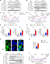
Figure 1.. Van Gogh-like 2 (Vangl2) ablation promotes inflammation during LPS treatment.
(A) Transcription levels of Vangl2 in PBMCs from healthy volunteers (healthy control, HC) and sepsis patients were analyzed by real-time PCR (n = 8). (B) Vangl2 mRNA in different organs from mice treated with or without LPS (n ≥ 3). (C) The survival rates of wild-type (WT) and Vangl2ΔM mice treated with high dosage of LPS (30 mg/kg, intraperitoneally [i.p.]) (n ≥ 3). (D–F) WT and Vangl2ΔM mice (n ≥ 3) were treated with LPS (30 mg/kg, i.p.). Splenocytes were collected at 9 hr after LPS treatment. Cell lysates of CD11b+ sorted splenocytes were analyzed by immunoblotting with the indicated antibodies (n ≥ 3) (D). RNAs from splenocytes were isolated and used for expression analysis of Il1b, Tnfa, and Il6 using qPCR (n ≥ 3) (E). Sera were collected at indicated times post LPS treatment and subjected to enzyme-linked immunosorbent assay (ELISA) analysis of IL-1β, tumor necrosis factor-α (TNF-α), and IL-6 (n ≥ 3) (F). PBMCs, peripheral blood mononuclear cells; Unsti, unstimulation; LPS, lipopolysaccharide; LN, lymph node; SP, spleen. Data are representative of three independent experiments and are plotted as the mean ± standard deviation (SD). Two-tailed Student's t test for A. Multiple t tests for B, E and F. Log-rank (Mantel-Cox) test for survival curve. *p < 0.05, **p < 0.01, ***p < 0.001 vs. corresponding control.
Figure 1—figure supplement 1.
Figure 1—figure supplement 1.. Expression of Van Gogh-like 2 (Vangl2) during sepsis and lipopolysaccharide (LPS) treatment.
(A) White blood cell (WBC) count, acute C-reactive protein (CRP), and IL-6 level in the peripheral blood mononuclear cell (PBMC) of healthy volunteers (HC) and sepsis patients (n ≥ 3). And WBC count, CPR, and IL-6 level in the PBMC of low Vangl2 group and high Vangl2 group (n ≥ 3). (B) Vangl2 expression in spleen of healthy volunteers and sepsis patients was analyzed by GEO (GSE69063, GSE145227, and GSE46955) analysis (n ≥ 5). (C–E) The mRNA and protein levels of Vangl2 in BMDMs, Neu, or pMac from wild-type (WT) mice after LPS treatment for the indicated times were detected (n ≥ 3). (F) Genotyping of transgenic mice by PCR and agarose gel electrophoresis. (G) Gross images of lymph nodes (LN) and spleens (SP) of WT and Vangl2ΔM mice. (H, I) Flow cytometry analysis of myeloid cell populations in the spleens of WT and Vangl2ΔM mice (n ≥ 3). BMDM, bone marrow-derived macrophage; Neu, neutrophil; pMac, peritoneal macrophage; Mon, monocyte. Data are representative of three independent experiments and are plotted as the mean ± standard deviation (SD). Two-tailed Student's t test for A and B. Multiple t tests for C, D, E, H and I. *p < 0.05, **p < 0.01, ***p < 0.001 vs. corresponding control.
Figure 2.
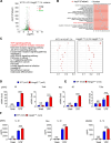
Figure 2.. Van Gogh-like 2 (Vangl2) negatively regulates lipopolysaccharide (LPS)-induced NF-κB activation and proinflammatory cytokines.
Wild-type (WT) and Vangl2-deficient (n ≥ 3) pMAC (A, C) or neutrophils (B, D) were stimulated with LPS (100 ng/ml) for the indicated times. Immunoblot analysis of total and phosphorylated p65, IKKα/β (A, B), and analysis of gray intensity was shown (C, D) (n ≥ 3). (E, F) WT and Vangl2-deficient (n ≥ 3) pMAC or neutrophils were stimulated with LPS (100 ng/ml) for 6 hr. mRNA levels of Il6 and Tnfa were measure by qPCR (E). IL-6 and tumor necrosis factor-α (TNF-α) secretion by WT and Vangl2-deficient bone marrow-derived macrophages (BMDMs) or neutrophils treated with or without LPS for 6 hr was measured by enzyme-linked immunosorbent assay (ELISA) (F) (n ≥ 3). (G, H) The WT and Vangl2-deficient (n ≥ 3) neutrophils were treated with LPS (1000 ng/ml) for 4 hr, and the nuclear translocation of p65 was detected by immunofluorescence (G) (p65, green; 4'-6-diamidino-2-phenylindole (DAPI), blue; scale bar, 50 μm). Percentages of p65 nuclear translocated cells in WT and Vangl2-deficient neutrophils were determined by counting 100–150 cells in non-overlapping fields (H) (n ≥ 3). (I, J) A549 cells were transfected with Flag-tagged Vangl2 plasmid or empty vector, then stimulated with LPS (100 ng/ml) for the indicated times. Immunoblot analysis of total and phosphorylated p65, IKKα/β (I) (representative image), and analysis of gray intensity was shown (J) (n ≥ 3). pMAC, peritoneal macrophage; Neu, neutrophil; EV, empty vector. Data are representative of three independent experiments and are plotted as the mean ± standard deviation (SD). Multiple t tests for C, D, E, F and J. Two-tailed Student's t test for H. *p < 0.05, ***p < 0.001 vs. corresponding control.
Figure 2—figure supplement 1.
Figure 2—figure supplement 1.. Van Gogh-like 2 (Vangl2) defection promotes lipopolysaccharide (LPS)-induced NF-κB activation and production of inflammatory cytokines.
Volcano plot (A), gene ontology (GO) analysis (B), and KEGG analysis (C) of differentially expressed genes in wild-type (WT) and Vangl2-deficient bone marrow-derived macrophages (BMDMs) after LPS treatment for 2 hr. (D, E) WT and Vangl2-deficient peritoneal macrophages and neutrophils were stimulated with LPS for 6 hr. mRNA levels of Il6 and Tnfa were measured by qPCR (n ≥ 3). (F) Enzyme-linked immunosorbent assay (ELISA) analysis of IL-1β in the supernatants of WT and Vangl2-deficient BMDMs, neutrophils, or peritoneal macrophages stimulated with LPS for 6 hr and ATP for another 0.5 hr. Data are representative of three independent experiments and are plotted as the mean ± standard deviation (SD). Multiple t tests for D-F. ***p < 0.001 vs. corresponding control.
Figure 3.
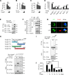
Figure 3.. Van Gogh-like 2 (Vangl2) inhibits NF-κB signaling by interacting with p65.
Cho (A) or HEK293T cells (B, C) were co-transfected with a NF-κB and TK-Renilla reporter along with increasing amounts of Vangl2 for 18 hr, then treated the cells with or without lipopolysaccharide (LPS) (A, 250 ng/ml), IL-1β (B, 40 ng/ml), or tumor necrosis factor-α (TNF-α) (C, 20 ng/ml) for 6 hr. NF-κB promoter-driven luciferase activity was measured and normalized to the Renilla luciferase activity (n ≥ 3). (D) Luciferase activity in HEK293T transfected with plasmids encoding an NF-κB luciferase reporter and TK-Renilla reporter, together with a vector encoding MyD88, IRAK1, TRAF6, IKKα, IKKβ, or p65, along with or without Vangl2 plasmid, was measured at 24 hr after transfection and normalized to the Renilla luciferase activity (n ≥ 3). (E) HEK293T cells were transfected with plasmids encoding HA-tagged Vangl2 and Flag-tagged p65, followed by immunoprecipitation with anti-Flag beads and immunoblot analysis with anti-HA. Throughout was the immunoblot analysis of whole-cell lysates without immunoprecipitation. (F) Bone marrow-derived macrophages (BMDMs) were stimulated with LPS (100 ng/ml) for the indicated times. The cell lysates were subjected to immunoprecipitation with an anti-p65 antibody or control IgG, followed by immunoblotting with indicated antibodies. (G) The wild-type (WT) and Vangl2-deficient peritoneal macrophages were treated with LPS (1000 ng/ml) for 4 hr, and co-localization of p65 and Vangl2 was detected by immunofluorescence (p65, green; Vangl2, red; DAPI, blue; scale bar, 50 μm). (H) A structural diagram of Vangl2 as well as schematic representation of Myc-tagged truncation mutants of Vangl2 (top). HEK293T cells were transfected with Flag-tagged p65 and empty vector, Myc-tagged Vangl2 (FL) or Vangl2 truncation mutants. The cell lysates were subjected to immunoprecipitation with anti-Flag beads and immunoblotted with the indicated antibodies (bottom). (I) HEK293T cells were transfected with Flag-tagged p65 and HA-tagged Vangl2 FL or PkBD truncation. The cell lysates were subjected to immunoprecipitation with anti-Flag beads and immunoblotted with the indicated antibodies. (J) Luciferase activity in HEK293T cells transfected with an NF-κB luciferase reporter, together with a vector encoding p65, along with the empty vector or with vectors encoding Vangl2 or its truncation mutants (n ≥ 3). The results are presented relative to Renilla luciferase activity. IP, immunoprecipitation; WCL, whole-cell lysate. Data are representative of three independent experiments and are plotted as the mean ± standard deviation (SD). Multiple t tests for A-D, and J. *p < 0.05, **p < 0.01, ***p < 0.001 vs. corresponding control. NS, not significant.
Figure 3—figure supplement 1.
Figure 3—figure supplement 1.. Van Gogh-like 2 (Vangl2) interacts with p65 to inhibit NF-κB activation.
Luciferase activity in HEK293T transfected with plasmids encoding an NF-κB luciferase reporter and TK-Renilla reporter, together with a vector encoding MyD88 (A), IRAK1 (B), TRAF6 (C), IKKα (D), IKKβ (E), or p65 (F), along with an increasing amount of Vangl2 (0, 250, 500, and 1000 ng), was measured at 24 hr after transfection and normalized to the Renilla luciferase activity (n ≥ 3). HEK293T cells were transfected with plasmids encoding HA-tagged Vangl2 and Flag-tagged key proteins in NF-κB signaling (Flag-IKKα, Flag-p65, Flag-TRAF6, Flag-IRAK1, Flag-MyD88 (G) and Flag-IKKβ (H)), followed by immunoprecipitation with anti-Flag beads and immunoblot analysis with anti-HA. Throughout was the immunoblot analysis of whole-cell lysates (WCL) without immunoprecipitation. (I) Bone marrow-derived macrophages (BMDMs) were stimulated with lipopolysaccharide (LPS) (100 ng/ml) for the indicated times. The cell lysates were subjected to immunoprecipitation with an anti-Vangl2 antibody or control IgG, followed by immunoblotting with the indicated antibodies. (J) Interacting domains of Vangl2 and p65 predicted by ZDOCK server. (K) Quantification of co-localization of p65 and Vangl2 in peritoneal macrophages (n ≥ 3). (L) THP-1 were stimulated with LPS (100 ng/ml) for 8 hr and the cells were isolated to cytoplasm and membrane fractions by kits. The cell lysates were subjected to immunoprecipitation with an anti-p65 antibody or control IgG, followed by immunoblotting with the indicated antibodies. (M) HEK293T cells were transfected with Flag-tagged p65 and empty vector, Myc-tagged Vangl2 (FL) or Vangl2 truncation mutants. The cell lysates were subjected to immunoprecipitation with anti-Flag beads and immunoblotted with the indicated antibodies. IP, immunoprecipitation; WCL, whole-cell lysate. Data are representative of three independent experiments. Multiple t tests for A-F. Two-tailed Student's t test for K. *p<0.05, **p<0.01, ***p<0.001 vs. corresponding control.
Figure 4.
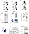
Figure 4.. Van Gogh-like 2 (Vangl2) promotes the autophagic degradation of p65.
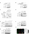
(A) Immunoblot analysis of HEK293T cells transfected with Flag-p65 and increasing amounts of the vector encoding HA-Vangl2 (0, 250, 500, and 1000 ng) (n ≥ 3). (B) Total RNA from HEK293T cells as in (A) was isolated and measured by semi-quantitative PCR. (C) HEK293T cells transfected with Flag-p65 and increasing amounts of the vector encoding HA-Vangl2, and the expressions of p65 in nuclear or cytoplasm were detected by immunoblot. (D) Wild-type (WT) and Vangl2-deficient bone marrow-derived macrophages (BMDMs) were treated with lipopolysaccharide (LPS) for the indicated times, and the expressions of p65 and Vangl2 were detected by immunoblot (n ≥ 3). (E) HEK293T cells were transfected with Flag-p65 and HA-Vangl2 plasmids, and treated with DMSO, MG132 (10 μM), CQ (50 μM), 3-MA (10 mM), or Baf-A1 (0.2 μM) for 6 hr. The cell lysates were analyzed by immunoblot (n ≥ 3). (F) HEK293T cells were transfected with empty vector (EV) or Flag-Vangl2 plasmid, and treated with rapamycin for the indicated times. The cell lysates were analyzed by immunoblot with indicated antibodies (n ≥ 3). (G) WT, ATG5 knockout (KO), and Beclin1 KO HEK293T cells were transfected with Flag-p65, together with or without HA-Vangl2 plasmids, and then the cell lysates were analyzed by immunoblot with indicated antibodies. Luciferase activity in WT, ATG5 KO (H) and Beclin1 KO (I) HEK293T cells transfected with plasmids encoding an NF-κB luciferase reporter and TK-Renilla reporter, together with p65 plasmid along with increasing amounts of Vangl2, was measured at 24 hr after transfection and normalized to the Renilla luciferase activity (n ≥ 3). CHX, cycloheximide; 3-MA, 3-methyladenine; CQ, chloroquine; Baf A1, bafilomycin A1. Data are representative of three independent experiments and are plotted as the mean ± standard deviation (SD). Multiple t tests for A, D, E, F, H and I. ***p < 0.001 vs. corresponding control. NS, not significant.
Figure 4—figure supplement 1.
Figure 4—figure supplement 1.. Van Gogh-like 2 (Vangl2) promotes p65 degradation by autophagic pathway.
(A) Immunoblot analysis of HEK293T cells transfected with HA-p65 (wild-type [WT] or S536A) and Flag-Vangl2. (B) Immunoblot analysis of HEK293T cells transfected with HA-p65 and Flag-Vangl2 (S82/84A). (C) HEK293T cells were transfected with Flag-tagged Vangl2 and empty vector, HA-tagged p65. The cells were isolated to cytoplasm, membrane, and nucleus fractions by kits. Cell lysates were subjected to immunoprecipitation with anti-Flag beads and immunoblotted with the indicated antibodies. (D) WT and Vangl2-deficient bone marrow-derived macrophages (BMDMs) were pretreated with lipopolysaccharide (LPS), then treated with cycloheximide (CHX) for the indicated times, and the expressions of p65 and Vangl2 were detected by immunoblot. (E) HEK293T cells were transfected with Flag-p65, together with or without HA-Vangl2 plasmids, and treated with DMSO, MG132 (10 μM), chloroquine (CQ) (50 μM), 3-methyladenine (3-MA) (10 mM), VX-765 (10 mM), or Z-VAD (0.2 μM) for 6 hr. The cell lysates were analyzed by immunoblot with indicated antibodies. WT and ATG5 knockout (KO) (F) or Beclin1 KO (G) HEK293T cells were treated with CHX for the indicated times, and then the cell lysates were analyzed by immunoblot with indicated antibodies. (H) Confocal microscopy of WT BMDMs treated with phosphate-buffered saline (PBS) and LPS. Statistics shown refer to the puncta formation by LC3-p65 in the indicated samples (n ≥ 3). Scale bar, 50 μm. (p65, green; LC3, red; DAPI, blue). Data are representative of three independent experiments and are plotted as the mean ± standard deviation (SD). Two-tailed Student's t test for H. ***p < 0.001 vs. corresponding control.
Figure 5.
Figure 5.. Van Gogh-like 2 (Vangl2) enhances the recognition of p65 by cargo receptor NDP52.
(A) HEK293T cells transfected with a vector expressing HA-Vangl2 along with the empty vector or vector encoding Flag-p62/NDP52/NBR1/Nix. The cell lysates were subjected to immunoprecipitation with anti-Flag beads and immunoblotted with the indicated antibodies. (B) HEK293T cells transfected with a vector expressing HA-p65 along with the empty vector or vector encoding Flag-p62/NDP52/NBR1/Nix. The cell lysates were subjected to immunoprecipitation with anti-Flag beads and immunoblotted with the indicated antibodies. (C) HEK293T cells were transfected with HA-p65 together with Flag-NDP52 or Flag-p62, as well as with empty vector or Myc-Vangl2. The cell lysates were subjected to immunoprecipitation with anti-Flag beads and immunoblotted with the indicated antibodies. (D) Wild-type (WT), NDP52 knockout (KO), and p62 KO HEK293T cells were transfected with a vector expressing HA-p65 along with the empty vector or vector encoding Flag-Vangl2. The cell lysates were immunoblotted with the indicated antibodies (n ≥ 3). (E) WT and Vangl2-deficient bone marrow-derived macrophages (BMDMs) were stimulated with lipopolysaccharide (LPS) (100 ng/ml) for the indicated times. The cell lysates were subjected to immunoprecipitation with an anti-p65 antibody or control IgG and immunoblotted with the indicated antibodies. (F) WT and NDP52 KO HEK293T cells were transfected with a vector expressing HA-p65 along with the empty vector or vector encoding Flag-Vangl2. The cell lysates were subjected to immunoprecipitation with anti-Flag beads and immunoblotted with the indicated antibodies. (G) Luciferase activity in WT and NDP52 KO HEK293T cells transfected with plasmids encoding NF-κB luciferase reporter and TK-Renilla reporter, together with p65 plasmid along with increasing amounts of Vangl2 plasmid, was measured at 24 hr after transfection (n ≥ 3). (H) WT and NDP52 KO HEK293T were treated with cycloheximide (CHX) for the indicated times. The cell lysates were immunoblotted with the indicated antibodies (n ≥ 3). Data are representative of three independent experiments and are plotted as the mean ± standard deviation (SD). Multiple t tests for D, G and H. ***p < 0.001 vs. corresponding control. NS, not significant.
Figure 5—figure supplement 1.
Figure 5—figure supplement 1.. Van Gogh-like 2 (Vangl2) promoted autophagic degradation of p65 is not mediated by cargo receptor p62.
(A) Wild-type (WT) and p62 knockout (KO) HEK293T cells transfected with a vector expressing HA-p65 along with the empty vector or vector encoding Flag-Vangl2. The cell lysates were subjected to immunoprecipitation with anti-Flag beads and immunoblotted with the indicated antibodies. (B) Luciferase activity in WT and p62 KO HEK293T cells transfected with plasmids encoding an NF-κB luciferase reporter and TK-Renilla reporter, together with p65 plasmid along with increasing amounts of Vangl2 plasmid, was measured at 24 hr after transfection (n ≥ 3). (C) WT and p62 KO HEK293T were treated with cycloheximide (CHX) for the indicated times. The cell lysates were immunoblotted with the indicated antibodies. Data are representative of three independent experiments and are plotted as the mean ± standard deviation (SD). Multiple t tests for B. ***p < 0.001 vs. corresponding control.
Figure 6.
Figure 6.. Van Gogh-like 2 (Vangl2) increases the K63-linked ubiquitination of p65.
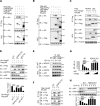
(A) Wild-type (WT) and Vangl2-deficient bone marrow-derived macrophages (BMDMs) were stimulated with lipopolysaccharide (LPS) (100 ng/ml) for the indicated times. The cell lysates were subjected to immunoprecipitation with an anti-p65 antibody or control IgG and immunoblotted with the indicated antibodies. (B) HEK293T cells were transfected with Flag-p65, Myc-Vangl2, HA-Ub, or HA-K63 plasmids with the indicated combinations for 24 hr and then treated with chloroquine (CQ) for 8 hr. The cell lysates were subjected to immunoprecipitation with anti-Flag beads and immunoblotted with the indicated antibodies. (C) HEK293T cells were transfected with Flag-p65, Myc-Vangl2, and HA-Ub/K63/K11/K27/K33/K48 plasmids with the indicated combinations for 24 hr and then treated with CQ and Baf-A1 for 8 hr. The cell lysates were subjected to immunoprecipitation with anti-Flag beads and immunoblotted with the indicated antibodies. (D) HEK293T cells were transfected with a vector expressing Flag-p65 and HA-K63 along with Scramble or Vangl2 small interfering RNA (siRNA). The cell lysates were subjected to immunoprecipitation with anti-Flag beads and immunoblotted with the indicated antibodies. Data are representative of three independent experiments.
Figure 7.
Figure 7.. Van Gogh-like 2 (Vangl2) recruits PDLIM2 to ubiquitinate p65.
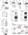
(A) HEK293T cells were transfected with the indicated small interfering RNA (siRNA), NF-κB reporter plasmids together with HA-Vangl2, Flag-p65, or the control vector as indicated for 24 hr, and then subjected to luciferase assay and immunoblotting analysis (n ≥ 3). Bone marrow-derived macrophages (BMDMs) were transfected with Pdlim2 or Scramble siRNA along with the empty vector or vector encoding Flag-Vangl2, stimulated with lipopolysaccharide (LPS) (100 ng/ml) for 6 hr, then analyzed by qPCR for Il6 (B) and Il1b (C) expression (n ≥ 3). (D) HEK293T cells transfected with HA- PDLIM2 along with the empty vector or vector encoding Flag-Vangl2. The cell lysates were subjected to immunoprecipitation with anti-Flag beads and immunoblotted with the indicated antibodies. (E) HEK293T cells were transfected with Flag-p65, HA-PDLIM2 and Myc-Vangl2 plasmids with the indicated combinations for 24 hr and then treated with chloroquine (CQ) and Baf-A1 for 8 hr. The cell lysates were subjected to immunoprecipitation with anti-Flag beads and immunoblotted with the indicated antibodies. (F) HEK293T cells were transfected with Flag-p65, HA-PDLIM2, and Myc-Vangl2 plasmids with the indicated combinations for 24 hr. The cell lysates were immunoblotted with the indicated antibodies (n ≥ 3). (G) HEK293T cells were transfected with Flag-p65, HA-K63, and Myc-Vangl2 plasmids, the expression of E3 ubiquitin ligase was interfered with Pdlim2 siRNA and then treated with CQ and Baf-A1 for 8 hr. The cell lysates were subjected to immunoprecipitation with anti-Flag beads and immunoblotted with the indicated antibodies. (H) HEK293T cells were transfected with Pdlim2 or Scramble siRNA, along with or without HA-PDLIM2, then treated with CQ and Baf-A1 for 8 hr. The cell lysates were subjected to immunoprecipitation with anti-Flag beads and immunoblotted with the indicated antibodies. (I) A schematic model to illustrate how Vangl2-PDLIM2-NDP52-p65 axis negatively regulates NF-κB activation. During LPS stimulation, Vangl2 expression is upregulated, thus constituting a negative feedback loop to regulate NF-κB activation. In detail, Vangl2 functions as an adaptor protein to recruit an E3 ubiquitin ligase PDLIM2 to increase K63-linked ubiquitination of p65 and promotes NDP52-mediated p65 degradation through selective autophagy, resulting in ameliorating sepsis and suppressing production of proinflammatory cytokines. Data are representative of three independent experiments and are plotted as the mean ± standard deviation (SD). Multiple t tests for A-C, and F. ***p < 0.001 vs. corresponding control. NS, not significant.
Figure 7—figure supplement 1.
Figure 7—figure supplement 1.. Expression of candidate E3 ubiquitin ligases in wild-type (WT) and Van Gogh-like 2 (Vangl2)-deficient bone marrow-derived macrophages (BMDMs) after lipopolysaccharide (LPS) treatment.
(A) Heatmap view of mRNA variations of E3 ubiquitin ligases in WT and Vangl2-deficient BMDMs treated with or without LPS. mRNA levels of Pdlim2 (B), Usp7 (C), and Trim21 (D) in the spleens from LPS-treated WT and Vangl2△M mice were detected by qPCR (n ≥ 3). mRNA levels of Pdlim2 (E), Usp7 (F), and Trim21 (G) in HEK293T after transfection with Pdlim2, Usp7, and Trim21 small interfering RNA (siRNA) (n ≥ 3). Data are representative of three independent experiments and are plotted as the mean ± standard deviation (SD). Multiple t tests for B-D. Two-tailed Student's t test for E-G. **p < 0.01, ***p < 0.001 vs. corresponding control. NS, not significant.
Author response image 1.
Author response image 1.. Vangl2 degrades p65 through a selective autophagic pathway, but not by the PCP pathway.
HEK293T cells were transfected with Flag-p65 and HA-Vangl2 plasmids, and treated with DMSO, CQ (50 mM) for 6 h, SP600125 (20 mM) for 1 h or FH535 (30 mM) for 6 h. The cell lysates were analyzed by immunoblot.
Author response image 2.
Author response image 2.. Vangl2 deficiency promotes NF-kB activation.
(A) The survival rates of WT, Vangl2ΔM/ΔM and Vangl2ΔM/WT mice treated with high-dosage of LPS (30 mg/kg, i.p.) (n≥4). (B) IL-6 and TNF-a secretion by WT and Vangl2-deficient BMDMs treated with LPS for 6 h was measured by ELISA. IL-1β secretion by WT, Vangl2ΔM/ΔM and Vangl2ΔM/WT BMDMs treated with LPS for 6 h and ATP for 30 min was measured by ELISA.

Update of

  • doi: 10.1101/2023.03.30.534894
  • doi: 10.7554/eLife.87935.1
  • doi: 10.7554/eLife.87935.2
  • doi: 10.7554/eLife.87935.3

Similar articles

Cited by

References

    1. Ashrafi G, Schwarz TL. The pathways of mitophagy for quality control and clearance of mitochondria. Cell Death and Differentiation. 2013;20:31–42. doi: 10.1038/cdd.2012.81. - DOI - PMC - PubMed
    1. Bailly E, Walton A, Borg JP. The planar cell polarity Vangl2 protein: From genetics to cellular and molecular functions. Seminars in Cell & Developmental Biology. 2018;81:62–70. doi: 10.1016/j.semcdb.2017.10.030. - DOI - PubMed
    1. Bartuzi P, Hofker MH, van de Sluis B. Tuning NF-κB activity: a touch of COMMD proteins. Biochimica et Biophysica Acta. 2013;1832:2315–2321. doi: 10.1016/j.bbadis.2013.09.014. - DOI - PubMed
    1. Brunt L, Greicius G, Rogers S, Evans BD, Virshup DM, Wedgwood KCA, Scholpp S. Vangl2 promotes the formation of long cytonemes to enable distant Wnt/β-catenin signaling. Nature Communications. 2021;12:2058. doi: 10.1038/s41467-021-22393-9. - DOI - PMC - PubMed
    1. Cao Y, Sun Y, Chang H, Sun X, Yang S. The E3 ubiquitin ligase RNF182 inhibits TLR-triggered cytokine production through promoting p65 ubiquitination and degradation. FEBS Letters. 2019;593:3210–3219. doi: 10.1002/1873-3468.13583. - DOI - PubMed

MeSH terms