Veillonella parvula as an anaerobic lactate-fermenting bacterium for inhibition of tumor growth and metastasis through tumor-specific colonization and decrease of tumor's lactate level
- PMID: 39251652
- PMCID: PMC11385575
- DOI: 10.1038/s41598-024-71140-9
Veillonella parvula as an anaerobic lactate-fermenting bacterium for inhibition of tumor growth and metastasis through tumor-specific colonization and decrease of tumor's lactate level
Abstract
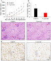
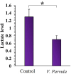
High tumor's lactate level directly associates with high tumor growth, metastasis, and patients' poor prognosis. Therefore, many studies have focused on the decrease of tumor's lactate as a novel cancer treatment. In the present study for the first time, a strictly anaerobic lactate-fermenting bacterium, Veillonella parvula, was employed for the decrease of tumor's lactate level. At first, 4T1 breast tumor-bearing BALB/c mice were administered with 106 V. parvula bacteria intravenously, orally, intraperitoneally, and intratumorally. Then, the bacteria biodistribution was evaluated. The best administration route according to tumor colonization was selected and its safety was assessed. Then, the therapeutic effect of V. parvula administration through the best route was investigated according to 4T1 murine breast tumor's growth and metastasis in vivo. In addition, histopathological and immunohistochemistry evaluations were done to estimate microscopic changes at the inner of the tumor and tumor's lactate level was measured after V. parvula administration. V. parvula exhibited considerable tumor-targeting and colonization efficacy, 24 h after intravenous administration. Normal organs were free of the bacteria after 72 h and no side effect was observed. Tumor colonization by V. parvula significantly decreased the tumors' lactate level for about 46% in comparison with control tumors which caused 44.3% and 51.6% decline (P < 0.05) in the mean tumors' volume and liver metastasis of the treatment group in comparison with the control group, respectively. The treatment group exhibited 35% inhibition in the cancer cell proliferation in comparison with the control according to the Ki-67 immunohistochemistry staining. Therefore, intravenous administration of V. parvula is a tumor-specific and safe treatment which can significantly inhibit tumors' growth and metastasis by decreasing the tumor lactate level.
Keywords: Veillonella parvula; Bacteria therapy; Breast cancer; Lactate.
© 2024. The Author(s).
Conflict of interest statement
The authors declare no competing interests.
Figures


Similar articles
-
Spirulina extract enriched for Braun-type lipoprotein (Immulina®) for inhibition of 4T1 breast tumors' growth and metastasis.Phytother Res. 2020 Feb;34(2):368-378. doi: 10.1002/ptr.6527. Epub 2019 Nov 5. Phytother Res. 2020. PMID: 31691383
-
Inflammation-associated nitrate facilitates ectopic colonization of oral bacterium Veillonella parvula in the intestine.Nat Microbiol. 2022 Oct;7(10):1673-1685. doi: 10.1038/s41564-022-01224-7. Epub 2022 Sep 22. Nat Microbiol. 2022. PMID: 36138166 Free PMC article.
-
Autotransporters Drive Biofilm Formation and Autoaggregation in the Diderm Firmicute Veillonella parvula.J Bacteriol. 2020 Oct 8;202(21):e00461-20. doi: 10.1128/JB.00461-20. Print 2020 Oct 8. J Bacteriol. 2020. PMID: 32817093 Free PMC article.
-
A new treatment of sepsis caused by veillonella parvula: A case report and literature review.J Clin Pharm Ther. 2017 Oct;42(5):649-652. doi: 10.1111/jcpt.12559. Epub 2017 May 23. J Clin Pharm Ther. 2017. PMID: 28543519 Review.
-
Epidural abscess caused by Veillonella parvula: Case report and review of the literature.J Microbiol Immunol Infect. 2016 Oct;49(5):804-808. doi: 10.1016/j.jmii.2014.05.002. Epub 2014 Jul 25. J Microbiol Immunol Infect. 2016. PMID: 25066704 Review.
References
-
- Bisoyi, P. A brief tour guide to cancer disease. In Understanding Cancer 1–20 (Elsevier, 2022).
MeSH terms
Substances
Grants and funding
LinkOut - more resources
Full Text Sources

