. 2024 Sep 5;19(1):77.
doi: 10.1186/s13062-024-00524-8.

GALNT9 enrichment attenuates MPP+-induced cytotoxicity by ameliorating protein aggregations containing α-synuclein and mitochondrial dysfunction

Affiliations

GALNT9 enrichment attenuates MPP+-induced cytotoxicity by ameliorating protein aggregations containing α-synuclein and mitochondrial dysfunction

Yuanwen Peng et al. Biol Direct. .

Abstract

Background: GALNTs (UDP-GalNAc; polypeptide N-acetylgalactosaminyltransferases) initiate mucin-type O-GalNAc glycosylation by adding N-GalNAc to protein serine/threonine residues. Abnormalities in O-GalNAc glycosylation are involved in various disorders such as Parkinson's disease (PD), a neurodegenerative disorder. GALNT9 is potentially downregulated in PD patients.

Methods: To determine whether GALNT9 enrichment ameliorates cytotoxicity related to PD-like variations, a pcDNA3.1-GALNT9 plasmid was constructed and transfected into SH-SY5Y cells to establish a GALNT9-overexpressing cell model.

Results: Downregulation of GALNT9 and O-GalNAc glycosylation was confirmed in our animal and cellular models of PD-like variations. GALNT9 supplementation greatly attenuated cytotoxicity induced by MPP+ (1-Methyl-4-phenylpyridinium iodide) since it led to increased levels of tyrosine hydroxylase and dopamine, reduced rates of apoptosis, and significantly ameliorated MPP+-induced mitochondrial dysfunction by alleviating abnormal levels of mitochondrial membrane potential and reactive oxygen species. A long-lasting mPTP (mitochondrial permeability transition pores) opening and calcium efflux resulted in significantly lower activity in the cytochrome C-associated apoptotic pathway and mitophagy process, signifying that GALNT9 supplementation maintained neuronal cell health under MPP+ exposure. Additionally, it was found that glycans linked to proteins influenced the formation of protein aggregates containing α-synuclein, and GALNT9 supplement dramatically reduced such insoluble protein aggregations under MPP+ treatment. Glial GALNT9 predominantly appears under pathological conditions like PD-like variations.

Conclusions: GALNT9 enrichment improved cell survival, and glial GALNT9 potentially represents a pathogenic index for PD patients. This study provides insights into the development of therapeutic strategies for the treatment of PD.

Keywords: GALNT; Glycosylation; Mitochondrial dysfunction; Parkinson’s disease; Α-synuclein.


Conflict of interest statement

The authors declare no competing interests.

Figures

Fig. 1
Fig. 1
Evaluation of levels of GALNT9 and its catalytic products by western blot assay. Examinations were performed in triplicate at least. M, PageRuler prestained protein ladder (Fermentas); *, P < 0.05; **, P < 0.01; ***, P < 0.001. (A) Representative images illustrating levels of DBA-recognized proteins. The probe was biotinylated DBA. (B) Representative images illustrating GALNT9 expression. The probe was a monoclonal anti-GALNT9 antibody. (C) Statistical analysis of DBA-recognized proteins normalized to GAPDH abundance. (D) Statistical analysis of soluble GALNT9 normalized to GAPDH levels
Fig. 2
Fig. 2
Identification of overexpressed GALNT9 in SH-SY5Y cells. Examinations were performed in triplicate at least. M, PageRuler prestained protein ladder (Fermentas). *, P < 0.05; **, P < 0.01; ***, P < 0.001. (A) Representative images illustrating cellular glycoproteins with GalNAc moiety by western blot assays. The probe was biotinylated DBA. (B) Statistical analysis of cellular glycoproteins with GalNAc moiety normalized to GAPDH levels. (C) Representative images illustrating GALNT9 expression by western blot assays. The probe was anti-GALNT9 monoclonal antibody. (D) Statistical analysis of GALNT9 normalized to GAPDH abundance. (E) Identification of plasmid pcDNA3.1-GALNT9. The plasmid was digested with XhoI and HindIII. (F) Abundance analysis of GALNT9 in SH-SY5Y cells. The probe was anti-GALNT9 monoclonal antibody. (G) Statistical analysis of GALNT9 abundance in SH-SY5Y cells normalized to GAPDH levels. (H) Abundance analysis of glycoproteins with GalNAc moiety in SH-SY5Y cells. The probe was biotin-labeled DBA. (I) Statistical analysis of glycoproteins with GalNAc moiety in SH-SY5Y cells normalized to GAPDH levels
Fig. 3
Fig. 3
Influence of GALNT9 on cytotoxicity in SH-SY5Y cells. Examinations were performed in triplicate at least. M, PageRuler prestained protein ladder (Fermentas); Black asterisks represent comparison between MPP+-treated and untreated groups; Red asterisks indicate comparison between GALNT9-related groups and controls. *, P < 0.05; **, P < 0.01; ***, P < 0.001. (A) Abundance analysis of TH in SH-SY5Y cells using western blot assays. Polyclonal anti-TH antibody was used as a probe. (B) Statistical analysis of intracellular TH levels in SH-SY5Y cells. (C) Statistical analysis of intracellular DA abundance in SH-SY5Y cells. (D) Apoptosis analysis of SH-SY5Y cells using an Annexin V-FITC/PI apoptosis detection kit
Fig. 4
Fig. 4
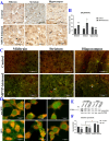
Influence of GALNT9 on protein aggregates containing DBA-recognized proteins and α-synuclein. Examinations were performed in triplicate at least. *, P < 0.05; **, P < 0.01; ***, P < 0.001. (A) Analysis of total GALNT9 using IHC assays. An anti-GALNT9 monoclonal antibody was used as the probe. The black arrows indicate GALNT9 in glial cells. The micrograph was 558.8 mm × 558.8 mm (B) Statistical analysis of total GALNT9 detected by IHC. (C) Dual immunofluorescence staining of DBA-recognized glycoproteins with α-synuclein in vivo. The micrograph was 1731.43 mm × 1151.47 mm. Red signals corresponded to DBA-recognized glycoproteins. Green signals corresponded to α-synuclein. Colocalization of DBA-recognized glycoproteins with α-synuclein is depicted as the juxtaposed images in green and red. Dual immunolabelling of DBA-recognized glycoproteins/α-synuclein visible as bright orange or yellow indicated by white tips. (D) Dual immunofluorescence staining of DBA-recognized glycoproteins with α-synuclein in vitro. The micrograph was 357.72 mm × 357.72 mm (E) Abundance analysis of cellular α-synuclein by western blot assay. The probe was a monoclonal antibody against α-synuclein. M, PageRuler prestained protein ladder (Fermentas). (F) Statistical analysis of soluble α-synuclein levels
Fig. 5
Fig. 5
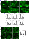
Influence of GALNT9 on energy generation. Examinations were performed in triplicate at least. (A) Representative micrographs of intracellular ROS determined by fluorescence microscopy. The images were 1731.43 mm × 1151.47 mm. Bright green signals represent positive areas of ROS. (B) Evaluation of ΔΨm by flow cytometry. JC-1 staining was used. The rates of FITC-positive cells were calculated to indicate a low ΔΨm. (C) Representative micrographs of cytoskeletal actin filaments and statistical analysis of fluorescence intensity. The dimension of micrographs was 529.17 mm × 529.17 mm. The cytoskeleton was stained with FITC-phalloidin (green), and images were captured using fluorescence a microscope. *, P < 0.05; **, P < 0.01; ***, P < 0.001
Fig. 6
Fig. 6
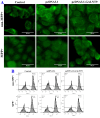
Evaluation of GALNT9 on mitochondrial membrane permeability. (A) Representative images of mPTPs measured by fluorescence microscopy. A fluorogenic dye of calcein-AM/cobalt was used. The dimension of images was 429.33 mm × 429.33 mm. Examinations were performed in triplicate at least. The marked circular distribution of bright green fluorescent signals indicates mPTPs in a closed configuration. (B) Representative images of cytosolic Ca2+ measured by flow cytometry. Examinations were performed in quadruplicate. Fluo-4 AM was used as a probe. The rates of FITC-positive cells (which indicate leakage of Ca2+) are shown
Fig. 7
Fig. 7
Evaluation of mitophagy and PCD involved in CytC-associated apoptotic pathway. (A) Evaluation of molecules in CytC-associated apoptotic pathway by western blot assay. M, PageRuler prestained protein ladder (Yamei WJ103). Protein bands were normalized to GAPDH levels. Examinations was performed in triplicate at least. (B) Statistical analysis of expression of molecules including CytC, Apaf1, CASP9, and CASP3, respectively. Red asterisks represent comparison between GALNT9-regulated groups and controls, and black asterisks represent comparison between MPP+-treated and -untreated groups. *, P < 0.05; **, P < 0.01; ***, P < 0.001. (C) Dual immunofluorescence staining of mitophagy. The dimension of micrographs was 1,731.43 mm × 1,151.47 mm. Green signals correspond to lysosomes. Red signals correspond to mitochondria. Mitochondrial fission is depicted as the juxtaposed images of green and red: dual immunolabelling of mitochondria/lysosomes is visible as orange or yellow indicated by white tips

Similar articles

Cited by

References

    1. Wandall HH, Nielsen MAI, King-Smith S, de Haan N, Bagdonaite I. Global functions of O‐glycosylation: promises and challenges in O‐glycobiology. FEBS J. 2021;288(24):7183–212. 10.1111/febs.16148 - DOI - PubMed
    1. Brockhausen I, Stanley P. O-GalNAc glycans–essentials of glycobiology. Cold Spring Harbor (NY):Cold Spring Harbor Lab Press. 2017;3rd edition:Chap10.
    1. Mereiter S, Balmaña M, Campos D, Gomes J, Reis CA. Glycosylation in the era of cancer-targeted therapy: where are we heading? Cancer Cell. 2019;36(1):6–16. 10.1016/j.ccell.2019.06.006 - DOI - PubMed
    1. Bennett EP, Mandel U, Clausen H, Gerken TA, Fritz TA, Tabak LA. Control of mucin-type O-glycosylation: a classification of the polypeptide GalNAc-transferase gene family. Glycobiology. 2011;22(6):736–56. 10.1093/glycob/cwr182 - DOI - PMC - PubMed
    1. Bloem BR, Okun MS, Klein C. Parkinson’s disease. Lancet. 2021;397(10291):2284–303. 10.1016/S0140-6736(21)00218-X - DOI - PubMed

MeSH terms

Substances

LinkOut - more resources