Review
. 2024 Sep 4;17(1):81.
doi: 10.1186/s13045-024-01601-1.

Heat shock proteins as hallmarks of cancer: insights from molecular mechanisms to therapeutic strategies

Affiliations
Review

Heat shock proteins as hallmarks of cancer: insights from molecular mechanisms to therapeutic strategies

Wei-Fang Zuo et al. J Hematol Oncol. .

Abstract

Heat shock proteins are essential molecular chaperones that play crucial roles in stabilizing protein structures, facilitating the repair or degradation of damaged proteins, and maintaining proteostasis and cellular functions. Extensive research has demonstrated that heat shock proteins are highly expressed in cancers and closely associated with tumorigenesis and progression. The "Hallmarks of Cancer" are the core features of cancer biology that collectively define a series of functional characteristics acquired by cells as they transition from a normal state to a state of tumor growth, including sustained proliferative signaling, evasion of growth suppressors, resistance to cell death, enabled replicative immortality, the induction of angiogenesis, and the activation of invasion and metastasis. The pivotal roles of heat shock proteins in modulating the hallmarks of cancer through the activation or inhibition of various signaling pathways has been well documented. Therefore, this review provides an overview of the roles of heat shock proteins in vital biological processes from the perspective of the hallmarks of cancer and summarizes the small-molecule inhibitors that target heat shock proteins to regulate various cancer hallmarks. Moreover, we further discuss combination therapy strategies involving heat shock proteins and promising dual-target inhibitors to highlight the potential of targeting heat shock proteins for cancer treatment. In summary, this review highlights how targeting heat shock proteins could regulate the hallmarks of cancer, which will provide valuable information to better elucidate and understand the roles of heat shock proteins in oncology and the mechanisms of cancer occurrence and development and aid in the development of more efficacious and less toxic novel anticancer agents.

Keywords: Cancer; Combination strategy; Dual inhibitors; Hallmarks of cancer; Heat shock protein; Target therapy.


Conflict of interest statement

The authors declare no competing interests.

Figures

Fig. 1
Fig. 1
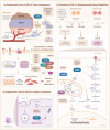
HSPs have been implicated in the hallmarks of cancer and exploited for developing targeted therapeutic strategies. In 2000, Hanahan and Weinberg delineated six hallmarks of cancer, including sustaining proliferative signaling, evading growth suppressors, resisting cell death, enabling replicative immortality, inducing angiogenesis, and activating invasion and metastasis, which are described as the acquired capabilities that allow cancer cells to form malignant tumors (depicted in the figure as the "original hallmarks"). Subsequently, they introduced two emerging hallmarks in 2011 and 2022, respectively, which include reprogramming energy metabolism and evading immune destruction, as well as the acquisition of unlocking phenotypic plasticity and senescence. Among these, the roles of unlocking phenotypic plasticity and senescence in cancer are still under validation and thus not represented here. HSPs have been demonstrated to play a significant role in modulating original and emerging hallmarks
Fig. 2
Fig. 2
Classification and structural characteristics of HSPs. a Structure of sHSPs (PDB ID: 2YGD) is exemplified by the monomeric structure of the human 24-meric eye lens chaperone alphaB-crystallin, which includes the C-terminal extension (red), the α-crystallin domain (green), and the N-terminal domain (NTD) (blue). b Structure of HSP40 illustrated by the Thermus thermophilus type B Hsp40 (PDB ID: 6PSI), comprising the J-domain (blue), the G/F-rich region (green), the C-terminal binding domain (yellow and red), and the dimerization domain (purple). c Structure of HSP60 (PDB ID: 7AZP) illustrated by the monomeric structure of the human mitochondrial HSPD1, consisting of the apical domain (blue), the intermediate domain (yellow), and the equatorial domain (red). d Structure of HSP70 (PDB ID: 4B9Q) is composed of the N-terminal nucleotide-binding domain (NBD) (blue), the C-terminal substrate-binding domain (SBD) (red), and a linker (yellow). The SBD is subdivided into a β-sheet-rich base and an α-helix-rich lid. e Structure of the HSP90 dimer (PDB ID: 5FWK) primarily comprises the NTD (red), the middle domain (yellow), and the C-terminal domain (CTD) (blue). f Structure of HSP110 (PDB ID: 6GFA) mainly consists of the NBD and an incompletely characterized SBD, with the NBD containing four subdomains: IA (red), IB (blue), IIA (purple), and IIB (green)
Fig. 3
Fig. 3
Diagram of HSPs in the core proliferative signaling pathway in cancer. Upon ligand binding, receptor tyrosine kinases (RTKs) activate Ras through the adaptor proteins Grb2 and SOS, facilitating the transition of Ras from its GDP-bound to GTP-bound state. Subsequently, Raf kinase is activated, leading to the activation of MEK and ERK1/2. Ultimately, ERK translocates to the nucleus to activate transcription factors, promoting cell proliferation. The phosphatidylinositol 3-kinase (PI3K) pathway also plays a critical role in cell proliferation and is activated by signals from RTKs and G-protein coupled receptors (GPCRs), etc. Activation of PI3K results in the conversion of PIP2 to PIP3, which, in turn, activates AKT, leading to transcriptional activation and cell proliferation. Additionally, signaling pathways such as JAK/STAT and Wnt/β-Catenin activate transcription, promoting tumor growth. Cyclin-dependent kinases (CDKs) form Cyclin-CDK complexes with cyclins to regulate the cell cycle. HSP90 promotes tumor cell proliferation by stabilizing and functionally regulating various client proteins, including RAF, AKT, HER2, STAT3, etc. HSP90 also interacts with Cdc37 to co-regulate the function of multiple proteins. Moreover, HSP110 is involved in the transduction of proliferation signals in tumor cells by promoting the phosphorylation of STAT3
Fig. 4
Fig. 4
Diagram of HSPs in the core apoptotic signaling pathway in cancer. Extrinsic and intrinsic pathways primarily regulate apoptosis. In the extrinsic pathway, upon ligand activation, death receptors recruit critical proteins such as FADD and procaspase-8, forming the death-inducing signaling complex (DISC). Subsequently, caspase-8 and downstream caspases-3/7 are activated, ultimately inducing apoptosis. The intrinsic pathway, also known as the mitochondrial pathway, is receptor-independent. When cells encounter DNA damage or oxidative stress, the balance of Bcl-2 family proteins shifts, with pro-apoptotic proteins (e.g., Bax and Bak) upregulated, and anti-apoptotic proteins (e.g., Bcl-2 and Bcl-xL) downregulated. These changes further lead to mitochondrial outer membrane permeabilization (MOMP) and cytochrome c release. Released cytochrome c binds to apoptotic protease activating factor-1 (Apaf-1), forming the apoptosome, activating the caspase cascade, leading to apoptosis. Additionally, mitochondrial-released proteins such as SMAC/DIABLO and Htra2/Omi can inhibit the activity of XIAP, thereby reducing the apoptotic inhibition caused by XIAP's inhibition of caspase-9. There is also a crosstalk between the intrinsic and extrinsic pathways. Caspase-8 can also cleave Bid into tBid to directly participate in mitochondria-dependent apoptosis. Furthermore, MAPK signaling, PI3K signaling, and endoplasmic reticulum stress can also induce apoptosis. HSPs have been shown to participate in the regulation of apoptotic signaling pathways through various mechanisms. HSP90 maintains the stability and function of many proteins, such as Bcl-xL and PI3K, thereby strengthening the signal transduction of its pathway and inhibiting tumor apoptosis. HSP70 regulates apoptosis through multiple pathways by modulating XIAP, caspases-3/-7/-9, Fas, Bim, and endoplasmic reticulum stability. Moreover, HSP27 has been shown to inhibit apoptosis induced by both intrinsic and extrinsic pathways by blocking the pro-apoptotic actions of Fas and Bax. Additionally, HSP60 and mortalin interact with survivin and p53 to inhibit mitochondria-dependent apoptosis, thereby promoting tumor progression
Fig. 5
Fig. 5
Illustration of HSPs in the Other Hallmarks of Cancer. a HSPs in inducting angiogenesis. Under hypoxic conditions, hypoxia-inducible factor degradation is inhibited. Accumulated HIF-α translocates to the nucleus, activating transcription that leads to the secretion of multiple pro-angiogenic factors (e.g., VEGF, IGF) and downstream signaling pathways, thereby promoting vascular growth. HSP90 contributes to angiogenesis by enhancing the protein stability of HIF-α, preventing its degradation, and modulating the activity of proteins such as STAT3 and AKT. Additionally, GRP78 has been implicated in vascular growth. b HSPs in activating tumor invasion and metastasis. Signaling pathways (e.g., EGF, FGF) activate downstream transcription factors regulating EMT, leading to loss of epithelial polarity and increased mobility. Multiple HSPs, including HSP90, HSP70, HSP40, and HSP27, promote the EMT process by modulating transmembrane receptor tyrosine kinases (e.g., EGFR, FGFR), transcription factors (e.g., TWIST1, Slug), and calcium-dependent adhesion proteins (e.g., E-cadherin, N-cadherin). c HSPs in reprogramming energy metabolism. Under aerobic conditions, normal cells metabolize glucose via glycolysis in the cytoplasm to produce pyruvate, which then undergoes oxidative phosphorylation in the mitochondria to generate energy. Under anaerobic conditions, oxidative phosphorylation in normal cells decreases, and glycolysis produces energy. In tumor cells, regardless of oxygen, there is a tendency to produce energy through glycolysis, known as aerobic glycolysis. HSPs regulate critical factors involved in the metabolism of tumor cells. Indeed, HSP90 achieves metabolic reprogramming in tumor cells by interacting with various proteins, including PKM2, HK2, and PFKP. TRAP1 stabilizes HIF-1α by inhibiting SDH, promoting tumor progression. HSP60 supports tumor metabolism via DLST and mitochondrial integrity. d HSP90 in evading immune destruction. Dendritic cells take up extracellular antigens in the cell. The internalized antigens bind with HSP90 and are then released into the cytoplasm, ultimately degraded through the proteasome pathway

Similar articles

References

    1. Goldenzweig A, Fleishman SJ. Principles of protein stability and their application in computational design. Annu Rev Biochem. 2018;87:105–29. 10.1146/annurev-biochem-062917-012102 - DOI - PubMed
    1. Taverna DM, Goldstein RA. Why are proteins marginally stable? Proteins Struct Funct Bioinform. 2002;46(1):105–9.10.1002/prot.10016 - DOI - PubMed
    1. Dobson CM, Šali A, Karplus M. Protein folding: a perspective from theory and experiment. Angew Chem Int Ed. 1998;37(7):868–93.10.1002/(SICI)1521-3773(19980420)37:7<868::AID-ANIE868>3.0.CO;2-H - DOI - PubMed
    1. Louros N, Schymkowitz J, Rousseau F. Mechanisms and pathology of protein misfolding and aggregation. Nat Rev Mol Cell Biol. 2023;24(12):912–33. 10.1038/s41580-023-00647-2 - DOI - PubMed
    1. Cheng MY, Hartl FU, Martin J, Pollock RA, Kalousek F, Neuper W, et al. Mitochondrial heat-shock protein hsp60 is essential for assembly of proteins imported into yeast mitochondria. Nature. 1989;337(6208):620–5. 10.1038/337620a0 - DOI - PubMed

MeSH terms

LinkOut - more resources