[Preprint]. 2024 Aug 23:2024.08.22.609173.
doi: 10.1101/2024.08.22.609173.

Transmembrane channel-like 4 and 5 proteins at microvillar tips are potential ion channels and lipid scramblases

Affiliations

Transmembrane channel-like 4 and 5 proteins at microvillar tips are potential ion channels and lipid scramblases

Seham Ebrahim et al. bioRxiv. .

Abstract

Microvilli-membrane bound actin protrusions on the surface of epithelial cells-are sites of critical processes including absorption, secretion, and adhesion. Increasing evidence suggests microvilli are mechanosensitive, but underlying molecules and mechanisms remain unknown. Here, we localize transmembrane channel-like proteins 4 and 5 (TMC4 and 5) and calcium and integrin binding protein 3 (CIB3) to microvillar tips in intestinal epithelial cells, near glycocalyx insertion sites. We find that TMC5 colocalizes with CIB3 in cultured cells and that a TMC5 fragment forms a complex with CIB3 in vitro. Homology and AlphaFold2 models reveal a putative ion permeation pathway in TMC4 and 5, and molecular dynamics simulations predict both proteins can conduct ions and perform lipid scrambling. These findings raise the possibility that TMC4 and 5 interact with CIB3 at microvillar tips to form a mechanosensitive complex, akin to TMC1 and 2, and CIB2 and 3, within the mechanotransduction channel complex at the tips of inner ear stereocilia.


Figures

Figure 1:
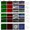
Figure 1:. TMC4 and 5 localize to the distal tips and base of enterocyte microvilli.
(a) Schematic showing the luminal side of the intestinal tract covered by a monolayer of epithelial cells, predominantly enterocytes, that are organized in repeating units of intestinal villi and crypts. The apical surface of each enterocyte is covered in arrays of microvilli- actin-based, membrane bound protrusions, known as the “brush border”. (b) Brush border microvilli are interconnected via several “lateral links”. (c) Confocal image of a cryosection of adult human small intestinal epithelium showing F-actin-based microvilli (green) and immunofluorescence staining of TMC4 (magenta), which is localized at the microvillar distal tips and also present in the cell cytoplasm. (d) Western blot of TMC4 and TMC5 on isolated mouse brush border. (e) A villus from the small intestine of a knockin mouse at P12 expressing TMC4-GFP (green) and TMC5-mCherry (red), stained with phalloidin to label F-actin (white). Both TMC4 and TMC5 are enriched at the apical surface of enterocytes. (f) TMC4-GFP (green) and TMC5-mCherry are localized to microvillar tips and base. (g) En face view of IECs from mouse expressing TMC4-GFP and TMC5-mCherry.
Figure 2:
Figure 2:. Microvillar localization of TMC4 and 5 shows spatio-temporal variation.
(a and b) TMC4-GFP (green) and TMC5-mCherry (red) are enriched at the distal tips and base of microvilli (white) early postnatal (P1) in the small intestine (a), and only at microvillar tips in the large intestine (b). Enrichment along the microvilli varies with age in both intestinal segments. (c) TMC4-GFP (green) and TMC5-mCherry (red) are also enriched at the apical surface of enterocytes in intestinal crypts. Panels on right depict fluorescence intensity of TMC4-GFP (green), TMC5-mCherry (red) and actin (blue), along microvilli. The direction of the arrow in (a) depicts the direction of the x-axes, with the first fluorescence intensity (FI) peak corresponding to microvillar tip, and the second peak corresponding to the microvillar base.
Figure 3:
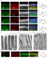
Figure 3:. TMC4 and 5 localization at the distal tip of microvilli is spatially distinct from the microvillar Usher interactome.
(a) Localization of TMC4-GFP (green) and TMC5-mCherry or various members of the IMAC (red) along microvilli (white) of small intestinal IECs from P12 knockin mice. Fluorescence intensity line scans along the microvillar length shown on the right (F-actin- blue, TMC4- green, TMC5 or IMAC proteins-red). The direction of the arrow in (a) depicts the direction of the x-axes, with the first FI peak corresponding to microvillar tip, and the second peak corresponding to the microvillar base. (b) Freeze-substitution thin section electron microscopy of microvilli from P4 mice showing inter-microvillar lateral links (white arrows). (c and d) Immuno-gold electron microscopy localization (black arrows) of Harmonin A (c) and MYO7B (d) in microvilli. (e) Western blot of CIB3 on mouse brush border. (f) Immunofluorescence staining of CIB3 (red) along microvilli (actin, green) of mouse enterocytes. Fluorescence intensity line scans along the microvillar length shown on the right.
Figure 4:
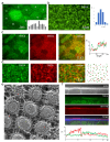
Figure 4:. TMC4 and 5 expression levels exhibit cellular and microvillar-level variability.
(a) En face view of enterocytes from knockin mouse at P12 expressing TMC4-GFP, with individual cells numbers, and average fluorescence intensity of GFP per cell plotted (inset). (b) TMC4-GFP at the tips of individual microvilli within a single cell localized by centroids (green dots), with fluorescence intensities plotted (right), RF= relative fluorescence and N = number of puncta. (c and d) Distribution of fluorescent TMC4-GFP (green) and TMC5-mCherry puncta show incidences of co-localization and also distinct localization. (e) Freeze-etch electron microscopy of microvillar tips from P4 mouse small IECs showing variable numbers of membrane proteins in this region. (f) Inverse relationship between TMC4-GFP (green) and TMC5-mCherry (red) concentration at microvillar tips (in FI) is often observed.
Figure 5:
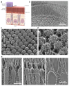
Figure 5:. Microvillar tips are sites for glycocalyx insertion.
(a) Schematic illustrating the glycocalyx network that is secreted from the tips of enterocyte microvilli. (b) Electron micrograph of a freeze-etch replica of mouse small intestine showing the stratified organization of the microvilli-rich brush border and glycocalyx layers. (c) Freeze-etching cross-fracture view of the microvilli tips at the point of insertion of the glycocalyx filaments on the membrane. (d) Close-up view of the area indicated by the rectangle in c showing the anchoring points of the glycocalyx filaments. (e) Left- The glycocalyx filaments emerging from the tips of the microvilli are distinct from the lateral links between microvilli. Right and inset- inflections of the glycocalyx filaments are observed at their connecting points to the microvillar membrane.
Figure 6:
Figure 6:
Diagram illustrating the localization of the TMC4-TMC5-CIB3 complex at the tips if IEC microvilli.
Figure 7:
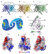
Figure 7:. Structural homology models for TMC4 and 5.
(a-c) Ribbon representation for the TMEM16-based models of TMC4 (a), TMC5 (b), and TMC1 (c). Chains A and B are indicated and colored in different tones. Two black lines indicate the approximated localization of the plasma membrane with intracellular (IC) and extracellular (EC) locations indicated. (d) TMC topology. (e) Schematic representation of TMC transmembrane helices and pore. (f) Conservation of the TMC residues lining the pore estimated by ConSurf. TM4-7 are indicated. Two spheres of planes indicate the approximated localization of the membrane bilayer calculated by OPM. (g-i) The charge surface at the pore of TMC4 (g), TMC5 (h), and TMC1 (i) depicts the electrostatic differences in this region. TM4 and TM6 lining the pore are indicated.
Figure 8:
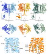
Figure 8:. AF2-based structural models for TMC4 and 5.
(a-b) AF2-based mm TMC4 and mm TMC5 dimers with side (top) and top-down (bottom) views. (c) Side and top views of dimeric ce TMC-1 cryo-EM structure. Insets show TM10 swaps in each system. (d-e) Cl binding residues in TMC4 and TMC5, respectively.
Figure 9.
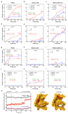
Figure 9.. Ion conduction and lipid scrambling in TMC4 and 5 proteins.
(a-f) Number of ion crossings vs time for TMC4 (a,d), TMC4-CIB3 (b,e), and TMC4-CIB3-Ca2+ (c,f) carried out at short (200 ns; a-c) and longer (480 ns – Anton2; d-f) timescales using −0.5 V. (g-l) Number of ion crossing vs time for TMC5 (g,j), TMC5-CIB3 (h,k), and TMC5-CIB3-Ca2+ (i,l) carried out at short (200 ns; g-i) and longer (480 ns – Anton2; j-l) timescales using −0.5 V. In all these cases monomer A is in closed conformation and monomer B was opened using TMD. (m) Full trajectory of a flipping lipid (555) from equilibration to the end of a voltage simulation of TMC5 system. Snapshots of lipid conformations are shown at indicated times. Some panels are also in Fig. S7.

Similar articles

References

    1. Delacour D., Salomon J., Robine S. & Louvard D. Plasticity of the brush border — the yin and yang of intestinal homeostasis. Nat. Rev. Gastroenterol. Hepatol. 13, 161–174 (2016). - PubMed
    1. Miura S., Sato K., Kato-Negishi M., Teshima T. & Takeuchi S. Fluid shear triggers microvilli formation via mechanosensitive activation of TRPV6. Nat. Commun. 6, 8871 (2015). - PMC - PubMed
    1. Kim S. W. et al. Shear stress induces noncanonical autophagy in intestinal epithelial monolayers. Mol. Biol. Cell 28, 3043–3056 (2017). - PMC - PubMed
    1. Müller T. et al. MYO5B mutations cause microvillus inclusion disease and disrupt epithelial cell polarity. Nat. Genet. 40, 1163–1165 (2008). - PubMed
    1. Ruemmele F. M. et al. Loss-of-function of MYO5B is the main cause of microvillus inclusion disease: 15 novel mutations and a CaCo-2 RNAicell model. Hum. Mutat. 31, 544–551 (2010). - PubMed

Publication types

LinkOut - more resources