[Preprint]. 2024 Aug 22:2024.08.21.609023.
doi: 10.1101/2024.08.21.609023.

Drosophila Clueless ribonucleoprotein particles display novel dynamics that rely on the availability of functional protein and polysome equilibrium

Affiliations

Drosophila Clueless ribonucleoprotein particles display novel dynamics that rely on the availability of functional protein and polysome equilibrium

Hye Jin Hwang et al. bioRxiv. .

Abstract

The cytoplasm is populated with many ribonucleoprotein (RNP) particles that post-transcriptionally regulate mRNAs. These membraneless organelles assemble and disassemble in response to stress, performing functions such as sequestering stalled translation pre-initiation complexes or mRNA storage, repression and decay. Drosophila Clueless (Clu) is a conserved multi-domain ribonucleoprotein essential for mitochondrial function that forms dynamic particles within the cytoplasm. Unlike well-known RNP particles, stress granules and Processing bodies, Clu particles completely disassemble under nutritional or oxidative stress. However, it is poorly understood how disrupting protein synthesis affects Clu particle dynamics, especially since Clu binds mRNA and ribosomes. Here, we capitalize on ex vivo and in vivo imaging of Drosophila female germ cells to determine what domains of Clu are necessary for Clu particle assembly, how manipulating translation using translation inhibitors affects particle dynamics, and how Clu particle movement relates to mitochondrial association. Using Clu deletion analysis and live and fixed imaging, we identified three protein domains in Clu, which are essential for particle assembly. In addition, we demonstrated that overexpressing functional Clu disassembled particles, while overexpression of deletion constructs did not. To examine how decreasing translation affects particle dynamics, we inhibited translation in Drosophila germ cells using cycloheximide and puromycin. In contrast to stress granules and Processing bodies, cycloheximide treatment did not disassemble Clu particles yet puromycin treatment did. Surprisingly, cycloheximide stabilized particles in the presence of oxidative and nutritional stress. These findings demonstrate that Clu particles have novel dynamics in response to altered ribosome activity compared to stress granules and Processing bodies and support a model where they function as hubs of translation whose assembly heavily depends on the dynamic availability of polysomes.


Conflict of interest statement

Conflict of interest: The authors declare that they have no conflicts of interest with the contents of this article.

Figures

Fig 1.
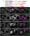
Fig 1.. Clu forms abundant cytoplasmic particles in Drosophila nurse cells.
(A-B’) Clu protein visualized in (A, A’) fixed follicles and (B, B’) still frames from live-imaged follicles that have large cytoplasmic particles in well-fed flies (A, B). Particles are disassembled with starvation (A’, B’). Clu protein is reduced in the oocyte (A, A’, B, arrows) compared to the nurse cells, and particles are absent. Fixed images were obtained using a Zeiss 700 confocal laser scanning microscope (Carl Zeiss Microscopy LLC, White Plains, NY, USA), and live images were obtained using a Nikon Eclipse Ti2 spinning disk microscope at 100x (Nikon Corporation, Tokyo, Japan). (C) Schematic showing Clu protein domains. clu06604 has an in-frame Green Fluorescent Protein (GFP) inserted in the endogenous clu locus, resulting in a GFP fusion protein (Clu::GFP). ms = melanogaster specific, ßGF = beta Grasp Fold, DUF = Domain of unknown function, M = Middle domain, TPR = Tetratricopeptide repeat. (D, E) Schematics depicting Drosophila oogenesis. Female Drosophila have a pair of ovaries (D) composed of strings of developing follicles called ovarioles (E). (E) Ovaries from well-fed females contain all the developing follicle stages (Stages 2-14). Follicles are composed of 15 nurse cells (yellow) and one oocyte (blue) surrounded by somatic follicle cells (green). Vitellogenesis starts at stage 8 when the polarity of the oocyte’s microtubule (MT) cytoskeleton changes. Analysis and images presented in this study are predominantly stages 6 & 7 (dashed box, S1-3 Tables). (A-B’) Stage 7 follicles. White = anti-Clu antibody (A, A’) and GFP (B, B’). Scale bar = 20 μm in B’ for A-B’.
Fig 2.
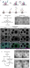
Fig 2.. DUF, Clu, and TPR domains are required for Clu particle association.
(A) Cartoon of full-length (FL) and domain deletion (ΔDUF, ΔClu, and ΔTPR) constructs of ectopic Clu tagged with the fluorescence tag mScarlet. (B-B”) Still-images from S1 Movie of a follicle from a cluCA06604 /+; nanos (nos) GAL4/UASp-FLclu::mScarlet female. Particles from endogenous Clu::GFP (B) and ectopic FLClu::mScarlet (B’) co-localize in germ cells (B”) as arrow heads indicate. 71% of nurse cells expressing mScarlet showed colocalization of Clu::GFP and mScarlet (n=14 follicles, see S1 Table for details). (C-E”) Still-images from ectopic Clu deletion constructs (S2-4 Movies). Endogenous Clu::GFP (C, D, E) forms particles. However, ΔDUF (C’), ΔClu (D’), and ΔTPR (E’) constructs do not form particles (C’, D’, E’) and cannot associate with endogenous Clu particles (C”, D”, E”, See S1 Table for details). Still-images from (C-C”) S2 Movie of a follicle from cluCA06604/+; nosGAL4/UASp-cluΔDUF::mScarlet, (D-D’’) S3 Movie of a follicle from cluCA06604/+; nosGAL4/UASp-cluΔClu::mScarlet, and (E-E’’) S4 Movie of a follicle from cluCA06604/+; nosGAL4/UASp-cluΔTPR::mScarlet females. (B-E”) Stage 7 egg chamber follicles expressing Clu::GFP and various ectopic Clu tagged with mScarlet were imaged with a 200 μg/mL insulin-containing Complete Schneider’s (CS) media in a time-lapse course at a single plane (see S1-4 Movies for details). The focal plane was selected by ensuring more than three nurse cells having nuclear and cytosolic area clearly were visible, with approximately 25 % depth from the top surface of each follicle (See Materials and Methods for details). Live images were obtained using a Nikon A1 plus Piezo Z Drive Confocal microscope at 60x (Nikon Corporation, Tokyo, Japan). The follicle stages analyzed (n) in each genotype: cluCA06604 /+; nanos (nos) GAL4/UASp-FLclu::mScarlet, stage 5 (2), stage 6 (3), stage 7 (8), stage 8 (1); cluCA06604/+; nosGAL4/UASp-cluΔDUF::mScarlet, stage 6 (2), stage 7 (4), stage 8 (2); cluCA06604/+; nosGAL4/UASp-cluΔClu::mScarlet, stage 5 (1), stage 6 (4), stage 7 (5), stage 8 (1); cluCA06604/+;nosGAL4/UASp-cluΔTPR::mScarlet, stage 6 (3), stage 7 (5), stage 8 (3). More details, including the number of nurse cells expressing mScarlet and having Clu particles, the number of nurse cells showing colocalization of endogenous GFP and ectopic mScarlet, and the number of dissected animals, are described in S1 Table. (B, C, D, E) White = endogenous Clu::GFP. (B’, C’, D’, E’) White = mScarlet. (B”, C”, D”, E”, merge) Green = Clu::GFP, magenta = Scarlet. Scale bar = 10 μm in B” for B-E”.
Fig 3.
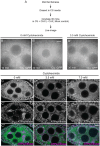
Fig 3.. High levels of functional Clu disassemble bliss particles.
(A-A”) Immunostaining of a follicle from a daughterless (da) GAL4/UASp-FLclu::mScarlet female. High levels of ectopic FLClu (A’) disrupt particle formation (A, A”). (B-B”) Immunostaining of a follicle from a daGAL4/UASp-cluΔDUF::mScarlet female. (C-C”) Immunostaining of a follicle from a daGAL4/UASp-cluΔTPR::mScarlet female. (D-D”) Immunostaining of a follicle fromdaGAL4/UASp-mCherry::cpb female. High levels of ΔDUF (B’), ΔTPR (C’) or CPB (D’) do not interfere with endogenous Clu particle formation (B, B” for ΔDUF, C, C” for ΔTPR or D, D” for CPB). 95% (ΔDUF), 90% (ΔTPR), and 94% (CPB) of nurse cells expressing each ectopic construct from the observed follicles showed Clu particles (See S1 Table for details). (A-D”) Stage 7 egg chamber follicles were imaged with a 1.2 μm thickness of z-stacks with an interval of 0.42 μm. The focal plane was selected by ensuring at least three to four nuclei were clearly visible in nurse cells, but also to avoid dim fluorescence signals due to deeper depth (See Materials and Methods for details). Images were obtained using a Zeiss LSM 980 confocal laser scanning microscope (Carl Zeiss Microscopy LLC, White Plains, NY, USA). The follicle stages analyzed (n) for each genotype by immunostaining: daGAL4/UASp-FLclu::mScarlet, stage 5 (1), stage 6 (3), stage 7 (4), stage 8 (1); daGAL4/UASp-cluΔDUF::mScarlet, stage 5 (1), stage 6 (1), stage 7 (1), stage 8 (1); daGAL4/UASp-cluΔTPR::mScarlet, stage 5 (1), stage 6 (1), stage 7 (2), stage8 (1); daGAL4/UASp-mCherry::cpb, stage5 (1), stage 6 (2), stage 7 (4), stage 8 (4). More details, including the number of nurse cells having Clu particles and the number of dissected animals, are described in S1 Table. (A, B, C, D) White = anti-Clu, (A’, B’, C’) white = anti-Scarlet, (D’) white = anti-mCherry. (A”, B”, C”, merge) Green = anti-Clu, magenta = anti-Scarlet. (D”, merge) Green = anti-Clu, magenta = anti- mCherry. For A, A”, B, B”, C, C”: Note: anti-Clu antibody also recognizes the mScarlet transgene. Scale bar = 10 μm in A” for A-D”.
Fig 4.
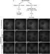
Fig 4.. The translation inhibitor puromycin disassembles Clu bliss particles.
(A) Schematic demonstrating the mechanism of action for the translation inhibitor, puromycin (PUR). PUR blocks nascent polypeptide chain elongation, thereby causing premature translation termination, disassembly of the ribosomal complex, and decreased polysomes. (B) Workflow for the experiment. Ovarioles dissected from Well- fed cluCA06604 females were treated with puromycin, then live-imaged (S5 Movie, C, C’). (C) The 1st still frame (at time zero after adding 10 μM PUR) of stage 6 follicle from S5 Movie showing Clu particles. (C’) The 22nd still-image (at seven minutes) of the same follicle demonstrating disassembled bliss particles by PUR (n=20/20 follicles, see S2 Table for details). The focal plane was selected by ensuring at least three to four nuclei were clearly visible in nurse cells, with approximately 25 % depth from the top surface of each follicle (See Materials and Methods for details). Live images were obtained using a Nikon Eclipse Ti2 spinning disk microscope at 100x (Nikon Corporation, Tokyo, Japan). The follicle stages analyzed (n): stage 5 (2), stage 6 (8), stage 7 (5), stage 8(5). Scale bar = 10 μm in C’ for C and C’.
Fig 5.
Fig 5.. The translation inhibitor cycloheximide maintains Clu bliss particles, but blocks insulin-induced assembly.
(A) Schematic demonstrating the mechanism of action for the translation inhibitor, cycloheximide (CHX). CHX blocks the 60S ribosome exit site, thereby stalling translation and increasing polysome densities. (B) Workflow for the CHX treatment showing in C and C’. Ovarioles from well-fed cluCA06604 females were incubated with CHX for 20 minutes then live-imaged (S6 and 7 Movies, C, C’). (C) Still-image of a stage 7 follicle after 20-minute incubation without CHX demonstrating the presence of Clu bliss particles (n=14/14 follicles, see S2 Table for details). (C’) Still-image of a stage 7 follicle after 20-minute incubation with 3.5 mM CHX demonstrating the assembled bliss particles are still present (n=20/20 follicles, see S2 Table for details). Follicles were imaged in a time-lapse course for 3 minutes at a single plane at the end of 20 minutes. The focal plane was selected by ensuring at least three to four nuclei were clearly visible in the nurse cells, with approximately 25 % depth from the top surface of each follicle (See Materials and Methods for details). Follicle stages analyzed (n): mock treatment, stage 5 (3), stage 6 (3), stage 7 (7), stage 8 (1); 3.5 mM CHX treatment, stage 5 (3) stage 6 (4), stage 7 (10), stage 8 (3). (D-F”) Immunostaining of stage 7 follicles from well-fed w1118 females fed for 24 hours with wet yeast paste containing (D-D”) 0 mM, (E-E”) 3.5 mM, and (F-F”) 7 mM CHX. None of CHX feeding disassemble bliss particles (D, E, F) and normal mitochondrial morphology and localization is maintained (D’, E’, F’). Observed bliss particles: 91% (0 mM CHX), 94% (3.5 mM CHX), and 96% (7 mM CHX) (See S2 Table for details) of nurse cells showed. Images are 2 μm projections assembled 0.42 μm sections. The focal plane was selected to show at least 3~4 nuclei but also to avoid dim fluorescence signals due to deeper depth (See Methods for details). The total number of follicles analyzed (n) by immunostaining: 0 mM CHX, stage 6 (1), stage 7 (5), stage 8 (6); 3.5 mM CHX, stage 5 (6), stage 6 (5), stage 7 (6), stage 8 (4); 7 mM CHX, stage (7), stage 6 (6), stage 7 (10), stage 8 (6). (G) Workflow for the CHX experiment showing in (H-I’). Well-fed cluCA06604 females were starved for 3 hours with water only, then ovarioles dissected from starved animals were incubated in insulin (H, H’, control) or CHX followed by insulin (I, I’). (H) The 1st still frame (at time zero after adding 100 μg/mL insulin) of stage 7 follicle from S8 Movie showing no Clu particles. (H’) The 46th still-image (at 15 minutes) of the same follicle from S5 Movie demonstrating the recovery of bliss particles by insulin as previously showed (Sheard, 2020) (n=4/4 follicles, see S2 Table for details). (I) The 1st still frame (at time zero after adding 100 μg/mL insulin) of stage 7 follicle starved and treated with CHX from S9 Movie. (I’) The 46th still frame (at 15 minutes) of the same follicle showing no recovery of bliss particle by insulin following CHX treatment (n=11/11 follicles, see S2 Table for details). The focal plane was selected by ensuring at least three to four nuclei were clearly visible in the nurse cells, with approximately 25 % depth from the top surface of each follicle (See Materials and Methods for details). Follicles stages analyzed (n): insulin only treated, stage 5 (1), stage 7 (2), stage 8 (1); insulin following 3.5 mM CHX, stage 5 (3) stage 6 (4), stage 7 (4). More details, including the number of nurse cells having Clu particles and the number of dissected animals, are described in S2 Table. Live images were obtained using a Nikon Eclipse Ti2 spinning disk microscope at 100x (Nikon Corporation, Tokyo, Japan). Immunostaining images were obtained using a Zeiss LSM 980 confocal laser scanning microscope (Carl Zeiss Microscopy LLC, White Plains, NY, USA). (D, E, F) White = anti-Clu. (D’, E’, F’) White = anti-ATP synthase. (D”, E”, F”, merge) Green = anti-Clu, magenta = anti-ATP synthase. Scale bar = 10 μm in B’ for B-C’, in F” for D-F”, and in H’ for H-I’.
Fig 6.
Fig 6.. Cycloheximide maintains bliss particles in the presence of nutritional stress.
(A) Workflow for the experiment. Ovarioles from well-fed cluCA06604 females were dissected without insulin, then treated with 3.5 mM CHX (B”) or not (mock treatment, B, B’). (B) Still-image of stage 7 follicle from well-fed females maintains Clu particles without insulin in 10 minutes (n= 5 follicles, 85% of the nurse cells having Clu::GFP particles, see S2 Table for details), (B’) but lost Clu particles in 30 minutes after dissection (n= 5 follicles, 42% of nurse cells having Clu::GFP particles, see S2 Table for details). (B”) Still images of stage 7 follicle treated with 3.5 mM CHX for 30 minutes demonstrating CHX treatment does not cause particle dispersion even without insulin (n=10 follicles, 98% of nurse cells having Clu::GFP particles, see S2 Table for details). The focal plane was selected to show at least three to four nuclei were clearly visible in the nurse cells, with approximately 25% depth from the top surface of each follicle (See Materials and Methods for details). The follicle stages analyzed (n): no CHX in 10 mins, stage 5 (3), stage 6 (0), stage 7 (2); no CHX in 30 mins, stage 6 (1), stage7 (3), stage8 (1); 3.5 mM CHX, stage 5 (3). Stage 6 (3), stage 7 (4). More details, including the number of nurse cells having Clu particles and the number of dissected animals, are described in S2 Table. (C-E”) Immunostaining of stage 7 follicles from well-fed w1118 females subsequently starved for 5 hours after feeding for 24 hours with wet yeast paste containing (C-C”) 0 mM, (D-D”) 3.5 mM, and (E-E”) 7 mM. Starvation disrupts particles (C) and causes mitochondrial clustering (C’) as we previously showed (Sheard 2020). (D-D”) 3.5 mM and (E-E”) 7 mM CHX feeding does not disperse Clu bliss particles (D, E) nor cause mitochondrial clump (D’,E’) even with starvation. 87% (3.5 mM CHX following starvation, n=15 follicles), and 94% (7 mM CHX following starvation, n=14 follicles) of nurse cells from the observed follicles had Clu particles (See S2 Table for details). Images are 2 μm projections assembled from 0.42 μm sections. The focal plane was chosen to show at least more than three nurse cells having clear visibility for nulear and cytoplasmic area but also to avoid dim fluorescence signals due to deeper depth (See Materials and Methods for details). The follicle stages examined (n) for each condition: no CHX-5 hour starvation, stage 5 (2), stage 6 (6), stage 7 (3), stage 8 (3); 3.5 mM CHX-5 hour starvation, stage 5 (3), stage 6 (2), stage 7 (6), stage8 (4); 7 mM CHX-5 hour starvation, stage5 (1), stage 6 (3), stage 7 (4), stage 8 (2). More details, including the number of nurse cells having Clu particles and clumped mitochondria and the number of dissected animals, are described in S2 Table. (C, D, E) White = anti-Clu. (C’, D’, E’) White = anti-ATP synthase. (C”, D”, E”, merge) Green = anti-Clu, magenta = anti-ATP synthase. Scale bar = 10 μm in B” for B-B” and in E” for C-E”.
Fig 7.
Fig 7.. Cycloheximide maintains bliss particles in the presence of oxidative stress.
(A) Workflow for the experiment. Ovarioles dissected from well-fed cluCA06604 females were treated with CHX, then exposed to 2 mM hydrogen peroxide (C-D’”). (B-B”’) Still-images of stage 7 follicle from S10 Movie showing the addition of 2 mM hydrogen peroxide disperses bliss particles as we previously showed (Sheard 2020). 93% of nurse cells (n=9 follicles, see S2 Table for details, 2020) clearly showed dispersion of bliss particles after hydrogen peroxide treatment. (C-C”’) Still-image of stage 7 follicle from S11 and (D-D”’) S12 Movies showing CHX treatment protects bliss particles from oxidative stress-induced dispersion. None of the nurse cells pre-treated with 3.5 mM CHX for 20 minutes (n=9/9 follicles) showed dispersion of bliss particles after hydrogen peroxide treatment, and 13% of the nurse cells pre-treated with 7 mM CHX for 20 minutes (n=7 follicles, see S2 Table for details) showed dispersion of CluGFP particles after hydrogen peroxide treatment. The focal plane was chosen to show at least three to four nurse cells having clear visibility of nuclear and cytoplasmic area, with approximately 25 % depth from the top surface of each follicle (See Materials and Methods for details). Follicle stages analyzed (n): no CHX-hydrogen peroxide, stage 5 (3), stage 6 (2), stage 7 (3), stage 8 (1); 3.5 mM CHX-hydrogen peroxide, stage 5 (1) stage 6 (1), stage 7 (5), stage 8 (2); 7 mM CHX-hydrogen peroxide, stage 5 (2), stage6 (3), stage7 (2). S2 Table. Scale bar = 10 μm in B”’ for B-D”’.
Fig 8.
Fig 8.. Cycloheximide does not affect velocity of Clu particle.
(A) Representative still-image of Clu::GFP particles from S6 Movie. This still-image is Fig 5C, inverted in LUT. (B) Three-minute S6 movie was stacked to find the orientations of Clu particles. The thin white box shows the area used to make a kymograph in (B’). (B’) Representative kymographs of Clu particles. The white arrows indicate a directed movement of Clu particles and the velocity was measured by applying a straight line using ImageJ. More details for velocity calculation are described in S2 Table. (C) No effect of cycloheximide on the average velocity of bliss particles. Dots in the graph represent the velocity of each particle. Six particles were analyzed in each follicle. The graph was generated using GraphPad Prism. Follicle stages analyzed (n): no CHX (control), stage 6 (1), stage 7(2), stage 8 (1); 3.5 mM CHX, stage 6 (1), stage 7 (3). Scale bar = 10 μm in A for A and B. Scale bar 2 μm in B’.
Fig 9.
Fig 9.. Clu particle associated with mitochondria moves slowly.
(A) Still-image (S13 Movie) of a follicle from a Clu GFPTrap cluCA06604 female labeled with 50 nM TMRE. (B) Representative kymographs of Clu particles (A, arrows). Kymographs (lower panels) were obtained from the z-stacked image of frames 70-85 (for 1 minute) of S13 Movie by applying segmented lines (upper panels). Representative graph of (C) speed and (D) size of Clu particles with or without mitochondrial association. A bar graph represents an arithmetic mean. Unpaired t-test was performed for statistical significance. (E) Representative graph of Clu particles plotted by speed, size, and mitochondrial association. Pearson correlation coefficient was obtained to measure the relationship between size and speed. (F, G) Simple logistic regression analysis of (E). This predicts a probability of mitochondrial association depending on particle speed (F) or size (G). The value 1 of the y-axis represents the mitochondrially-associated particle, and 0 represents the particles free from mitochondria. The solid line indicates the mean of the probability, and the dotted line indicates a 95% confidence interval. Logistic regression tests were performed using GraphPad Prism. The total number of particles (n) = 35. The number of follicles examined is as follows: stage 5 (2), stage 6 (4), and stage 7(3). More details, including the number of particles in each follicle, particle area, particle speed, and particle binding are described in S3 Table. Red = mitochondria-associated particles, blue = mitochondria-unassociated particles. (A, B) Green = Clu, magenta = TMRE. Scale bar = 10 μm in A. Scale bar = 2 μm in B for upper panels.

Similar articles

References

    1. Alberti S., Gladfelter A., & Mittag T. (2019). Considerations and Challenges in Studying Liquid-Liquid Phase Separation and Biomolecular Condensates. Cell, 176(3), 419–434. 10.1016/j.cell.2018.12.035 - DOI - PMC - PubMed
    1. Anderson P., & Kedersha N. (2006). RNA granules. J Cell Biol, 172(6), 803–808. 10.1083/jcb.200512082 - DOI - PMC - PubMed
    1. Aviner R. (2020). The science of puromycin: From studies of ribosome function to applications in biotechnology. Comput Struct Biotechnol J, 18, 1074–1083. 10.1016/j.csbj.2020.04.014 - DOI - PMC - PubMed
    1. Bauer K. E., de Queiroz B. R., Kiebler M. A., & Besse F. (2023). RNA granules in neuronal plasticity and disease. Trends Neurosci, 46(7), 525–538. 10.1016/j.tins.2023.04.004 - DOI - PubMed
    1. Bennett C. F., Latorre-Muro P., & Puigserver P. (2022). Mechanisms of mitochondrial respiratory adaptation. Nat Rev Mol Cell Biol, 23(12), 817–835. 10.1038/s41580-022-00506-6 - DOI - PMC - PubMed

Publication types