Review
. 2024 Sep 2;5(9):e669.
doi: 10.1002/mco2.669. eCollection 2024 Sep.

Posttransplant complications: molecular mechanisms and therapeutic interventions

Affiliations
Review

Posttransplant complications: molecular mechanisms and therapeutic interventions

Xiaoyou Liu et al. MedComm (2020). .

Abstract

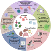
Posttransplantation complications pose a major challenge to the long-term survival and quality of life of organ transplant recipients. These complications encompass immune-mediated complications, infectious complications, metabolic complications, and malignancies, with each type influenced by various risk factors and pathological mechanisms. The molecular mechanisms underlying posttransplantation complications involve a complex interplay of immunological, metabolic, and oncogenic processes, including innate and adaptive immune activation, immunosuppressant side effects, and viral reactivation. Here, we provide a comprehensive overview of the clinical features, risk factors, and molecular mechanisms of major posttransplantation complications. We systematically summarize the current understanding of the immunological basis of allograft rejection and graft-versus-host disease, the metabolic dysregulation associated with immunosuppressive agents, and the role of oncogenic viruses in posttransplantation malignancies. Furthermore, we discuss potential prevention and intervention strategies based on these mechanistic insights, highlighting the importance of optimizing immunosuppressive regimens, enhancing infection prophylaxis, and implementing targeted therapies. We also emphasize the need for future research to develop individualized complication control strategies under the guidance of precision medicine, ultimately improving the prognosis and quality of life of transplant recipients.

Keywords: T cell; infection; malignancy; organ transplantation; posttransplant complications; rejection.


Conflict of interest statement

The authors declare that they have no conflict of interests.

Figures

FIGURE 1
FIGURE 1
Mechanisms of complications associated with immune overactivation following transplantation. (A) Three pathways of allorecognition. In the direct pathway, donor antigen‐presenting cells (APCs) interact directly with recipient T cells. In the indirect pathway, recipient APCs present processed donor alloantigen peptides to recipient T cells, resembling a typical immune response. In the semi‐direct pathway, recipient APCs acquire donor HLA molecules that directly present peptides to recipient T cells. (B) Mechanisms of immune activation associated with transplantation. Tissue damage and the release of damage‐associated molecular patterns (DAMPs), such as ATP, uric acid, IL‐33, and HMGB‐1, can result from ischemia–reperfusion injury and surgical trauma during allogeneic solid organ transplantation, as well as preoperative chemotherapy and radiotherapy for hematopoietic stem cell transplantation. Intestinal barrier damage can also lead to alterations in gut microbiota and the release of pathogen‐associated molecular patterns (PAMPs). These DAMPs and PAMPs serve as danger signals that interact with the recipient's innate immune cell surface and intracellular pattern recognition receptors (PRRs), initiating and sustaining innate immune responses. Upon APC stimulation, naïve CD8+ T cells differentiate into cytotoxic T cells, which can destroy donor organs or host cells through the release of granules and perforin. Following APC stimulation, naïve CD4+ T cells differentiate into effector T (Teff) cells. Teff cells enhance the cytotoxic activity of natural killer (NK) cells and cytotoxic T lymphocytes (CTLs) by secreting proinflammatory cytokines, including IL‐6, IL‐12, and TNF‐α. B cells can function as specific antigen‐presenting cells and modulate CD4+ T cell activation via indirect allorecognition pathways. Furthermore, activated B cells can differentiate into plasma cells that produce antibodies, potentially leading to antibody‐mediated rejection (ABMR) through mechanisms such as antibody‐dependent cell‐mediated cytotoxicity (ADCC) and direct cytotoxic effects. Substantial quantities of donor‐specific antibodies produced by recipient B cells can bind to the vascular endothelium of the graft, rapidly inducing inflammatory infiltration and vascular damage. This figure was created using tools provided by Biorender.com (accessed July 2, 2024).
FIGURE 2
FIGURE 2
Mechanisms of complications associated with immunosuppression following transplantation. (A) Under normal circumstances, antigen‐presenting cells (APCs) stimulate the differentiation of naïve CD8+ T cells into cytotoxic T cells, which can eliminate infected or malignant cells through the release of granules and perforin. However, the excessive use of immunosuppressive agents can disrupt this process. Immunosuppressants can inhibit the activation, differentiation, and cytotoxic function of naïve CD8+ T cells. Moreover, immunosuppressants can activate regulatory T cells (Tregs), which may hinder the development of naïve CD8+ T cells and the production of interferon‐γ (IFN‐γ) by cytotoxic T cells. Furthermore, reduced IFN‐γ secretion may elevate the risk of Epstein–Barr virus (EBV) infection in B cells, consequently promoting the development of EBV‐associated posttransplant lymphoproliferative disorder (PTLD). Additionally, the suppression of cytotoxic T cell function against EBV further increases the risk of posttransplant malignancies. (B) When the body's antitumor immune system is functioning optimally, APCs stimulate the differentiation of naïve CD4+ T cells into effector T (Teff) cells. Teff cells enhance the anti‐infective and antitumor effects of natural killer (NK) cells and cytotoxic T lymphocytes (CTLs) by secreting proinflammatory cytokines, including IL‐6, IL‐12, and TNF‐α. However, the overuse of immunosuppressive agents can disrupt this process by inhibiting the differentiation of naïve CD4+ T cells and impairing the ability of Teff cells to secrete proinflammatory cytokines. Moreover, excessive immunosuppression promotes the competitive binding of cytotoxic T‐lymphocyte‐associated protein 4 (CTLA‐4) on Teff cells to CD80/86 on APCs, thereby inhibiting the activation of naïve CD4+ T cells. Furthermore, Tregs suppress Teff cell function and subsequent antitumor effects by secreting immunomodulatory metabolites (e.g., indoleamine 2,3‐dioxygenase [IDO] and tryptophan 2,3‐dioxygenase [TDO]) and anti‐inflammatory cytokines (e.g., IL‐10 and IL‐35), as well as by inhibiting IL‐2 production. This figure was created using tools provided by Biorender.com (accessed July 2, 2024). GzmB, granzyme B; IDO, indoleamine 2,3‐dioxygenase; PRF, perforin; TDO, tryptophan 2,3‐dioxygenase.
FIGURE 3
FIGURE 3
Diagnostic biomarkers and therapeutic strategies for posttransplant cancers. (A) Interleukins. IL‐27 can promote the anticancer response of T cells and has an inhibitory effect on Treg activity. IL‐22 released by T cells is considered to be associated with the occurrence and development of posttransplant cancers originating from epithelial tissues. Both of these cytokines have potential value as diagnostic biomarkers. (B) Cell surface biomarkers. CD45 on T cells can promote the activation of naïve T cells by APCs, but transplant‐related factors may inhibit CD45, affecting T‐cell function. CD200, expressed on the cell surface in some hematopoietic system cancers, can activate Tregs, thereby inhibiting the anticancer effects of effector T cells. CD57 is expressed on late‐stage T cells, which lack proliferative capacity, resulting in weakened anticancer effects. These markers have the potential to serve as diagnostic biomarkers. (C) Adoptive T‐cell therapy and Treg depletion therapy hold significant value for treating posttransplant cancers. ACT includes CAR‐T cell therapy and antiviral ACT. CAR‐T cell therapy involves the genetic modification of patient T cells by introducing CAR genes, allowing them to recognize cancer antigens with high specificity. The modified CAR‐T cells are expanded in vitro and then reinfused into the patient's body to enhance the immune system's attack on cancers. Antiviral ACT involves the in vitro expansion of posttransplant cancer‐specific T cells, which are then reinfused into the patient's body to rebuild virus‐specific immunity, thereby inhibiting cancer development. Treg depletion therapy primarily utilizes drugs targeting CTLA‐4, such as ipilimumab, drugs targeting CD25, such as basiliximab, and denileukin diftitox, which can inhibit the procancer effects of Tregs, thereby enhancing the anticancer effects of the immune system. This figure was created using tools provided by Biorender.com (accessed July 2, 2024).
FIGURE 4
FIGURE 4
Future research directions regarding posttransplant complications. This figure was created using tools provided by Biorender.com (accessed July 24, 2024).

Similar articles

References

    1. Datta RR, Schran S, Persa OD, et al. Post‐transplant malignancies show reduced T‐cell abundance and tertiary lymphoid structures as correlates of impaired cancer immunosurveillance. Clin Cancer Res. 2022;28(8):1712‐1723. - PubMed
    1. Grinyó JM. Why is organ transplantation clinically important? Cold Spring Harb Perspect Med. 2013;3(6). - PMC - PubMed
    1. Conrad SA, Chhabra A, Vay D. Long‐term follow‐up and complications after cardiac transplantation. J La State Med Soc. 1993;145(5):217‐220. 223–5. - PubMed
    1. Sen A, Callisen H, Libricz S, Patel B. Complications of solid organ transplantation: cardiovascular, neurologic, renal, and gastrointestinal. Crit Care Clin. 2019;35(1):169‐186. - PubMed
    1. Li Q, Lan P. Activation of immune signals during organ transplantation. Signal Transduct Target Ther. 2023;8(1):110. - PMC - PubMed

LinkOut - more resources