. 2024 Aug 16:15:1419881.
doi: 10.3389/fphar.2024.1419881. eCollection 2024.

Protective effects of Ganoderma lucidum spores on estradiol benzoate-induced TEC apoptosis and compromised double-positive thymocyte development

Affiliations

Protective effects of Ganoderma lucidum spores on estradiol benzoate-induced TEC apoptosis and compromised double-positive thymocyte development

Jihong Yang et al. Front Pharmacol. .

Abstract

Backgroud: Thymic atrophy marks the onset of immune aging, precipitating developmental anomalies in T cells. Numerous clinical and preclinical investigations have underscored the regulatory role of Ganoderma lucidum spores (GLS) in T cell development. However, the precise mechanisms underlying this regulation remain elusive. Methods: In this study, a mice model of estradiol benzoate (EB)-induced thymic atrophy was constructed, and the improvement effect of GLS on thymic atrophy was evaluated. Then, we employs multi-omics techniques to elucidate how GLS modulates T cell development amidst EB-induced thymic atrophy in mice. Results: GLS effectively mitigates EB-induced thymic damage by attenuating apoptotic thymic epithelial cells (TECs) and enhancing the output of CD4+ T cells into peripheral blood. During thymic T cell development, sporoderm-removed GLS (RGLS) promotes T cell receptor (TCR) α rearrangement by augmenting V-J fragment rearrangement frequency and efficiency. Notably, biased Vα14-Jα18 rearrangement fosters double-positive (DP) to invariant natural killer T (iNKT) cell differentiation, partially contingent on RGLS-mediated restriction of peptide-major histocompatibility complex I (pMHCⅠ)-CD8 interaction and augmented CD1d expression in DP thymocytes, thereby promoting DP to CD4+ iNKT cell development. Furthermore, RGLS amplifies interaction between a DP subpopulation, termed DPsel-7, and plasmacytoid dendritic cells (pDCs), likely facilitating the subsequent development of double-negative iNKT1 cells. Lastly, RGLS suppresses EB-induced upregulation of Abpob and Apoa4, curbing the clearance of CD4+Abpob+ and CD4+Apoa4+ T cells by mTECs, resulting in enhanced CD4+ T cell output. Discussion: These findings indicate that the RGLS effectively mitigates EB-induced TEC apoptosis and compromised double-positive thymocyte development. These insights into RGLS's immunoregulatory role pave the way for its potential as a T-cell regeneration inducer.

Keywords: Ganoderma lucidum spores; T cell development; T cell receptor gene rearrangement; proteomics; single-cell RNA sequencing; thymic atrophy.


Conflict of interest statement

The authors declare that the research was conducted in the absence of any commercial or financial relationships that could be construed as a potential conflict of interest. The author(s) declared that they were an editorial board member of Frontiers, at the time of submission. This had no impact on the peer review process and the final decision.

Figures
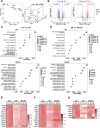
FIGURE 1
FIGURE 1
Proteomics analysis of thymus tissues. (A) Principal component analysis (PCA) of proteomic data. (B) Differential expression analysis of proteomic data. (C,D) Gene Ontology (GO) and Kyoto Encyclopedia of Genes and Genomes (KEGG) terms enriched by differentially expressed proteins. (E) Effect of RGLSH treatment on the expression of cell cycle-related proteins in the thymus tissue. (F) Effect of RGLSH treatment on the expression of ribonucleoproteins in the thymus tissue. (G) Effect of RGLSH treatment on the expression of apoptosis-related proteins in the thymus tissue.
FIGURE 2
FIGURE 2
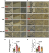
The co-localization of cytokeratin (CK) and Casp3 were analyzed using immunohistochemistry (IHC) staining in thymus tissues. (A,B) The co-localization of CK8 and Casp3 (A) and relative quantitative analysis (B). (C,D) The co-localization of CK5 and Casp3 (C) and relative quantitative analysis (D). Scale bar = 500 μm (40 ×), 50 μm (400 ×), n = 6.
FIGURE 3
FIGURE 3
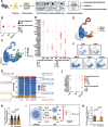
Single-cell RNA sequencing analysis of thymocytes. (A) Flowchart of single-cell RNA sequencing. (B) Gene signature analysis of T cell differentiation in thymus. (C) Cluster analysis of thymocytes and visualization in a two-dimensional uniform manifold approximation and projection (UMAP). (D) Cell-type-specific marker gene expression. (E) Cluster analysis of DP thymocytes and visualization in a two-dimensional UMAP. (F) CD4, CD8a, Mki67, Rag1, and Itm2a marker genes projected onto UMAP plots. (G) Effect of RGLSH treatment on the number of DPblas, DPres, and DPsels thymocytes and their proportion to DP thymocytes. (H) Effect of RGLSH treatment on the expression of Rag1 mRNA in DPres thymocytes. (I) Schematic diagram of the developmental pattern of DPsels thymocytes. (J) Gene signature analysis of CD4+ T cell differentiation in thymus. (K) Effect of RGLSH treatment on the expression of Aire protein in the thymus tissue.
FIGURE 4
FIGURE 4
TCRα sequencing analysis of thymocytes. (A) Schematic diagram of the TCRα sequencing. (B) Bubble map of TCRα clone. (C–E) Effect of RGLSH treatment on the clonality diversity (C), clonality efficiency (D), and clonality index (E) of TCRα. (F) Effect of RGLSH treatment on the combination of different sequences TRAV11 and TRAJ18. (G) Circos map of TCR V fragment and J fragment combination. (H) Effect of RGLSH treatment on the total combination of TRAV11 and TRAJ18.
FIGURE 5
FIGURE 5
The co-localization of CK5 and Aire were analyzed using IHC staining in thymus tissues, and relative quantitative analysis. Scale bar = 500 μm (40 ×), 50 μm (400 ×), n = 6.
FIGURE 6
FIGURE 6
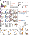
RGLSH facilitates the differentiation of DPsel-7 thymocytes into CD4+ iNKT cells. (A) Cluster analysis of DP thymocytes and visualization in a two-dimensional UMAP. (B) Expression levels of Mki67, Rag1, and Itm2a in different cell populations. (C) Effect of RGLSH treatment on the differentiation trajectories of thymocytes (Monocle3). (D) Effect of RGLSH treatment on the expression of CD44 and CD24a in CD4 SP cells. (E) Effect of RGLSH treatment on the expression of CD44, CD24a, and Klrb1c in CD4 SP-2 cells. (F) Effect of RGLSH treatment on the expression of CD44 and CD24a in DPsels thymocytes. (G) Effect of RGLSH treatment on the expression of cell cycle-related regulator in DPsel-7 thymocytes. (H) Effect of RGLSH treatment on the differentiation trajectories of thymocytes (Monocle2). (I) Effect of RGLSH treatment on the expression of CD44, CD24a, and Klrb1c in DN cells. (J,L) Effect of RGLSH treatment on the number of iNKT1 cells (J) and DN iNKT1 cells (L) in the thymus tissues. (K,M) Effect of RGLSH treatment on the proportion of iNKT1 cells (K) and DN iNKT1 cells (M) to thymocytes.
FIGURE 7
FIGURE 7
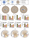
RGLSH restricts pMHC-CD8 communication between DPsel-7 thymocytes and DP thymocytes. (A,B) Effect of RGLSH treatment on the number and intensity of interactions between thymic stromal cells. (C) Effect of RGLSH treatment on the expression of CD1d in DP thymocytes. (D) Effect of RGLSH treatment on the expression of H2-Q1-pMHCⅠ in DP thymocytes.
FIGURE 8
FIGURE 8
Effect of EB (A) and RGLSH (B) treatment on the ligand-receptor interaction between DPsel-7 thymocytes and DP thymocytes.
FIGURE 9
FIGURE 9
RGLSH facilitates the differentiation of DPsel-7 thymocytes into DN iNKT1 cells. (A–C) Effect of RGLSH treatment on the number (A, C) and intensity (B,C) of DPsel-7 thymocytes’ interactions with pDCs. (D) Ligand-receptor interaction analysis between DPsel-7 thymocytes and pDCs. (E) Effect of RGLSH treatment on the expression of CD226 mRNA in DPsel-7 thymocytes and DN iNKT1 cells.
FIGURE 10
FIGURE 10
The protective effect of GLS on EB-induced thymic atrophy and T cell development and the underlying mechanism. On the one hand, GLS treatment significantly protected EB-induced thymic atrophy by inhibiting apoptosis of TECs. On the other hand, GLS facilitates thymocyte development from DN to DP stage and DPres thymocytes into iNKT cells, which was mediated by the Vα14-Jα18 TCRα rearrangement, DP-DP thymocyte interactions, and DP thymocyte-pDCs interactions.

Similar articles

References

    1. Alawam A. S., Anderson G., Lucas B. (2020). Generation and regeneration of thymic epithelial cells. Front. Immunol. 11, 858. 10.3389/fimmu.2020.00858 - DOI - PMC - PubMed
    1. Bennstein S. B. (2017). Unraveling natural killer T-cells development. Front. Immunol. 8, 1950. 10.3389/fimmu.2017.01950 - DOI - PMC - PubMed
    1. Bishop K. S., Kao C. H., Xu Y., Glucina M. P., Paterson R. R., Ferguson L. R. (2015). From 2000years of Ganoderma lucidum to recent developments in nutraceuticals. Phytochemistry 114, 56–65. 10.1016/j.phytochem.2015.02.015 - DOI - PubMed
    1. Bonamichi B., Lee J. (2017). Unusual suspects in the development of obesity-induced inflammation and insulin resistance: NK cells, iNKT cells, and ILCs. Diabetes Metab. J. 41, 229–250. 10.4093/dmj.2017.41.4.229 - DOI - PMC - PubMed
    1. Cao J., Spielmann M., Qiu X., Huang X., Ibrahim D. M., Hill A. J., et al. (2019). The single-cell transcriptional landscape of mammalian organogenesis. Nature 566, 496–502. 10.1038/s41586-019-0969-x - DOI - PMC - PubMed

Grants and funding

The author(s) declare that financial support was received for the research, authorship, and/or publication of this article. The work was supported by the Young Scientists Fund of the National Natural Science Foundation of China (Grant No. 82304824).

LinkOut - more resources