. 2024 Aug 29;24(1):886.
doi: 10.1186/s12879-024-09807-x.

TgVax452, an epitope-based candidate vaccine targeting Toxoplasma gondii tachyzoite-specific SAG1-related sequence (SRS) proteins: immunoinformatics, structural simulations and experimental evidence-based approaches

Affiliations

TgVax452, an epitope-based candidate vaccine targeting Toxoplasma gondii tachyzoite-specific SAG1-related sequence (SRS) proteins: immunoinformatics, structural simulations and experimental evidence-based approaches

Hamidreza Majidiani et al. BMC Infect Dis. .

Erratum in

Abstract

Background: The highly expressed surface antigen 1 (SAG1)-related sequence (SRS) proteins of T. gondii tachyzoites, as a widespread zoonotic parasite, are critical for host cell invasion and represent promising vaccine targets. In this study, we employed a computer-aided multi-method approach for in silico design and evaluation of TgVax452, an epitope-based candidate vaccine against T. gondii tachyzoite-specific SRS proteins.

Methods: Using immunoinformatics web-based tools, structural modeling, and static/dynamic molecular simulations, we identified and screened B- and T-cell immunodominant epitopes and predicted TgVax452's antigenicity, stability, safety, adjuvanticity, and physico-chemical properties.

Results: The designed protein possessed 452 residues, a MW of 44.07 kDa, an alkaline pI (6.7), good stability (33.20), solubility (0.498), and antigenicity (0.9639) with no allergenicity. Comprehensive molecular dynamic (MD) simulation analyses confirmed the stable interaction (average potential energy: 3.3799 × 10⁶ KJ/mol) between the TLR4 agonist residues (RS09 peptide) of the TgVax452 in interaction with human TLR4, potentially activating innate immune responses. Also, a dramatic increase was observed in specific antibodies (IgM and IgG), cytokines (IFN-γ), and lymphocyte responses, based on C-ImmSim outputs. Finally, we optimized TgVax452's codon adaptation and mRNA secondary structure for efficient expression in E. coli BL21 expression machinery.

Conclusion: Our findings suggest that TgVax452 is a promising candidate vaccine against T. gondii tachyzoite-specific SRS proteins and requires further experimental studies for its potential use in preclinical trials.

Keywords: Toxoplasma Gondii; Dynamic simulations; SRS proteins.


Conflict of interest statement

The authors declare no competing interests.

Figures

Fig. 1
Fig. 1
T. gondii life cycle. (1) Unsporulated oocysts are shed in the cat’s faeces, as definitive hosts of the infection. (2) The sporulated and infective oocysts are formed in the environment. The intermediate host stage includes humans and other warm-blooded animals such as rodents, birds, and livestock. (3) Animals become infected after ingesting contaminated food or water or by direct contact with the cat’s infected faeces. The oocysts release sporozoites that invade cells in various tissues such as muscle, brain, and eyes. Then sporozoites differentiate into tachyzoites, and so as the immune system of the host responds to the infection, some tachyzoites differentiate into bradyzoites and form tissue cysts. (4) Cats can be infected via a prey-predator transmission system after consuming intermediate hosts like birds and/or rodents or directly by ingestion of sporulated oocysts. (5) Next, the tissue cysts are ingested and the sexual phase of T. gondii’s life cycle begins again. (6) Humans can become infected after eating undercooked meat of livestock harbouring bradyzoite cysts. (7) Moreover, humans can become infected after ingestion of sporulated oocysts in the environment. (7.1–7.3) T. gondii can also be transmitted vertically from mother to fetus during pregnancy or through organ transplantation or blood transfusion. Created with BioRender.com
Fig. 2
Fig. 2
The crystal structure (PDB 1ynt) of the monomeric form of T. gondii tachyzoite-derived SAG1 surface antigen complexed to a human Fab fragment. (a) The tachyzoite-derived SAG1 antigen is bound to a human Fab fragment. The antibody’s ligand binding site is highlighted. (b & c) This panel highlighted the SAG1 amino acid residues that are involved in H-bond interactions with the Fab. (d) The tachyzoite-specific SRS antigen SRS29B (A0A125YP09) is aligned with chains D and C of the Fab fragment. The alignment shows the Fab interacting rsidues of SRS29B (red coloured residues) are conserved and can be used as template for the validation of in silico predicted epitopes and so selection of the most B-cell immunodominant residues. (e) B-cell epitopes of the SAG1 are represented as CPK style and using BIOVIA Discovery Studio visualizer v21.1.0.20298
Fig. 3
Fig. 3
The amino acid sequence of the TgVax452, its three-dimensional (3D) model with various components, and 3D modeling validation plots. (a) Different components of TgVax452 as an amino acid sequence and three-dimensional (3D) model. (b) The residue-based ERRAT values of the 3D model are represented using the ERRAT method (top left). The ERRAT plot shows how well the predicted 3D model fits into its electron density map. The red-coloured regions of the ERRAT plot indicate areas with high errors that require further refinement. The initial model’s Ramachandran plot (top right). The verify3D’s 3D/1D plot (bottom left). The x-axis represents the residue number, while the y-axis represents the Verify3D score. The Verify3D score is a measure of how well the model fits into its environment, based on factors such as side-chain packing, backbone conformation, and solvent accessibility. (c) The consensus B-cell epitopes of TgVax452 are visualized using the Discovery Studio’s CPK (Corey-Pauling-Koltun) tool as an atom colouring scheme
Fig. 4
Fig. 4
Protein-protein docking analysis between the vaccine construct and TLR4. (a) The antigen-interacting residues of the human TLR4 (green molecule) were identified based on the PDB ID 3fxi. (b) To prepare the TLR4 molecule for docking, the additional molecules were removed using the UCSF Chimera software. (c) The 3D-modeled vaccine is visualized using the UCSF Chimera software. (d) The docked complex of the vaccine-TLR4 is visualized on the right-hand panel. The 3D interacting interface of the vaccine model and the TLR4 is exhibited on the left-hand panel. The H-bond surfaces are selected to display the ligand H-bond interacting residues
Fig. 5
Fig. 5
Dynamics and thermodynamics of TgVax452:TLR4 complex. The variation plots in temperature (a), potential energy (b), and pressure (c) experienced by the TgVax452:TLR4 complex throughout the simulation. The plots were generated within the Spyder IDE 5.2.2 environment, running Python v3.9.13
Fig. 6
Fig. 6
Analysis of RMSD, RMSF, and radius of gyration for the TgVax452:TLR4 complex. (a) RMSD plots indicate the stability of the TgVax452 model and TLR4 structure over a 250 ns duration. (b) Residue-based local RMSF profiles are illustrated for the vaccine and TLR4 structures. The vaccine model displays some significant peaks above the threshold (average RMSF of 0.264), indicating regions of heightened fluctuation. (c) Radius of gyration (Rg) profiles of TLR4. (d) Radius of gyration (Rg) profiles of the TgVax452. The plots were generated within the Spyder IDE 5.2.2 environment, running Python v3.9.13
Fig. 7
Fig. 7
Distribution of the hydrogen bond and solvent accessible surface area (SASA) curves related to the TgVax452 and TLR4. (a) the profile of the number of hydrogen bonds per 25,000 (ps) timeframe within TLR4, out of a potential 733,562 bonds. (b) the profile of the number of hydrogen bonds per timeframe (25,000 ps) within TgVax452, out of a potential 309,650 bonds. (c) Count of atom pairs within a 0.35 nm cutoff distance within TLR4, reflecting the proximity and potential for interactions. (d) Count of atom pairs within a 0.35 nm cutoff distance within TgVax452, indicating the spatial arrangement and potential interactions between atoms. (e) The SASA plot for TLR4. The curve indicates relatively constant fluctuations throughout the simulation, with a minor deviation observed within the initial 260 ps. (f) The SASA plot for TgVax452. After approximately 45 ns, the SASA curve for TgVax452 reaches an equilibrium state, reflecting a stabilized protein conformation. The plots were generated within the Spyder IDE 5.2.2 environment, running Python v3.9.13
Fig. 8
Fig. 8
Principal component analysis (PCA) and free energy landscape (FEL) analysis of TgVax452:TLR4 complex in 2D and 3D graphical presentation. (a) Variance of eigenvectors computed from the covariance matrix of the TLR4 structure. (b) Distribution of conformations or states sampled during MD simulations’ trajectory projection onto the first two eigenvectors for TLR4 (c) FEL plot of TLR4 in 2D (left) and 3D (right) graphical presentation corresponding to energy minimized structure. (d) Variance of eigenvectors computed from the covariance matrix of TgVax452 structure. (e) Projection of the motion for the vaccine along the PC1 and PC2. (f) FEL plot of the TgVax452 in 2D (left) and 3D (right) graphical formats corresponding to energy-minimized structure. The plots were generated within the Spyder IDE 5.2.2 environment, utilizing the Matplotlib library of Python v3.9.13
Fig. 9
Fig. 9
In silico immune simulation profile of the multi-epitope vaccine construct using C-ImmSim server. A single dose of vaccine injection demonstrated significant rises in IgG + IgM antibodies, followed by IFN-γ upsurge (over 400000 ng/ml), substantial increase in CTL, memory HTL and memory B-cells
Fig. 10
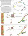
Fig. 10
The codon-optimized DNA sequence of the vaccine construct, its DNA-to-RNA transcribed format, subcloning into the pET-22b(+) vector, and mRNA secondary structure. (a) The protein-to-DNA back-translated and codon-optimized sequence of the vaccine construct. (b) The DNA sequence of the vaccine is subcloned between XhoI and XbaI restriction sites of the pET-22b(+) expression vector. (c) The DNA-to-RNA transcribed sequence of the vaccine. The mRNA secondary structure of the vaccine is mapped as positional entropy and base-pairing possibilities. (d) The minimum free energy (MFE) secondary structure is shown as positional entropy probability. (e) The MFE secondary structure is shown as a positional base-pairing probability

Similar articles

Cited by

References

    1. Dubey JP. Outbreaks of clinical toxoplasmosis in humans: five decades of personal experience, perspectives and lessons learned. Parasites Vectors. 2021;14(1):1–12. - PMC - PubMed
    1. Amouei A, Sarvi S, Sharif M, Aghayan SA, Javidnia J, Mizani A, et al. A systematic review of Toxoplasma gondii genotypes and feline: geographical distribution trends. Transbound Emerg Dis. 2020;67(1):46–64. - PubMed
    1. Fallahi S, Rostami A, Shiadeh MN, Behniafar H, Paktinat S. An updated literature review on maternal-fetal and reproductive disorders of Toxoplasma Gondii infection. J Gynecol Obstet Hum Reprod. 2018;47(3):133–40. - PubMed
    1. Foroutan-Rad M, Majidiani H, Dalvand S, Daryani A, Kooti W, Saki J, et al. Toxoplasmosis in blood donors: a systematic review and meta-analysis. Transfus Med Rev. 2016;30(3):116–22. - PubMed
    1. Dard C, Marty P, Brenier-Pinchart M-P, Garnaud C, Fricker-Hidalgo H, Pelloux H, et al. Management of toxoplasmosis in transplant recipients: an update. Expert Rev Anti-Infective Therapy. 2018;16(6):447–60. - PubMed

MeSH terms

LinkOut - more resources