. 2024 Aug 18;25(16):8980.
doi: 10.3390/ijms25168980.

NK Cell Degranulation Triggered by Rituximab Identifies Potential Markers of Subpopulations with Enhanced Cytotoxicity toward Malignant B Cells

Affiliations

NK Cell Degranulation Triggered by Rituximab Identifies Potential Markers of Subpopulations with Enhanced Cytotoxicity toward Malignant B Cells

Marta Wlodarczyk et al. Int J Mol Sci. .

Abstract

A promising strategy in cancer immunotherapy is to restore or enhance the cytotoxicity of NK cells, among others, by activating the mechanism of antibody-dependent cellular cytotoxicity (ADCC). Monoclonal antibodies targeting tumor antigens, such as rituximab (targeting CD20), induce NK cell-mediated ADCC and have been used to treat B cell malignancies, such as non-Hodgkin lymphoma, but not always successfully. The aim of this study was to analyze the gene expression profile of the NK cells involved in the cytolytic response stimulated by rituximab. NK cells were co-cultured with rituximab-opsonized Raji cells. Sorting into responder and non-responder groups was based on the presence of CD107a, which is a degranulation marker. RNA-seq results showed that the KIT and TNFSF4 genes were strongly down-regulated in the degranulating population of NK cells (responders); this was further confirmed by qRT-PCR. Both genes encode surface proteins with cellular signaling abilities, namely c-KIT and the OX40 ligand. Consistent with our findings, c-KIT was previously reported to correlate inversely with cytokine production by activated NK cells. The significance of these findings for cancer immunotherapy seems essential, as the pharmacological inhibition of c-KIT and OX40L, or gene ablation, could be further tested for the enhancement of the anti-tumor activity of NK cells in response to rituximab.

Keywords: Burkitt lymphoma; CD16; NK cells; OX40L; c-KIT; mAbs; rituximab.


Conflict of interest statement

The authors declare no conflicts of interest.

Figures

Figure 1
Figure 1
Mechanisms of rituximab-mediated cell death. RTX-coated malignant B cells are eliminated by three major mechanisms. (A) binding of RTX to CD20 on the B cell surface causes the activation of the complement cascade, which generates the membrane attack complex (MAC), directly inducing target cell lysis by complement-dependent cytotoxicity (CDC). (B) The binding of RTX to the target cell allows interaction with natural killer (NK) cells via the Fcγ receptor (FcγR) IIIa (CD16), which leads to antibody-dependent cellular cytotoxicity (ADCC). (C) The Fc portion of RTX and the deposited complement fragments allow recognition by both FcγRs and complement receptors on macrophages, which leads to antibody-dependent phagocytosis (ADP). Generated with BioRender.com, accessed on 9 July 2024.
Figure 2
Figure 2
Scheme illustrating the CD16 downstream signaling cascades in NK cells, which lead to cytotoxicity against RTX-coated target cells and cytokine production. The binding of CD16 to the Fc of RTX on CD20-positive target cells initiates a complex sequence of events through ITAM phosphorylation, which induces an activation cascade of Syk family kinases, PLCγ, and subsequently increases the intracellular Ca2+ levels, and calcineurin activation. This signaling cascade results in the rapid mobilization of granules to the immunologic synapse of NK with the target cell and merges with the plasma membrane. In the course of this process, the movement of granules containing granzymes and perforin is facilitated by CD107a/LAMP1, which appears on the cell surface. At this stage, degranulating NK cells, considered as fully activated NK cells, can be identified using flow cytometry analysis and CD107a-specific antibodies. The ITAM/SyK/PLCγ/Ca2+/calcineurin signaling axis also promotes the activation of transcription factors NFkB, NFAT, and AP-1 for enhancing the production of proinflammatory cytokines (such as TNFα and IFNγ), which play a role in engaging other immune cells for the anti-tumor immune response. Generated with BioRender.com, accessed on 9 July 2024.
Figure 3
Figure 3
Experimental design of CD16-mediated NK cell degranulation. Scheme explaining the design of experiments leading to the degranulation of NK cells. The buffy coats from human donors were used to isolate PBMCs and NK cells sequentially. The Burkitt lymphoma cell line, Raji, was preincubated with the therapeutic anti-CD20 antibody, rituximab (RTX), followed by co-incubation with isolated primary NK cells. After co-incubation, two populations of NK cells can be identified using flow cytometry—responders (cells positive for degranulation marker, CD107a/LAMP1) and non-responders (NK cells CD107a-negative). Generated with BioRender.com, accessed on 9 July 2024.
Figure 4
Figure 4
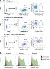
Primary NK cells exposed to Rituximab-coated Raji cells degranulate non-uniformly. (A) Examples of graphs generated using the FlowJo software (v10.7.1) depicting the flow cytometry analysis of primary NK cell degranulation. Dot plots with SSC and V500 channels (left panels) allowed the discrimination of viable cells (FVS510-negative events). Dot plots with SSC and PE/Cy7 channels (middle panels) enabled the detection of the NK cell population (CD56-PE/Vio770-positive events), followed by an analysis of NK cells for the presence of CD107a-FITC signal (right panels). The samples of NK cells alone (top row) exhibited the CD107a-negative events (non-responders) exclusively. The samples of NK cells co-incubated with Raji cells, without RTX (middle row), also showed the presence of mainly CD107a-negative events (non-responders). However, the samples of NK cells co-incubated with RTX-coated Raji cells (bottom row) exhibited both the CD107a-negative (non-responders) and the CD107a-positive (responders) populations. (B) Examples of graphs generated using the FlowJo software depicting the histogram plots of CD107a analysis in NK cells isolated from three healthy donors. Only the co-incubation with RTX-coated Raji cells induced the appearance of CD107a-positive events in NK cell populations.
Figure 5
Figure 5
The degranulating population of NK cells produces increased levels of TNFα. (A) Examples of graphs generated using the FlowJo software depicting the flow cytometry analysis and percentage of NK cells exhibiting low and high intracellular TNFα staining in the non-responder (CD107a-negative events) and responder populations (CD107a-positive events) of NK cells co-incubated with Raji in the absence of RTX (left panel) and in the presence of RTX (right panel). (B) The percentage of cells with low and high TNFα staining in NK cells deriving from three donors was summarized in the tables. (C) The graph presents the mean fluorescence intensity (MFI) of TNFα staining in the non-responders and the responders populations of NK cells co-incubated with Raji without RTX (−RTX) or with RTX-coated Raji (+RTX) ** <0.005; **** < 0.0001.
Figure 5
Figure 5
The degranulating population of NK cells produces increased levels of TNFα. (A) Examples of graphs generated using the FlowJo software depicting the flow cytometry analysis and percentage of NK cells exhibiting low and high intracellular TNFα staining in the non-responder (CD107a-negative events) and responder populations (CD107a-positive events) of NK cells co-incubated with Raji in the absence of RTX (left panel) and in the presence of RTX (right panel). (B) The percentage of cells with low and high TNFα staining in NK cells deriving from three donors was summarized in the tables. (C) The graph presents the mean fluorescence intensity (MFI) of TNFα staining in the non-responders and the responders populations of NK cells co-incubated with Raji without RTX (−RTX) or with RTX-coated Raji (+RTX) ** <0.005; **** < 0.0001.
Figure 6
Figure 6
The expression of a small number of genes is significantly changed in response to CD16 stimulation. (A) The volcano plot compares gene expression in the responder versus the non-responder groups of NK cells. Green dots represent genes with significant down-regulation (log2 fold change < −0.6), while the red dot represents a significantly up-regulated gene in responder NK cells. (B) The heatmap presents the hierarchical clustering of mRNAs exhibiting significant (p-value < 0.05) changes between the non-responder (“Non-resp.”) and responder (“Resp.”) populations. (C) The qRT-PCR analysis of KIT mRNA levels (left panel) and TNFSF4 mRNA levels (right panel) in sorted non-responder (NR) and responder (Resp) populations of primary NK cells deriving from healthy donors (Donor 1, Donor 2, and Donor 3). The Y axis presents the “Target per Reference” expression values, therefore providing normalization to reference genes, namely 18S rRNA and GAPDH.

Similar articles

References

    1. Imai K., Matsuyama S., Miyake S., Suga K., Nakachi K. Natural cytotoxic activity of peripheral-blood lymphocytes and cancer incidence: An 11-year follow-up study of a general population. Lancet. 2000;356:1795–1799. doi: 10.1016/S0140-6736(00)03231-1. - DOI - PubMed
    1. Veuillen C., Aurran-Schleinitz T., Castellano R., Rey J., Mallet F., Orlanducci F., Pouyet L., Just-Landi S., Coso D., Ivanov V., et al. Primary B-CLL resistance to NK cell cytotoxicity can be overcome in vitro and in vivo by priming NK cells and monoclonal antibody therapy. J. Clin. Immunol. 2012;32:632–646. doi: 10.1007/s10875-011-9624-5. - DOI - PubMed
    1. Pierpont T.M., Limper C.B., Richards K.L. Past, Present, and Future of Rituximab-The World’s First Oncology Monoclonal Antibody Therapy. Front. Oncol. 2018;8:163. doi: 10.3389/fonc.2018.00163. - DOI - PMC - PubMed
    1. Prica A., Baldassarre F., Hicks L.K., Imrie K., Kouroukis T., Cheung M., Members of the Hematology Disease Site Group of the Cancer Care Ontario Program in Evidence-Based Care Rituximab in Lymphoma and Chronic Lymphocytic Leukaemia: A Practice Guideline. Clin. Oncol. 2017;29:e13–e28. doi: 10.1016/j.clon.2016.09.004. - DOI - PubMed
    1. Salles G., Barrett M., Foa R., Maurer J., O’Brien S., Valente N., Wenger M., Maloney D.G. Rituximab in B-Cell Hematologic Malignancies: A Review of 20 Years of Clinical Experience. Adv. Ther. 2017;34:2232–2273. doi: 10.1007/s12325-017-0612-x. - DOI - PMC - PubMed

MeSH terms

LinkOut - more resources