The Exosomes of Stem Cells from Human Exfoliated Deciduous Teeth Suppress Inflammation in Osteoarthritis
- PMID: 39201248
- PMCID: PMC11354937
- DOI: 10.3390/ijms25168560
The Exosomes of Stem Cells from Human Exfoliated Deciduous Teeth Suppress Inflammation in Osteoarthritis
Abstract
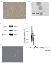
Hyaluronic acid injection is commonly used clinically to slow down the development of osteoarthritis (OA). A newly developed therapeutic method is to implant chondrocytes/stem cells to regenerate cartilage in the body. The curative effect of stem cell therapy has been proven to come from the paracrine of stem cells. In this study, exosomes secreted by stem cells from human exfoliated deciduous teeth (SHED) and hyaluronic acid were used individually to evaluate the therapeutic effect in slowing down OA. SHED was cultured in a serum-free medium for three days, and the supernatant was collected and then centrifuged with a speed difference to obtain exosomes containing CD9 and CD63 markers, with an average particle size of 154.1 nm. SW1353 cells were stimulated with IL-1β to produce the inflammatory characteristics of OA and then treated with 40 μg/mL exosomes and hyaluronic acid individually. The results showed that the exosomes successfully inhibited the pro-inflammatory factors, including TNF-α, IL-6, iNOS, NO, COX-2 and PGE2, induced by IL-1β and the degrading enzyme of the extrachondral matrix (MMP-13). Collagen II and ACAN, the main components of the extrachondral matrix, were also increased by 1.76-fold and 2.98-fold, respectively, after treatment, which were similar to that of the normal joints. The effect can be attributed to the partial mediation of SHED exosomes to the NF-κB pathway, and the ability of exosomes to inhibit OA is found not inferior to that of hyaluronic acid.
Keywords: SW1353 cells; exosome; hyaluronic acid; osteoarthritis (OA); stem cells from human exfoliated deciduous teeth (SHED).
Conflict of interest statement
The authors declare no conflicts of interest.
Figures


Similar articles
-
Protective effects of stem cells from human exfoliated deciduous teeth derived conditioned medium on osteoarthritic chondrocytes.PLoS One. 2020 Sep 4;15(9):e0238449. doi: 10.1371/journal.pone.0238449. eCollection 2020. PLoS One. 2020. PMID: 32886713 Free PMC article.
-
Exosomes of stem cells from human exfoliated deciduous teeth as an anti-inflammatory agent in temporomandibular joint chondrocytes via miR-100-5p/mTOR.Stem Cell Res Ther. 2019 Jul 29;10(1):216. doi: 10.1186/s13287-019-1341-7. Stem Cell Res Ther. 2019. PMID: 31358056 Free PMC article.
-
Effects of human umbilical cord mesenchymal stem cell-derived exosomes in the rat osteoarthritis models.Stem Cells Transl Med. 2024 Aug 16;13(8):803-811. doi: 10.1093/stcltm/szae031. Stem Cells Transl Med. 2024. PMID: 38913985 Free PMC article.
-
Mesenchymal stem cell-derived exosomes: a new therapeutic approach to osteoarthritis?Stem Cell Res Ther. 2019 Nov 21;10(1):340. doi: 10.1186/s13287-019-1445-0. Stem Cell Res Ther. 2019. PMID: 31753036 Free PMC article. Review.
-
Roles of Exosomes from Mesenchymal Stem Cells in Treating Osteoarthritis.Cell Reprogram. 2020 Jun;22(3):107-117. doi: 10.1089/cell.2019.0098. Epub 2020 May 4. Cell Reprogram. 2020. PMID: 32364765 Review.
References
MeSH terms
Substances
Grants and funding
LinkOut - more resources
Full Text Sources
Medical
Research Materials
Miscellaneous

