. 2024 Aug 21;12(8):1920.
doi: 10.3390/biomedicines12081920.

NeuroAiDTM-II (MLC901) Promoted Neurogenesis by Activating the PI3K/AKT/GSK-3β Signaling Pathway in Rat Spinal Cord Injury Models

Affiliations

NeuroAiDTM-II (MLC901) Promoted Neurogenesis by Activating the PI3K/AKT/GSK-3β Signaling Pathway in Rat Spinal Cord Injury Models

Anam Anjum et al. Biomedicines. .

Abstract

Traumatic damage to the spinal cord (SCI) frequently leads to irreversible neurological deficits, which may be related to apoptotic neurodegeneration in nerve tissue. The MLC901 treatment possesses neuroprotective and neuroregenerative activity. This study aimed to explore the regenerative potential of MLC901 and the molecular mechanisms promoting neurogenesis and functional recovery after SCI in rats. A calibrated forceps compression injury for 15 s was used to induce SCI in rats, followed by an examination of the impacts of MLC901 on functional recovery. The Basso, Beattie, and Bresnahan (BBB) scores were utilized to assess neuronal functional recovery; H&E and immunohistochemistry (IHC) staining were also used to observe pathological changes in the lesion area. Somatosensory Evoked Potentials (SEPs) were measured using the Nicolet® Viking Quest™ apparatus. Additionally, we employed the Western blot assay to identify PI3K/AKT/GSK-3β pathway-related proteins and to assess the levels of GAP-43 and GFAP through immunohistochemistry staining. The study findings revealed that MLC901 improved hind-limb motor function recovery, alleviating the pathological damage induced by SCI. Moreover, MLC901 significantly enhanced locomotor activity, SEPs waveform, latency, amplitude, and nerve conduction velocity. The treatment also promoted GAP-43 expression and reduced reactive astrocytes (GFAP). MLC901 treatment activated p-AKT reduced p-GSK-3β expression levels and showed a normalized ratio (fold changes) relative to β-tubulin. Specifically, p-AKT exhibited a 4-fold increase, while p-GSK-3β showed a 2-fold decrease in T rats compared to UT rats. In conclusion, these results suggest that the treatment mitigates pathological tissue damage and effectively improves neural functional recovery following SCI, primarily by alleviating apoptosis and promoting neurogenesis. The underlying molecular mechanism of this treatment mainly involves the activation of the PI3K/AKT/GSK-3β pathway.

Keywords: GAP-43; GFAP; MLC901 (NeuroAiDTM-II); calibrated forceps compression injury; rat mechanical spinal injury model; signaling pathway.


Conflict of interest statement

The authors declare no conflicts of interest in preparing this article.

Figures

Figure 1
Figure 1
Diagrammatic representation of the step-by-step procedure for creating a mechanical spinal cord injury (SCI) model using the calibrated forceps compression method. (a) Sublime Animal Position: The animal is positioned in a prone orientation on the surgical table, ensuring stability and access to the spinal region. (b) Marking T10, T12, and T13 vertebra: Identification of the vertebrae to be targeted and marked for precise surgical intervention. (c) subcutaneous cut: An incision is made through the skin to gain access to the underlying tissues. (d) Removing Muscles: The overlying muscles are carefully removed to expose the spinal column while minimizing damage to surrounding tissues. (e) Exposing Spinal Cord: The spinal column is accessed, providing visibility to the spinal cord. (f) Removing T12 Vertebrae: The T12 vertebra is removed to allow direct compression of the spinal cord. (g) Compression of the Spinal Cord for 15 Sec: The spinal cord is compressed using calibrated forceps for a precise duration to induce injury, the blue box indicates spinal cord exposure and the compression site. (h) Wound Closing (Suture of Tissue and Skin): The surgical wound is closed with sutures, including tissue layers and skin, to complete the procedure, n = 6: Indicates the number of animals used for this procedure.
Figure 2
Figure 2
Health evaluation after compression injury. (a) Changes in body weight following spinal cord injury: Injured rats initially lost more body weight on Days 3 and 7 than sham (H) rats, but later began gaining weight. Treated (T) rats lost less weight compared to untreated (UT) animals on Days 3 and 7 (mean ± standard deviation [SD]; one-way analysis of variance [ANOVA] with post hoc test, * p < 0.05, ** p < 0.01) (n = 6). (b) Urine volume per void: Manual bladder voiding by the experimenter was required for 1 week (Day 7) post-SCI, after which spontaneous recovery began. No significant difference was observed between UT and T rats. Data were analyzed using Student’s t-test for non-parametric and unpaired comparisons (n = 6). (c) Length of the incision area: The length of the incision area was measured up to Day 28 post-injury. Wound recovery continued until Day 28, with no significant difference observed between UT and T rats. Data were analyzed using Student’s t-test for unpaired comparisons (n = 6).
Figure 3
Figure 3
Assessment of locomotor function following MLC901 treatment (a) Basso, Beattie, and Bresnahan (BBB) scores, (b) distance traveled in the open field test (OFT), (c) performance in the running wheel test, (d) grid walk assessment, (e) grid distance traveled, (f) inverted grid (grip strength test), (g) total number of footsteps taken, and (h) fore- and hind-limb faults. Assessments were conducted on Day 0 (pre-injury) and Days 3, 7, 14, 21, and 28 post-injuries. Data are presented as means with error bars indicating the standard error of the mean (n = 6). Statistical analysis was performed using one-way ANOVA followed by post hoc tests. Treated (T) rats demonstrated significant recovery and improvement compared to controls. Statistical significance is indicated as * p < 0.05, ** p < 0.01, and *** p < 0.001 for comparisons among the three groups (sham [H], treated [T], and untreated [UT]).
Figure 4
Figure 4
Somatosensory Evoked Potential (SEP) Analysis. (a) Waveform: SEPs were obtained by stimulating the left hindlimb in sham (H), untreated (UT), and treated (T) rats with moderate compression injury on Day 0 (pre-injury), Day 14, and Day 28 post-injury. The waveform data show the characteristic peaks and changes over time. (b) Amplitude: Amplitude of SEPs was significantly reduced in UT rats compared to sham controls, with treated (T) rats showing a significant improvement. Statistical significance (p < 0.05) was observed between UT and T rats on Day 14 and Day 28. (c) Latency: The latency period of SEPs was significantly increased in injured rats compared to sham controls, with no significant differences between UT and T rats at Days 14 and 28. (d) Duration: The duration of SEPs, inversely related to nerve conduction velocity, was longer in UT rats compared to T rats. No significant differences were found between groups at Days 14 and 28. Data are expressed as mean ± standard deviation (SD) and were analyzed using one-way analysis of variance (ANOVA) followed by post hoc tests (n = 6). Statistical significance is indicated by * p < 0.05.
Figure 5
Figure 5
Histological analysis of spinal cord: (a) Transverse Section: Transverse spinal cord sections stained with hematoxylin and eosin (H&E) at 4 weeks post-injury. This view highlights the extent of tissue damage and demyelination in untreated (UT), treated (T), and sham (H) rats. Images are captured at 4× magnification with a scale bar of 500 µm. Significant differences in tissue integrity and lesion characteristics are evident between groups. Sham (H) rats show normal spinal cord morphology, while UT rats display extensive tissue damage and large cystic cavities. Treated (T) rats show reduced lesion size and less severe tissue degeneration compared to UT rats. (b) Longitudinal Section: Longitudinal spinal cord sections stained with H&E, provide a detailed view of demyelination and histopathological features across the length of the injury. Images are taken at 10× magnification with a scale bar of 500 µm. Similar to the transverse sections, UT rats exhibit pronounced tissue loss and hemorrhagic foci, while T rats demonstrate reduced tissue damage and smaller lesions. Sham (H) rats present with normal spinal cord structure. (c) Relative Tissue Loss: Quantitative analysis of tissue loss in the center of the lesion, normalized to spinal cord sections from sham (H) rats without lesions. Bars represent the means and standard deviation (SD) of tissue loss measurements. Significant differences are observed between treatment groups, with T rats showing less tissue loss compared to UT rats (p < 0.05). Data were analyzed by one-way analysis of variance (ANOVA) with the Dunnett post hoc test. (d) Lesion Size: Measurement of lesion size in the center of the injury site, comparing untreated (UT), treated (T), and sham (H) rats. Bars represent the means and standard deviation (SD) of lesion size measurements. Statistical Significance: Significant reduction in lesion size in T rats compared to UT rats (p < 0.05 *). Data were analyzed using one-way ANOVA with the Dunnett post hoc test. These findings confirm that MLC901 treatment effectively mitigates spinal cord damage and supports tissue repair following mechanical compression injury.
Figure 6
Figure 6
Comparison of GAP-43 and GFAP expression 28 days post-compression spinal cord injury with MLC901 treatment. (a,b) GAP-43 Expression: Immunofluorescence staining revealed that the intensity of Growth-associated protein 43 (GAP-43, green) was significantly higher in MLC901-treated (T) rats compared to untreated (UT) rats 28 days post-injury ((a,b); p < 0.05 *). This increased GAP-43 immunoreactivity indicates enhanced neurogenesis and axonal growth in the T group. (c,d) GFAP Expression: Conversely, the intensity of Glial fibrillary acidic protein (GFAP, red) immunoreactivity was notably lower in the T rats compared to the UT rats ((c,d); p < 0.05 *). Reduced GFAP staining suggests that MLC901 treatment effectively mitigates astrocytic scar formation and inflammation, contributing to a more favorable environment for neuroprotection and recovery. Nuclei Staining: Nuclei are stained with DAPI (blue), which helps to visualize cell bodies in the spinal cord sections. The higher magnification images (20×) on the right side of the figures showed morphological changes and detailed regions of demyelination with double staining for GAP-43-DAPI and GFAP-DAPI, highlighting the distinct areas of neurogenesis and astrocytic response. Scale bars are 1000 µm and 100 µm, providing context for the images’ magnification. Data are expressed as mean ± standard deviation (SD). Statistical significance was determined using Student’s t-test (n = 6). Significance is indicated as * p < 0.05. These findings support that MLC901 treatment enhances neurogenesis while reducing astrocytic scar formation, contributing to improved functional recovery following spinal cord injury.
Figure 7
Figure 7
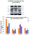
MLC901 activated the PI3K/AKT/GSK-3β signaling pathway. (a) Western Blot Analysis: To assess the relative expression levels of p-AKT, AKT, p-GSK-3β, and GAP-43 proteins in spinal cord tissues from sham (H), treated (T), and untreated (UT) rats with β-Tubulin as a loading control. (b) Mean Fluorescence Intensity (MFI): To measure the expression levels of p-AKT/AKT, p-GSK-3β, and GAP-43 (neurogenesis marker) in spinal cord tissues of sham (H), treated (T), and untreated (UT) rats. (i) p-AKT/AKT Ratio: T rats showed higher expression compared to UT rats, indicating enhanced activation of the PI3K/AKT pathway. (ii) p-GSK-3β expression: UT rats showed higher expression compared to T rats, suggesting that MLC901 treatment inhibits GSK-3β activity and reduces apoptosis. (iii) GAP-43 Expression: Significantly higher expression in T rats compared to UT rats (p < 0.05), indicating that MLC901 promotes neurogenesis. Statistical Analysis: Data are presented as mean ± standard deviation (SD) (n = 6/group). Statistical significance was determined using a one-way analysis of variance (ANOVA), followed by a post hoc test. * p < 0.05 indicates significant differences between T rats and the UT SCI group.
Figure 8
Figure 8
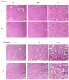
Histopathological Evaluation of Liver and Kidney Tissues. (a) Hepatic Tissue (Liver): Hematoxylin and eosin (H&E) staining of liver tissue sections to assess potential hepatotoxicity associated with MLC901 treatment, observed under different magnifications (10×, 20×, and 40×), using scale bars 50, 100, and 500 µm. The images showed no sign of sinusoidal dilatation (enlargement of the hepatic capillaries), necrosis, hemorrhage, or congestion observed in treated (T) rats associated with MLC901 treatment, in liver tissue. MLC901 treatment did not induce hepatotoxicity after 28 days, indicating the liver remained healthy under the treatment conditions, n = 6. (b) Renal Tissue (Kidney): Hematoxylin and eosin (H&E) staining of kidney tissue sections to evaluate potential nephrotoxicity associated with MLC901 treatment, observed under different magnifications (10×, 20×, and 40×), using scale bars 50, 100, and 500 µm. The images showed no vacuolization in tubular cells, focal necrosis, or hemorrhage observed in the kidney tissue of T rats. MLC901 treatment did not induce nephrotoxicity after 28 days, confirming that the kidney function remained intact, n = 6. Both liver and kidney tissues in T rats showed no adverse effects such as hepatotoxicity or nephrotoxicity, suggesting that MLC901 is safe for these organs after 28 days of treatment, UT represents untreated rats without MLC901 treatment and receiving normal saline.

Similar articles

References

    1. Hu X., Xu W., Ren Y., Wang Z., He X., Huang R., Ma B., Zhao J., Zhu R., Cheng L. Spinal cord injury: Molecular mechanisms and therapeutic interventions. Signal Transduct. Target. Ther. 2023;8:245. doi: 10.1038/s41392-023-01477-6. - DOI - PMC - PubMed
    1. Anjum A., Yazid M.D.i., Fauzi Daud M., Idris J., Ng A.M.H., Selvi Naicker A., Ismail O.H.R., Athi Kumar R.K., Lokanathan Y. Spinal cord injury: Pathophysiology, multimolecular interactions, and underlying recovery mechanisms. Int. J. Mol. Sci. 2020;21:7533. doi: 10.3390/ijms21207533. - DOI - PMC - PubMed
    1. Zheng B., Kuang Y., Yuan D., Huang H., Liu S. The research landscape of immunology research in spinal cord injury from 2012 to 2022. JOR Spine. 2023;6:e1261. doi: 10.1002/jsp2.1261. - DOI - PMC - PubMed
    1. Anjum A., Cheah Y.J., Yazid M.D.I., Daud M.F., Idris J., Ng M.H., Naicker A.S., Ismail O.H., Athi Kumar R.K., Tan G.C. Protocol paper: Kainic acid excitotoxicity-induced spinal cord injury paraplegia in Sprague–Dawley rats. Biol. Res. 2022;55:38. doi: 10.1186/s40659-022-00407-0. - DOI - PMC - PubMed
    1. Zhang W., Xu H., Li C., Han B., Zhang Y. Exploring Chinese herbal medicine for ischemic stroke: Insights into microglia and signaling pathways. Front. Pharmacol. 2024;15:1333006. doi: 10.3389/fphar.2024.1333006. - DOI - PMC - PubMed

LinkOut - more resources