. 2024 Aug 8;16(16):2791.
doi: 10.3390/cancers16162791.

SKF-96365 Expels Tyrosine Kinase Inhibitor-Treated CML Stem and Progenitor Cells from the HS27A Stromal Cell Niche in a RhoA-Dependent Mechanism

Affiliations

SKF-96365 Expels Tyrosine Kinase Inhibitor-Treated CML Stem and Progenitor Cells from the HS27A Stromal Cell Niche in a RhoA-Dependent Mechanism

Audrey Dubourg et al. Cancers (Basel). .

Abstract

Background: A major issue in Chronic Myeloid Leukemia (CML) is the persistence of quiescent leukemia stem cells (LSCs) in the hematopoietic niche under tyrosine kinase inhibitor (TKI) treatment.

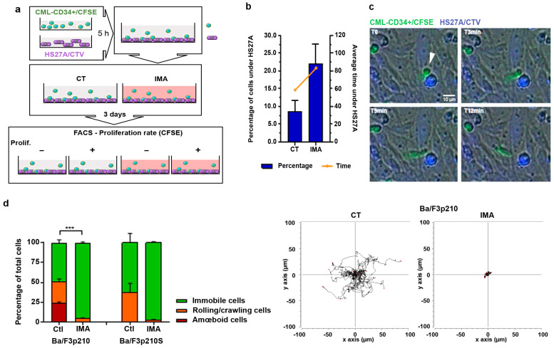
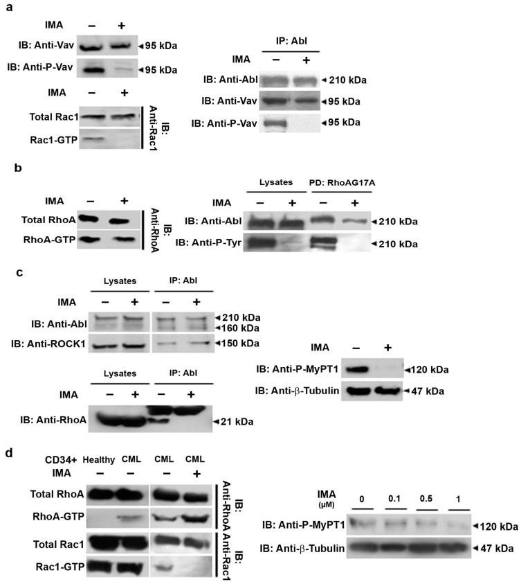
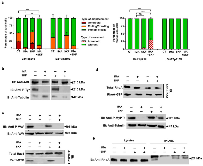
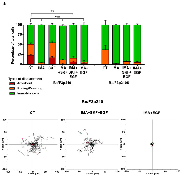
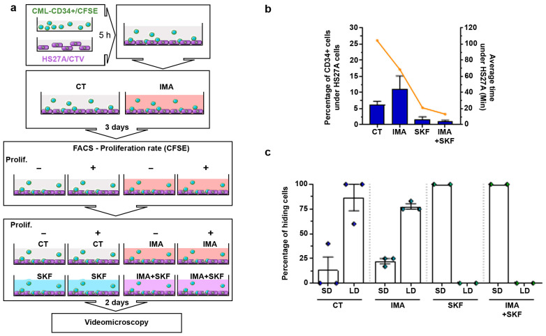
Results: Here, using CFSE sorting, we show that low-proliferating CD34+ cells from CML patients in 3D co-culture hide under HS27A stromal cells during TKI treatment-a behavior less observed in untreated cells. Under the same conditions, Ba/F3p210 cells lose their spontaneous motility. In CML CD34+ and Ba/F3p210 cells, while Rac1 is completely inhibited by TKI, RhoA remains activated but is unable to signal to ROCK. Co-incubation of Ba/F3p210 cells with TKI, SKF-96365 (a calcium channel inhibitor), and EGF restores myosin II activation and amoeboid motility to levels comparable to untreated cells, sustaining the activation of ROCK. In CFSE+ CD34+ cells containing quiescent leukemic stem cells, co-incubation of TKI with SKF-96365 induced the expulsion of these cells from the HS27A niche.

Conclusions: This study underscores the role of RhoA in LSC behavior under TKI treatment and suggests that SKF-96365 could remobilize quiescent CML LSCs through reactivation of the RhoA/ROCK pathway.

Keywords: Chronic Myeloid Leukemia; RhoGTPases; SKF-96365; leukemia niche; leukemia stem cells; tyrosine kinase inhibitors.


Conflict of interest statement

The authors declare no conflicts of interest. The funders had no role in the design of the study; in the collection, analyses, or interpretation of data; in the writing of the manuscript; or in the decision to publish the results.

Figures

Figure 1
Figure 1
Imatinib treatment modifies CML-patient CD34+ and Ba/F3p210 cell mobility. (a) CML patients’ CD34+ cells and HS27A cells were stained with 5 µM CFSE and 5 µM CTV, respectively. After 5 h, CFSE-stained CD34+ cells were added to adherent CTV-HS27A cells with or without 5 µM imatinib. After 3 days of co-culture, CFSE-CD34+ and CTV-HS27A were sorted according to CFSE intensity: low CFSE (high proliferation) and high CFSE (low proliferation). (b) The percentage of CD34+ cells hiding under HS27A and the average time spent under HS27A were determined through a time-lapse videomicroscopy analysis (N = 2 patients, n = 5). (c) Time-lapse images (t = 0; t = 3 min; t = 9 min and t = 12 min) extracted from JuliStage recordings of CFSE-stained CD34+ cells from CML patients seeded on CTV-stained HS27A stromal cells in imatinib (5 µM) conditions. Scale bar: 10 µm. Arrowhead shows CD34+ cell hiding under HS27A cells. (d) Cells (0.5 × 10⁵ Ba/F3p210 and Ba/F3p210S509A) were included in 2.5 mg/mL liquid Matrigel diluted in culture medium and cells were incubated with 5 µM imatinib. Each type of displacement (immobile cells, rolling/crawling, or amœboid displacement) was determined through time-lapse videomicroscopy analysis (n > 3). Right panel: spider graph showing the accumulated tracks of analyzed individual cell motility for Ba/F3p210 cells treated or not with imatinib 5 µM. t-test *** p ≤ 0.001.
Figure 2
Figure 2
RhoGTPase signaling in imatinib conditions. (a) Western blot analysis of phosphorylated and total Vav, activated (GTP-Rac1), and total Rac1 in Ba/F3p210 cells. Coimmunoprecipitation of Vav and P-Vav, with p210BCR-ABL. Ba/F3p210 cell lysates were immunoprecipitated using anti-ABL IgG and immunoblotted as indicated. Western blot analysis of activated (GTP-Rac1) and total Rac1 in Ba/F3p210 cells obtained via GST-pulldown. (b) Western blot analysis of activated (GTP-RhoA) and total RhoA in Ba/F3p210 cells obtained via GST-pulldown. Affinity-binding assay of phosphorylated and total p210BCR-ABL with negative dominant RhoA (RhoAG17A). Ba/F3p210 cell lysates were immunoprecipitated with sepharose-bound GST-fused with RhoA-G17A and immunoblotted as indicated. (c) Coimmunoprecipitation of ROCK1 and RhoA with p210BCR-ABL. Ba/F3p210 cell lysates were immunoprecipitated with anti-ABL IgG and immunoblotted as indicated. Western blot analysis of phosphorylated MYPT1 and tubulin in Ba/F3p210 cells. (d) Western blot analysis of activated (GTP-RhoA or GTP-Rac1) and total RhoA or Rac1 in CML patient CD34+ cells obtained via GST-pulldown. Western blot analysis of phosphorylated MYPT1 and β-tubulin in CML patient CD34+ cells. Each experiment was carried out 3 times. Ba/F3p210 and CD34+ patient cells were treated with 1 µM ((d) top), 5 µM imatinib (ac), or as indicated (d).
Figure 3
Figure 3
SKF-96365 induces amœboid movements in immobile imatinib-treated Ba/F3p210 cells. (a) Cells (0.5 × 10⁵ Ba/F3p210 and Ba/F3p210S509A) were included in 2.5 mg/mL liquid Matrigel diluted in culture medium and incubated with or without 5 µM imatinib and/or 40 µM SKF-96365. Each type of displacement (immobile cells, rolling/crawling, or amœboid displacement) was determined through time-lapse videomicroscopy analysis. The right panel shows the analysis of amoeboid contractions in immobile cells (n > 3). t-test ** p ≤ 0.01; *** p ≤ 0.001; ns: non-significant. (b) Western blot analysis of phosphorylated and total p210BCR-ABL in Ba/F3p210 cells during imatinib and SKF-96365 treatment. (c) Western blot analysis of phosphorylated and total Vav, activated (GTP-Rac1), and total Rac1 in Ba/F3p210 cells during imatinib and SKF-96365 treatment. (d) Upper panel: Western blot analysis of activated (GTP-RhoA) and total RhoA or Rac1 in Ba/F3p210 cells obtained via GST-pulldown. Lower panel: Western blot analysis of phosphorylated MYPT1 and β-tubulin in CML patient CD34+ cells. (e) Coimmunoprecipitation of RhoA with p210BCR-ABL. Ba/F3p210 cell lysates were immunoprecipitated with anti-ABL IgG and immunoblotted as indicated.
Figure 4
Figure 4
EGF in conjunction with SKF-96365 restores Ba/F3p210 cell motility inhibited by imatinib. (a) Cells (0.5 × 10⁵ Ba/F3p210 and Ba/F3p210S509A) were included in 2.5 mg/mL liquid Matrigel diluted in culture medium and incubated with or without 5 µM imatinib and/or 40 µM SKF-96365 and/or EGF 10 ng/mL. Each type of displacement (immobile cells, rolling/crawling, or amœboid displacement) was determined through time-lapse videomicroscopy analysis (n > 3). Right panel: spider graph showing the accumulated tracks of analyzed individual cell motility for Ba/F3p210 cells treated or not with imatinib 5 µM. t-test ** p ≤ 0.01; *** p ≤ 0.001. (b) Analysis of velocity (µm/s) (left) and accumulated distance (µm) (right) of Ba/F3p210 cells treated with SKF-96365 alone or imatinib+ SKF-96365 and EGF. Data were obtained using TrackMate plugins. * p < 0.05; ** p < 0.01; ns: non-significant. (c) Western blot analysis of phosphorylated p210BCR-ABL in Ba/F3p210 cells under the indicated conditions. (d) Upper panel: Western blot analysis of activated (GTP-RhoA) and total RhoA in Ba/F3p210 cells obtained via GST-pulldown. Lower panel: Western blot analysis of phosphorylated MYPT1 and actin in Ba/F3p210 cells. (e) Western blot analysis of activated (GTP-Rac1) and total Rac1, and phosphorylated and total Vav in Ba/F3p210 cells under the indicated conditions.
Figure 4
Figure 4
EGF in conjunction with SKF-96365 restores Ba/F3p210 cell motility inhibited by imatinib. (a) Cells (0.5 × 10⁵ Ba/F3p210 and Ba/F3p210S509A) were included in 2.5 mg/mL liquid Matrigel diluted in culture medium and incubated with or without 5 µM imatinib and/or 40 µM SKF-96365 and/or EGF 10 ng/mL. Each type of displacement (immobile cells, rolling/crawling, or amœboid displacement) was determined through time-lapse videomicroscopy analysis (n > 3). Right panel: spider graph showing the accumulated tracks of analyzed individual cell motility for Ba/F3p210 cells treated or not with imatinib 5 µM. t-test ** p ≤ 0.01; *** p ≤ 0.001. (b) Analysis of velocity (µm/s) (left) and accumulated distance (µm) (right) of Ba/F3p210 cells treated with SKF-96365 alone or imatinib+ SKF-96365 and EGF. Data were obtained using TrackMate plugins. * p < 0.05; ** p < 0.01; ns: non-significant. (c) Western blot analysis of phosphorylated p210BCR-ABL in Ba/F3p210 cells under the indicated conditions. (d) Upper panel: Western blot analysis of activated (GTP-RhoA) and total RhoA in Ba/F3p210 cells obtained via GST-pulldown. Lower panel: Western blot analysis of phosphorylated MYPT1 and actin in Ba/F3p210 cells. (e) Western blot analysis of activated (GTP-Rac1) and total Rac1, and phosphorylated and total Vav in Ba/F3p210 cells under the indicated conditions.
Figure 5
Figure 5
SKF-96365 expels low-proliferating CD34+ cells from the HS27A niche. (a) CML patient CD34+ cells and HS27A cells were stained with 5 µM CFSE and 5 µM CTV, respectively. After 5 h, CFSE-stained CD34+ cells were added to adherent CTV-HS27A cells with or without 5 µM imatinib. After 3 days of co-culture, CFSE-CD34+ and CTV-HS27A were sorted according to CFSE intensity: low CFSE (high proliferation) and high CFSE (low proliferation). CFSE-sorted CD34+ cells from CML patients were reseeded on previously cultured CTV-stained HS27A cells and cultured for 2 days under the indicated conditions. Then, videomicroscopy was performed using JuliStage in fluorescent mode during 6 h recording. (b) The percentage of CD34+ cells hiding under HS27A cells and the average time spent under HS27A cells were determined through time-lapse videomicroscopy analysis (N = 2 patients, n = 5). (c) Analysis of the hiding behavior of low-proliferating CD34+ cells from CML patients during the recording time. Two behaviors were identified corresponding to a short-duration stay under HS27A cells (SD; <30 min) and a long-duration stay (LD; 30 to >300 min) (see Supplementary Figure S5 for details).

Similar articles

References

    1. Osman A.E.G., Deininger M.W. Chronic Myeloid Leukemia: Modern therapies, current challenges and future directions. Blood Rev. 2021;49:100825. doi: 10.1016/j.blre.2021.100825. - DOI - PMC - PubMed
    1. Mahon F.X., Réa D., Guilhot J., Guilhot F., Huguet F., Nicolini F., Legros L., Charbonnier A., Guerci A., Varet B. Discontinuation of imatinib in patients with chronic myeloid leukaemia who have maintained complete molecular remission for at least 2 years: The prospective, multicentre Stop Imatinib (STIM) trial. Lancet Oncol. 2010;11:1029–1035. doi: 10.1016/S1470-2045(10)70233-3. - DOI - PubMed
    1. Chomel J.C., Bonnet M.L., Sorel N., Bertrand A., Meunier M.C., Fichelson S., Melkus M., Bennaceur-Griscelli A., Guilhot F., Turhan A.G. Leukemic stem cell persistence in chronic myeloid leukemia patients with sustained undetectable molecular residual disease. Blood. 2011;118:3657–3660. doi: 10.1182/blood-2011-02-335497. - DOI - PMC - PubMed
    1. Charaf L., Mahon F.X., Lamrissi-Garcia I., Moranvillier I., Beliveau F., Cardinaud B., Dabernat S., de Verneuil H., Moreau-Gaudry F., Bedel A. Effect of tyrosine kinase inhibitors on stemness in normal and chronic myeloid leukemia cells. Leukemia. 2017;31:65–74. doi: 10.1038/leu.2016.154. - DOI - PubMed
    1. Moreno-Lorenzana D., Avilés-Vazquez S., Sandoval Esquivel M.A., Alvarado-Moreno A., Ortiz-Navarrete V., Torres-Martínez H., Ayala-Sánchez M., Mayani H., Chavez-Gonzalez A. CDKIs p18INK4c and p57Kip2 are involved in quiescence of CML leukemic stem cells after treatment with TKI. Cell Cycle. 2016;15:1276–1287. doi: 10.1080/15384101.2016.1160976. - DOI - PMC - PubMed

LinkOut - more resources