. 2024 Jul 23;14(8):888.
doi: 10.3390/biom14080888.

FGF1 Suppresses Allosteric Activation of β3 Integrins by FGF2: A Potential Mechanism of Anti-Inflammatory and Anti-Thrombotic Action of FGF1

Affiliations

FGF1 Suppresses Allosteric Activation of β3 Integrins by FGF2: A Potential Mechanism of Anti-Inflammatory and Anti-Thrombotic Action of FGF1

Yoko K Takada et al. Biomolecules. .

Abstract

Several inflammatory cytokines bind to the allosteric site (site 2) and allosterically activate integrins. Site 2 is also a binding site for 25-hydroxycholesterol, an inflammatory lipid mediator, and is involved in inflammatory signaling (e.g., TNF and IL-6 secretion) in addition to integrin activation. FGF2 is pro-inflammatory and pro-thrombotic, and FGF1, homologous to FGF2, has anti-inflammatory and anti-thrombotic actions, but the mechanism of these actions is unknown. We hypothesized that FGF2 and FGF1 bind to site 2 of integrins and regulate inflammatory signaling. Here, we describe that FGF2 is bound to site 2 and allosterically activated β3 integrins, suggesting that the pro-inflammatory action of FGF2 is mediated by binding to site 2. In contrast, FGF1 bound to site 2 but did not activate these integrins and instead suppressed integrin activation induced by FGF2, indicating that FGF1 acts as an antagonist of site 2 and that the anti-inflammatory action of FGF1 is mediated by blocking site 2. A non-mitogenic FGF1 mutant (R50E), which is defective in binding to site 1 of αvβ3, suppressed β3 integrin activation by FGF2 as effectively as WT FGF1.

Keywords: FGF1; FGF2; anti-inflammatory action; anti-thrombotic action; integrin.


Conflict of interest statement

The authors declare no conflicts of interest.

Figures
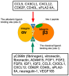
Figure 1
Figure 1
Binding of ligands to the classical-ligand binding site (site 1) and the allosteric binding site (site 2) of αvβ3. Fibrinogen γ-chain C-terminal peptide (γC399tr) and ADAM15 [20] specifically bind to the classical ligand binding site (site1) of αvβ3. FGF9 [21], the heparin-binding site of VEGF165 [22]. FGF1 [5], FGF2 [6], IGF1 [23], IGF2 [24], neuregulin [25], pro-inflammatory CX3CL1 [18], CCL5 [14], CD40L [25,26], CD62P binds to site 2 and activate αvβ3 [17]. Site 2-derived peptides bind to these ligands and suppress integrin activation [15].
Figure 2
Figure 2
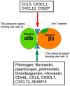
The binding of ligands to the Fibrinogen γ-chain C-terminal peptide (γC390-411) and ADAM15 [27] specifically occurs at the classical ligand binding site (site1) of αIIbβ3. Several inflammatory chemokines (CCL5, CX3CL1, CXCL12) [14] and pro-inflammatory CD62P bind to site 2 and activate αIIbβ3 [17]. Site 2-derived peptides bind to these ligands and suppress integrin activation [15].
Figure 3
Figure 3
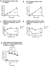
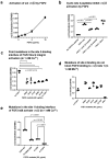
FGF2 and FGF1 bind to site 1 of αIIbβ3. (a) FGF2 binds to soluble αIIbβ3. Soluble αIIbβ3 was incubated with immobilized FGF2 in 1 mM Mn2+. Bound αIIbβ3 was quantified using anti-β3 and HRP-conjugated anti-mouse IgG. (b) FGF1 binds to soluble αIIbβ3. The binding of soluble αIIbβ3 to immobilized FGF1 was measured as in (a), except FGF1 was used instead of FGF2. (c,d) Inhibition of FGF1/FGF2 binding to soluble αIIbβ3 by ADAM15, another ligand to αIIbβ3. Wells of 96-well microtiter plate were coated with FGF2 (c) or FGF1 (d) and incubated with soluble αIIbβ3 in the presence of ADAM15 disintegrin domain fused to GST or control GST in 1 mM Mn2+. (e) Effect of FGF2 mutation that blocked binding to αvβ3 (site 1) on binding to αIIbβ3. FGF2 mutants defective in binding to αvβ3 (site 1) were tested for their ability to bind to αIIbβ3 in 1 mM Mn2+. The data are shown as means +/− SD in triplicate experiments.
Figure 4
Figure 4
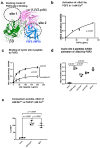
FGF2 binds to site 2 of integrin αIIbβ3 and activates αIIbβ3. (a) Docking simulation of the interaction between FGF2 (1BFG.pdb) and αvβ3 (closed headpiece form, 1JV2.pdb). (b) FGF2 activates αIIbβ3. The fibrinogen fragment (γC390-411) fused to GST, a specific ligand to αIIbβ3, was immobilized to wells of a 96-well microtiter plate and incubated with soluble αIIbβ3 (1 μg/mL) and bound αIIbβ3 was measured in 1 mM Ca2+ (to keep integrins inactive). (c) Binding of FGF2 to site 2 peptide of β3. FGF2 was immobilized to wells of a 96-well microtiter plate and incubated with cyclic site 2 peptide fused to GST or control scrambled β3 peptide, and the bound peptide was measured using anti-GST. (d) Cyclic site 2 peptides inhibit activation of αIIbβ3 by FGF2. Activation of αIIbβ3 was measured as described in (b). The concentrations used were 20 μg/mL (FGF2) and 100 μg/mL (site 2 peptides). Bound integrin was measured using anti-β3 mAb. (e) FGF2 activates soluble αIIbβ3 to an extent similar to that of 1 mM Mn2+. Activation of αIIbβ3 was measured as described in (b) using 1 mM Mn2+ or FGF2 (50 μg/mL). The data are normalized with 1 mM Mn2+ as 100%. The data are shown as means +/− SD in triplicate experiments. ANOVA using Prism 10 was used for statistical analysis (n = 3).
Figure 5
Figure 5
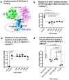
Point mutations in site 2-binding interface of FGF2 effectively reduce activation of integrin αIIbβ3 by FGF2. (a) Positions of amino acid residues involved in site 2 binding predicted by docking simulation. K119E/R120E and K125E mutations suppressed FGF2 binding to integrin site 1 of αvβ3 and thereby suppressed FGF2 mitogenicity [6]. Arg72, Lys77, Lys86, and Lys110 are in the predicted site 2-binding interface of FGF2. (b) Mutations in the site 2 binding interface of FGF2 blocked activation of αIIbβ3 in 1 mM Ca2+. (c) The point mutations in the predicted site 2-binding site of FGF2 did not affect FGF2 binding to site 1 in 1 mM Mn2+. The results indicate that site 1 and site 2-binding sites in FGF2 are distinct. (d) FGF2 mutants of K119E/R120E and K125E still activate αIIbβ3. The data are shown as means +/− SD in triplicate experiments.
Figure 6
Figure 6
FGF2 binds to site 2 of integrin and αvβ3 and activates integrins (act as an agonist). (a) FGF2 activated αvβ3. The fibrinogen fragment (γC399tr), a specific ligand to αvβ3, was immobilized to wells of a 96-well microtiter plate and incubated with soluble αvβ3 (1 μg/mL) in the presence of FGF2, and bound αvβ3 was measured using anti-β3 mAb. The data show that FGF2 activated αvβ3. (b) Cyclic site 2 peptides inhibit activation of αvβ3 by FGF2. Activation of αvβ3 was measured as described in (a). The concentrations used were 20 μg/mL (FGF2) and 100 μg/mL (site 2 peptides). Bound integrin was measured using anti-β3 mAb. Cyclic site 2 peptides from β1 or β3 suppressed αvβ3 activation by FGF2, but control β3 scrambled peptide did not. (c) FGF2 with point mutations in the predicted site 2-binding interface of FGF2 did not activate integrin αvβ3. (d) Point mutations in the predicted site 2-binding interface did not affect FGF2 binding to site 1 in 1 mM Mn2+. Wells of 96-well microtiter plate were coated with FGF2 WT and mutants and incubated with soluble αvβ3 in 1 mM Mn2+. Bound αvβ3 was quantified using anti-β3 and anti-mouse IgG conjugated with HRP. (e) Mutations in the site 1 binding interface of FGF2 still activate αvβ3 (in 1 mM Ca2+). Activation assays were performed as described in (a). The data are shown as means +/− SD in triplicate experiments.
Figure 7
Figure 7
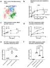
FGF1 binds to site 2 but suppresses FGF2-induced activation of αIIbβ3. (a) FGF1 is predicted to bind to site 2. Docking simulation of the interaction between FGF1 and site 2 of close headpiece form of αvβ3 (1JV2.pdb). (b) Binding of cyclic site 2 peptide to FGF1. (c) FGF1 does not activate soluble αIIbβ3. Wells of 96-well microtiter plate were coated with γC390-411, a specific ligand to αIIbβ3, and incubated with soluble αIIbβ3 in the presence of WT FGF2 or FGF1 in 1 mM Ca2. (d,e). FGF1 suppresses FGF2-induced activation of soluble αIIbβ3. Activation of soluble αIIbβ3 was assayed as described in (b). (f) Non-mitogenic FGF1 mutant (R50E) suppressed integrin activation by FGF2 at a level comparable to that of WT FGF1 in Ca2+.
Figure 8
Figure 8
FGF1 binds to site 2 but does not activate αvβ3. FGF1 suppressed integrin activation by FGF2. (a) FGF2 allosterically activated soluble αvβ3 in a dose-dependent manner, but FGF1 did not. Wells of 96-well microtiter plate were coated with γC399, a specific ligand for αvβ3, and incubated with soluble αvβ3 in 1 mM Ca2+. Bound αvβ3 was quantified using anti-β3. (b,c) FGF1 inhibits integrin activation by FGF2. Soluble αvβ3 (1 μg/mL) was incubated with immobilized ligand (γC399tr specific to αvβ3) in the presence of FGF2 and/or FGF1 in 1 mM Ca2+. (d) Non-mitogenic FGF1 mutant (R50E) suppressed integrin activation by FGF2 at a level comparable to that of WT FGF1 in Ca2+.
Figure 9
Figure 9
Agonistic action of FGF2 and antagonistic action of FGF1 to the allosteric site (site 2) of β3 integrins. The present study showed that FGF1 and FGF2 bind to the site 1 of β3 integrins. FGF2 (stored in platelet granules) binds to site 2 as well and induces allosteric activation of β3 integrins, leading to platelet aggregation and pro-inflammatory signals. In contrast, FGF1 binds to site 2 but suppresses β3 integrin activation induced by FGF2. This is a potential mechanism of anti-thrombotic action or anti-inflammatory action of FGF1. Non-mitogenic FGF1 R50E mutant is comparable to WT FGF1 in inhibiting FGF2-induced activation of β3 integrins. FGF1 R50E has potential as an anti-thrombotic and anti-inflammatory agent.

Similar articles

References

    1. Takada Y., Ye X., Simon S. The integrins. Genome Biol. 2007;8:215. doi: 10.1186/gb-2007-8-5-215. - DOI - PMC - PubMed
    1. Hynes R.O. Integrins: Bidirectional, allosteric signaling machines. Cell. 2002;110:673–687. doi: 10.1016/S0092-8674(02)00971-6. - DOI - PubMed
    1. Brooks P.C., Clark R.A., Cheresh D.A. Requirement of vascular integrin alpha v beta 3 for angiogenesis. Science. 1994;264:569–571. doi: 10.1126/science.7512751. - DOI - PubMed
    1. Cheresh D.A., Stupack D.G. Regulation of angiogenesis: Apoptotic cues from the ECM. Oncogene. 2008;27:6285–6298. doi: 10.1038/onc.2008.304. - DOI - PubMed
    1. Mori S., Wu C.-Y., Yamaji S., Saegusa J., Shi B., Ma Z., Kuwabara Y., Lam K.S., Isseroff R.R., Takada Y.K., et al. Direct binding of integrin alphavbeta3 to FGF1 plays a role in FGF1 signaling. J. Biol. Chem. 2008;283:18066–18075. doi: 10.1074/jbc.M801213200. - DOI - PMC - PubMed

Grants and funding

This work is partly supported by the UC Davis Comprehensive Cancer Center Support Grant (CCSG) awarded by the National Cancer Institute (NCI P30CA093373).