. 2024 Aug 14;13(16):1350.
doi: 10.3390/cells13161350.

Actin Cytoskeleton and Integrin Components Are Interdependent for Slit Diaphragm Maintenance in Drosophila Nephrocytes

Affiliations

Actin Cytoskeleton and Integrin Components Are Interdependent for Slit Diaphragm Maintenance in Drosophila Nephrocytes

Megan Delaney et al. Cells. .

Abstract

In nephrotic syndrome, the podocyte filtration structures are damaged in a process called foot process effacement. This is mediated by the actin cytoskeleton; however, which actins are involved and how they interact with other filtration components, like the basement membrane, remains poorly understood. Here, we used the well-established Drosophila pericardial nephrocyte-the equivalent of podocytes in flies-knockdown models (RNAi) to study the interplay of the actin cytoskeleton (Act5C, Act57B, Act42A, and Act87E), alpha- and beta-integrin (basement membrane), and the slit diaphragm (Sns and Pyd). Knockdown of an actin gene led to variations of formation of actin stress fibers, the internalization of Sns, and a disrupted slit diaphragm cortical pattern. Notably, deficiency of Act5C, which resulted in complete absence of nephrocytes, could be partially mitigated by overexpressing Act42A or Act87E, suggesting at least partial functional redundancy. Integrin localized near the actin cytoskeleton as well as slit diaphragm components, but when the nephrocyte cytoskeleton or slit diaphragm was disrupted, this switched to colocalization, both at the surface and internalized in aggregates. Altogether, the data show that the interdependence of the slit diaphragm, actin cytoskeleton, and integrins is key to the structure and function of the Drosophila nephrocyte.

Keywords: Drosophila; actin cytoskeleton; integrin; lacunar channel; nephrocyte; piezo; slit diaphragm.


Conflict of interest statement

The authors declare no conflict of interest.

Figures
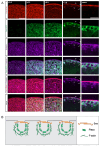
Figure 1
Figure 1
Actin cytoskeleton encloses Piezo below the SD filtration structure. (A) Shown are representative confocal images of Klf15-Gal4, sns-mRuby3/UAS-GFP-Piezo nephrocytes (1-day-old, female). The columns show the cortical and subcortical regions of z-stacks separated by 0.3 µm (+0.0; +0.3; +0.6) and the medial view (+3.0). Box indicates the magnified image (zoom) to show the plane at which actin (phalloidin), Piezo, and sticks and stones (Sns-mRuby3) intersect directly beneath the membrane surface. Sns-mRuby3 is in red, Piezo-GFP is in green, Phalloidin is in purple. Scale bars: (cortical/subcortical) 5 µm; (zoom) 2 µm. (B) Proposed model depicting the localization and interaction of the actin cytoskeleton (F-actin), the SD (Sns), and the lacunar channel (lined by Piezo) in a typical nephrocyte.
Figure 2
Figure 2
Knockdown of nephrocyte actin genes results in disrupted nephrocyte morphology and function. (A) Table displays RNA-seq data for actin genes in typical Drosophila nephrocytes (GSE168774; GSE266297), ranked by expression level in adult nephrocytes. DIOPT, DRSC integrated ortholog prediction tool (version 9.1) [49] to identify homologs among species. Embryonic nephrocyte expression levels given in counts per million (CPM). Adult nephrocyte expression levels provided in transcripts per million (TPM) and ranking out of 15,000 genes. (B) Quantification of nephrocyte number. Note: no nephrocytes were observed in Act5C-IR (RNAi) flies. Statistical analysis: Kruskal–Wallis test; ***, p < 0.001. Klf15-Gal4 was used as a control, which also applies in panels (C,D). (C) Quantification of nephrocyte size in 1-day-old female flies (see (C), for details on flies). Statistical analysis: Kruskal–Wallis test; **, p < 0.01; ****, p < 0.0001. Note, since no nephrocytes were left in Act5C-IR (RNAi) flies, cell size could not be determined. (D) Quantification of 10 kDa dextran uptake assays in nephrocytes from 1-day-old female flies (see Figure S1, for details on flies). Statistical analysis: Kruskal–Wallis test; ****, p < 0.0001.
Figure 3
Figure 3
Distinct roles for different actin genes in maintaining the cytoskeleton and SD structures. (A,A’,B,B’) Fluorescent confocal images of Control, Act42A-IR (RNAi), Act57B-IR, and Act87E-IR nephrocyte SD (Sns-mRuby) and actin cytoskeletons (phalloidin). Sns-mRuby3 is in red, Phalloidin is in green. Whole cell cortical views (A) alongside magnified cortical views (A’); whole cell medial views (B) alongside magnified medial views (B’). Asterisks (*) indicate aggregations, arrows indicate actin stress fibers. Scale bars: (A) 20 µm; (A’) 5 µm; (B) 20 µm; (B’) 5 µm. (C) Graph shows SD cortical density based on the average Sns-mRuby fluorescent peaks. Values reflect the number of SD lines per cortical region (µm2). Five flies were collected per genotype, in which 3 nephrocytes were analyzed. Statistical analysis: Kruskal–Wallis test; **, p < 0.01; ****, p < 0.0001. (D) Quantification of internalized sticks and stones (Sns-mRuby) based on the medial plane. Values represent percent nephrocytes with visible internalized Sns of total nephrocytes for that genotype. Nephrocyte numbers analyzed were 13 for control, 39 for Act42-RNAi, 52 for Act57B-RNAi, and 51 for Act87E-RNAi flies Statistical analysis: Kruskal–Wallis test; *, p < 0.05; **, p < 0.01; ***, p < 0.001. (E) Quantification of actin stress fibers based on the medial plane. Values represent the percent nephrocytes with visible actin stress fibers of total nephrocytes for that genotype. Nephrocyte numbers analyzed were 24 for control, 51 for Act42A-RNAi, 36 for Act57B-RNAi, and 33 for Act87E-RNAi flies. Statistical analysis: Kruskal–Wallis test; *, p < 0.05.
Figure 4
Figure 4
Actin from distinct protein sequence group can rescue the nephrocyte phenotype caused by deficient actin from a different group. (A) Representative images of heart tubes (dashed outline) and adjacent nephrocytes (1-day-old, female) from Klf15-Gal4, Hand-GFP> Act5C-IR flies (asterisks indicate missing nephrocytes; control) crossed with UAS-Act42A-GFP (group1) or UAS-Act87E-GFP (group 2). Hand-GFP, green fluorescence visualized nephrocytes. Scale bar: 50 µm. (B) Representative control nephrocyte with cortical and medial views. Phalloidin stains F-actin in green, Sns-mRuby3 is shown in red. Scale bars: 20 µm; (magnified) 5 µm. (C) Representative confocal images of the cortical surface of nephrocytes from the flies in (A). Phalloidin stains F-actin blue, Sns-mRuby3 is shown in red, GFP is in green. Scale bars: 20 µm; (zoom) 5 µm. (D) Representative confocal images of the medial plane of nephrocytes from the flies in (A). Phalloidin stains F-actin in blue, Sns-mRuby3 is shown in red, GFP is in green. Scale bars: 20 µm; (zoom) 5 µm.
Figure 5
Figure 5
SD structure and function depend on integrin. (A) Representative confocal images of cortical surface of nephrocytes from Klf15-Gal4, sns-mRuby3/+ flies (1-day-old, female). Boxed area shown magnified below (insets). Scale bars: 10 µm; (insets) 1 µm. (B) Representative confocal images of the medial plane of nephrocytes from Klf15-Gal4, sns-mRuby3/+ flies (1-day-old, female). Boxed area shown magnified below (insets). Arrow heads point to the dots that show Beta-integrin and Sns-mRuby3 fluorescence. Scale bars: 10 µm; (insets) 1 µm. (C) Representative confocal images of the magnified cortical surface of nephrocytes from control (Klf15-Gal4, sns-mRuby3/+), alpha-integrin-IR (mew RNAi), and beta-integrin-IR (mys RNAi) flies (1-day-old, females). Dashed line outlines the nephrocyte. Scale bar: 1 µm. (D) Representative confocal images of the magnified medial plane of nephrocytes from control (Klf15-Gal4, sns-mRuby3/+), alpha-integrin-IR (mew RNAi), and beta-integrin-IR (mys RNAi) flies (1-day-old, females). Dashed line outlines the nephrocyte. DAPI stain used to visualize the nucleus. Scale bar: 1 µm.
Figure 6
Figure 6
Actin cytoskeleton, integrin, and SD structures are interdependent. (A) Representative confocal images of magnified cortical surface of nephrocytes from control (Klf15-Gal4, sns-mRuby3/+) and Act87E-IR (RNAi) flies (1-day-old, female). Scale bars: 1 µm. (B) Representative confocal images of magnified medial view of nephrocytes from control (Klf15-Gal4, sns-mRuby3/+) and Act87E-IR (RNAi) flies (1-day-old, female). Scale bars: 1 µm. (C) Representative confocal images of magnified cortical surface of nephrocytes from control (Klf15-Gal4, sns-nRuby3/+), pyd-IR (RNAi), and sns-IR (RNAi) flies (1-day-old, female). Scale bars: 1 µm. (D) Representative confocal images of magnified medial views of nephrocytes from control (Klf15-Gal4, sns-mRuby3/+), pyd-IR (RNAi), and sns-IR (RNAi) flies (1-day-old, female). Scale bars: 1 µm.
Figure 7
Figure 7
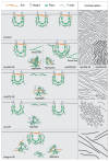
Model showing interdependence of slit diaphragm, integrin, and actin cytoskeleton when a single component is knocked down. This figure shows the normal distribution of the slit diaphragm (sns), the lacuna channel (piezo), integrin (alpha and beta subunits), and actin cytoskeleton (F-actin) in a control nephrocyte. It also models what appears to happen in the nephrocyte when either an actin, slit diaphragm, or integrin gene is no longer expressed, specifically how the remaining components accumulate cortically to create ring structures or subcortically to create aggregates or stress fibers. The left column shows a medial view while the right column shows a cortical view, with both the lines and the circles indicating the overall sns/integrin/actin interaction. Row 1 = Control, row 2 = split between Act42A-IR and Act57B-IR, row 3 = Act87E-IR, row 4 = sns-IR, and row 5 = integrin-IR.

Similar articles

References

    1. Pollak M.R., Quaggin S.E., Hoenig M.P., Dworkin L.D. The Glomerulus: The Sphere of Influence. Clin. J. Am. Soc. Nephrol. 2014;9:1461–1469. doi: 10.2215/CJN.09400913. - DOI - PMC - PubMed
    1. Weavers H., Prieto-Sánchez S., Grawe F., Garcia-López A., Artero R., Wilsch-Bräuninger M., Ruiz-Gómez M., Skaer H., Denholm B. The Insect Nephrocyte Is a Podocyte-like Cell with a Filtration Slit Diaphragm. Nature. 2009;457:322–326. - PMC - PubMed
    1. Welsh G.I., Saleem M.A. The Podocyte Cytoskeleton—Key to a Functioning Glomerulus in Health and Disease. Nat. Rev. Nephrol. 2011;8:14–21. doi: 10.1038/nrneph.2011.151. - DOI - PubMed
    1. Endlich K., Kliewe F., Endlich N. Stressed Podocytes-Mechanical Forces, Sensors, Signaling and Response. Pflugers Arch. 2017;469:937–949. doi: 10.1007/s00424-017-2025-8. - DOI - PubMed
    1. Blaine J., Dylewski J. Regulation of the Actin Cytoskeleton in Podocytes. Cells. 2020;9:1700. doi: 10.3390/cells9071700. - DOI - PMC - PubMed

LinkOut - more resources