

Ontogeny of Skin Stem Cells and Molecular Underpinnings
- PMID: 39194698
- PMCID: PMC11352238
- DOI: 10.3390/cimb46080481
Ontogeny of Skin Stem Cells and Molecular Underpinnings
Abstract
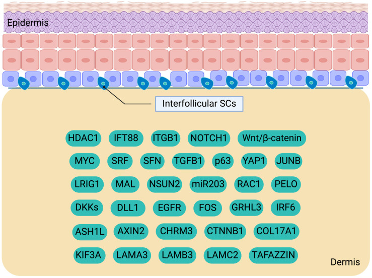
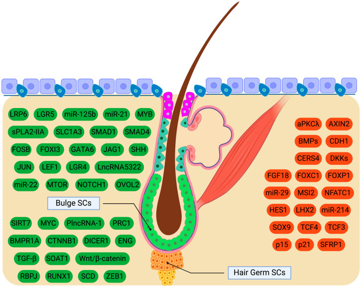
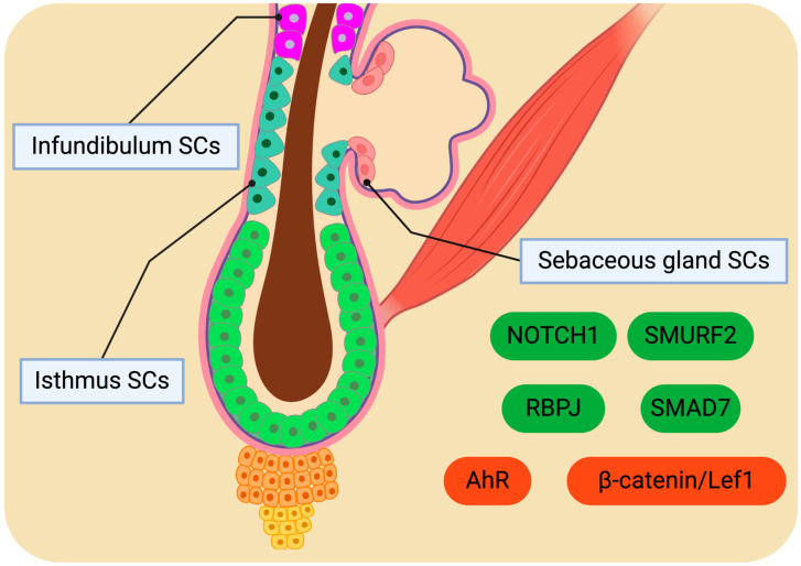
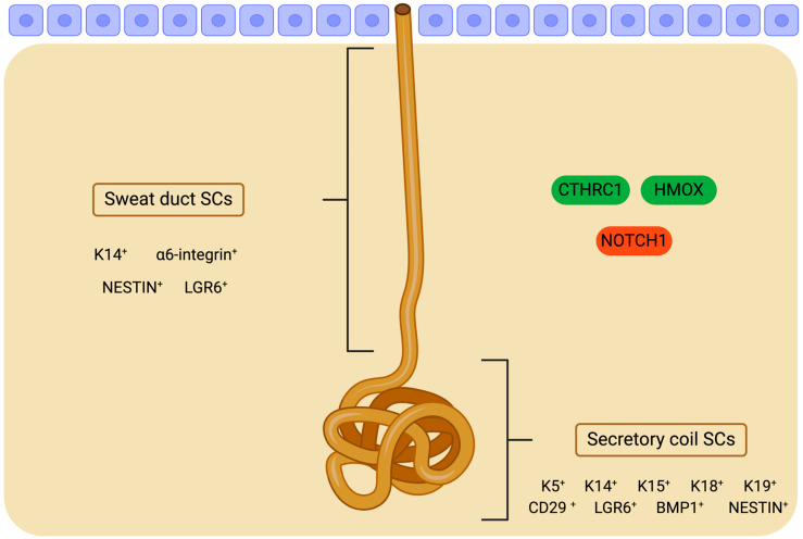
Skin stem cells (SCs) play a pivotal role in supporting tissue homeostasis. Several types of SCs are responsible for maintaining and regenerating skin tissue. These include bulge SCs and others residing in the interfollicular epidermis, infundibulum, isthmus, sebaceous glands, and sweat glands. The emergence of skin SCs commences during embryogenesis, where multipotent SCs arise from various precursor populations. These early events set the foundation for the diverse pool of SCs that will reside in the adult skin, ready to respond to tissue repair and regeneration demands. A network of molecular cues regulates skin SC behavior, balancing quiescence, self-renewal, and differentiation. The disruption of this delicate equilibrium can lead to SC exhaustion, impaired wound healing, and pathological conditions such as skin cancer. The present review explores the intricate mechanisms governing the development, activation, and differentiation of skin SCs, shedding light on the molecular signaling pathways that drive their fate decisions and skin homeostasis. Unraveling the complexities of these molecular drivers not only enhances our fundamental knowledge of skin biology but also holds promise for developing novel strategies to modulate skin SC fate for regenerative medicine applications, ultimately benefiting patients with skin disorders and injuries.
Keywords: epidermis; hair follicle; homeostasis; molecular cues; ontogeny; sebaceous glands; signaling; skin; stem cells; sweat glands.
Conflict of interest statement
The authors declare no conflicts of interest.
Figures
Similar articles
-
Human skin stem cells and the ageing process.Exp Gerontol. 2008 Nov;43(11):986-97. doi: 10.1016/j.exger.2008.09.001. Epub 2008 Sep 9. Exp Gerontol. 2008. PMID: 18809487 Review.
-
Stem cell dynamics and heterogeneity: implications for epidermal regeneration and skin cancer.Curr Med Chem. 2012;19(35):5984-92. Curr Med Chem. 2012. PMID: 22963565 Review.
-
Hair Follicle and Sebaceous Gland De Novo Regeneration With Cultured Epidermal Stem Cells and Skin-Derived Precursors.Stem Cells Transl Med. 2016 Dec;5(12):1695-1706. doi: 10.5966/sctm.2015-0397. Epub 2016 Jul 25. Stem Cells Transl Med. 2016. PMID: 27458264 Free PMC article.
-
Disruption of Smad4 in mouse epidermis leads to depletion of follicle stem cells.Mol Biol Cell. 2009 Feb;20(3):882-90. doi: 10.1091/mbc.e08-07-0731. Epub 2008 Dec 10. Mol Biol Cell. 2009. PMID: 19073883 Free PMC article.
-
Advances in a rapidly emerging field of hair follicle stem cell research.Coll Antropol. 2014 Mar;38(1):373-8. Coll Antropol. 2014. PMID: 24851645 Review.
References
-
- Gilaberte Y., Prieto-Torres L., Pastushenko I., Juarranz Á. Chapter 1—Anatomy and Function of the Skin. In: Hamblin M.R., Avci P., Prow T.W., editors. Nanoscience in Dermatology. Academic Press; Boston, FL, USA: 2016. pp. 1–14.
Publication types
Grants and funding
LinkOut - more resources
Full Text Sources
