Review
. 2024 Aug 22;14(8):407.
doi: 10.3390/bios14080407.

Recent Advances in Nanomaterials for Modulation of Stem Cell Differentiation and Its Therapeutic Applications

Affiliations
Review

Recent Advances in Nanomaterials for Modulation of Stem Cell Differentiation and Its Therapeutic Applications

Chang-Dae Kim et al. Biosensors (Basel). .

Abstract

Challenges in directed differentiation and survival limit the clinical use of stem cells despite their promising therapeutic potential in regenerative medicine. Nanotechnology has emerged as a powerful tool to address these challenges and enable precise control over stem cell fate. In particular, nanomaterials can mimic an extracellular matrix and provide specific cues to guide stem cell differentiation and proliferation in the field of nanotechnology. For instance, recent studies have demonstrated that nanostructured surfaces and scaffolds can enhance stem cell lineage commitment modulated by intracellular regulation and external stimulation, such as reactive oxygen species (ROS) scavenging, autophagy, or electrical stimulation. Furthermore, nanoframework-based and upconversion nanoparticles can be used to deliver bioactive molecules, growth factors, and genetic materials to facilitate stem cell differentiation and tissue regeneration. The increasing use of nanostructures in stem cell research has led to the development of new therapeutic approaches. Therefore, this review provides an overview of recent advances in nanomaterials for modulating stem cell differentiation, including metal-, carbon-, and peptide-based strategies. In addition, we highlight the potential of these nano-enabled technologies for clinical applications of stem cell therapy by focusing on improving the differentiation efficiency and therapeutics. We believe that this review will inspire researchers to intensify their efforts and deepen their understanding, thereby accelerating the development of stem cell differentiation modulation, therapeutic applications in the pharmaceutical industry, and stem cell therapeutics.

Keywords: cellular adhesion; external stimulation; intracellular regulation; nanomaterials; stem cell differentiation.


Conflict of interest statement

The authors declare no conflicts of interest.

Figures

Figure 2
Figure 2
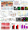
(A) AuNP treatment enhanced autophagic activity in inflammatory-conditioned periodontal ligament stem cells (I-PDLSCs) during the early phase of osteogenic differentiation represented by upregulated levels of LC3 II in the AuNP-treated I-PDLSCs and control PDLSCs. (B) Confocal images of accumulated FITC-LC3 puncta per cell in the AuNP-incubated I-PDLSCs and I-PDLSCs. (C) Knockdown of TFEB abrogated the AuNP-mediated rescue of the osteogenic potential of I-PDLSC. (D) Osteogenic protein expression in the I-PDLSCs represented by the decrease in RUNX2 expression. (E) Topographic images of different Ti substrates using SEM. (F) Quantitative analysis of the LC3 expression in MSCs treated with various samples in normal DMEM media at 1 and 7 days. (G) Related qualitative ALP activity on various sample surfaces at 7 and 14 days. (H) Relative mRNA expression of osteoclastogenesis-related genes in the RAW264.7 cells grown on different substrates. (I) quantitative TRAP activities after incubation for 1 and 4 days. (J) Confocal images of multinucleated cells on different substrates after culturing for 4 days. The asterisks and number signs indicate p-values *p and # p < 0.05, ** p and ## p < 0.01, and *** p < 0.001. Reprinted with permission from [94]. Copyright 2022, Elsevier; reprinted with permission from [97]. Copyright 2022, Elsevier.
Figure 4
Figure 4
(A) Schematic diagram of the characterization and (B) electrical stimulation process on graphene film. (C) Variations in electrical current or voltage intensity induced by diverse magnetic field (MF) strengths in graphene (upper panel) compared to vehicle control (lower panel). (D) Relative fluorescence intensity of MAP2 in the ADMSCs cultured in different substrates. (E) Schematic illustration of electrical delivery of mRNA to osteogenic differentiation of the MSCs on the polypyrrole–graphene oxide (PPy–GO)–mRNA hybrid platform. Examination of the electrical modulation of mRNA release using NaFl, based on factors such as (F) stimulation duration and (G) frequency. (H) Assessment of osteogenic differentiation in the MSCs with or without mRNA and electrical stimulation. (I) Quantitative analysis of the mineralization in the MSCs during osteogenic differentiation. The asterisks and number sign indicate p-values * p < 0.05, *** p < 0.001, **** p and #### p < 0.0001. Reprinted with permission from [122]. Copyright 2022, Wiley Online Library; reprinted with permission from [126]. Copyright 2022, Springer Nature.
Figure 1
Figure 1
Schematic illustrations of various nanomaterials to modulate stem cell functions and mechanisms.
Figure 3
Figure 3
(A) Schematic illustrations of Mn-Co3O4 utilized as antioxidant nanostructures for regulating stem cell fates. (B) Crystal model of MC-1.0 which displays the optimized catalytic ROS-scavenging activity. (C) X-ray diffraction (XRD) analysis for the Mn-Co3O4 crystal structures. (D) Electron energy-loss spectroscopy (EELS) exhibiting uniformly distributed Co and Mn atoms on the surface. (E) Scavenging activities of Mn3O4, Co3O4, and MC-1.0 for DPPH radical. (F) Quantitative analysis of cell proliferation after the H2O2 treatment. (G) Relative fluorescence intensity of osteogenic markers. (H) Schematic diagram of the preparation of SOD-modified gold nanospheres (SOD@AuNS). (I) TEM and elemental mapping images of SOD@AuNS. (J) Histological analysis of lipid accumulation (ORO) and calcium deposition (ARS) by staining the MSCs, with and without labeling, following adipogenic and osteogenic induction, respectively. (K) Quantitative analysis of each differentiation results. (L) Cell viabilities of the MSCs labeled with SOD@Au, SOD@AuNS, and MUA@AuNS. The asterisks indicate p-values * p < 0.05, ** p < 0.01, *** p < 0.001, **** p < 0.0001 and ns represents no significant difference. Reprinted with permission from [101]. Copyright 2022, Wiley Online Library; reprinted with permission from [103]. Copyright 2023, Elsevier.
Figure 5
Figure 5
(A) Immunofluorescence images for visualization of spiral ganglion neurons (SGNs) after 3 days of culture on TCPS and GelMA–SACNT substrates with the neuronal marker Tuj1. (B) Neurite lengths of SGNs cultured on the TCPs and GelMA–SACNT composite substrates. (C) Filopodia length and (D) growth cone size of SGNs cultivated on the TCPS and GelMA–SACNT substrates. (E) Fluorescent imaging of SGNs cultivated on the TCPS and GelMA–SACNT substrates, using the calcium-sensitive dye Fluo-4 AM to record calcium transients, taken after 10 days of culture. (F) Schematic of the fabrication and applications of a 3D NMJ system. (G) Confocal images of representative marker expression at day 1 and day 28 of neurogenesis in islet1, Tuj1, and Hoechst 33342. (H) Quantified results of the ChAT, islet1, and HB9 of the MNSs. (I) Confocal images on day 10 of coculture differentiation for muscle bundles with the 3D nano-biohybrid hydrogel using single/multi-MNSs. (J) Quantified results of CHRNA7 with 3D and 3D nano-biohybrid hydrogels using multi-MNSs (left panel). Contraction of muscle bundle in the 3D NMJ biosensing system using multi-MNSs upon electrical stimulation (1 Hz, 10 V) (right panel). (K) Quantified mRNA results of HB9, islet1, and ChAT on D35 related to MN differentiation between ALS–MNSs and H–MNSs (left panel). TDP-43, NEFL, and NEFM on D35 between ALS-MNSs and H-MNSs (right panel). (L) Confocal images of the NMJs treated with 100 μM bosutinib. The NMJs integrated a biosensor using a 3D ALS-nano-biohybrid hydrogel and multi-MNSs. The asterisks indicate p-values * p < 0.05, ** p < 0.01, **** p < 0.0001 and ns represents no significant difference. Reprinted with permission from [132]. Copyright 2022, Elsevier; reprinted with permission from [133]. Copyright 2023, Elsevier.
Figure 6
Figure 6
(A) Schematic of the delivery and uptake of miR-124 loaded onto Ca-MOF nanoparticles within NSCs. (B) Illustrations of the embedding strategy of miR-124 onto the surface of Ca-MOF by hydrogen bonds between the −NH2 and −OH groups and the rapid cleavage of the hydrogen bonds upon exposure to a low pH. (C) Protective effect of Ca-MOF@miR-124 nanoparticles from nuclease degradation. (D) The results of the quantitative PCR analysis performed to evaluate the expression of various neural markers in differentiated NSC groups cultured with Ca-MOF, miR-124, or Ca-MOF@miR-124 nanoparticles over different time points. (E) Schematic of the different differentiation mechanisms between the conventional supply method and autonomous stem cell differentiation (SMENA). (F) Immunocytochemical analysis of the neuronal differentiation for each treatment group at DIV 14 and (G) quantitative analysis and relative quantification of the protein expression. The asterisks indicate p-values ** p < 0.01, *** p < 0.001 and ns represents no significant difference. Reprinted with permission from [154]. Copyright 2022, American Chemical Society; reprinted with permission from [155]. Copyright 2022, American Association for the Advancement of Science.
Figure 7
Figure 7
(A) Schematic of the functionalized UCNP platform. This platform is designed for NIR light-controlled and real-time monitoring of osteogenic differentiation in MSCs. The platform facilitates precise tracking and modulation of osteogenic differentiation processes, aiding in the development of targeted therapies for osteoporosis. Characterizations of the UCNP platform. (B) TEM image of UCNP@mSiO2-peptide-BHQ3-ONA-CD and (C) spectrum upon irradiation at 980 nm with 1 W/cm2. (D) Quantification of the NIR-mediated release of ICA from the UCNP nanocomplex at different NIR power levels (0, 0.5, 1, and 2 W/cm2) and (E) irradiation durations at 1 W/cm2. (F) Immunofluorescence analysis of MSCs following 10 days of culture, evaluating the expression of proteins associated with osteogenic differentiation markers. (G) Schematic of the NIR stimulation triggering the release of ONA, enabling the control of cell adhesion, spreading, and multilineage differentiation of MSCs on the UCNP-based substrate under different NIR irradiation intensities. (H) TEM image and (I) fluorescence emission of the UCNP@SiO2-RGD-ONA nanoparticles under 980 nm NIR irradiation. (J) UV-vis absorption spectra of RGD, ONA, UCNP@SiO2, UCNP@SiO2-RGD, and UCNP@SiO2-RGD-ONA. (K) Histological analysis to evaluate the osteogenic and adipogenic differentiation of MSCs cultured on the UCNP substrate following exposure to NIR irradiation at different intensities, after 7 days of induced differentiation. (L) Quantification of positive cells for different powers of NIR. The asterisks indicate p-values * p < 0.05, ** p < 0.01, *** p < 0.001. Reprinted with permission from [159]. Copyright 2022, American Chemical Society; reprinted with permission from [160]. Copyright 2022, American Chemical Society.

Similar articles

References

    1. Cahan P., Li H., Morris S.A., Lummertz da Rocha E.L., Daley G.Q., Collins J.J. CellNet: Network biology applied to stem cell engineering. Cell. 2014;158:903–915. doi: 10.1016/j.cell.2014.07.020. - DOI - PMC - PubMed
    1. Hsu M.-N., Chang Y.-H., Truong V.A., Lai P.-L., Nguyen T.K.N., Hu Y.-C. CRISPR technologies for stem cell engineering and regenerative medicine. Biotechnol. Adv. 2019;37:107447. doi: 10.1016/j.biotechadv.2019.107447. - DOI - PubMed
    1. Kerativitayanan P., Carrow J.K., Gaharwar A.K. Nanomaterials for engineering stem cell responses. Adv. Healthc. Mater. 2015;4:1600–1627. doi: 10.1002/adhm.201500272. - DOI - PubMed
    1. Peerani R., Zandstra P.W. Enabling stem cell therapies through synthetic stem cell–niche engineering. J. Clin. Investig. 2010;120:60–70. doi: 10.1172/JCI41158. - DOI - PMC - PubMed
    1. Xu Y., Chen C., Hellwarth P.B., Bao X. Biomaterials for stem cell engineering and biomanufacturing. Bioact. Mater. 2019;4:366–379. doi: 10.1016/j.bioactmat.2019.11.002. - DOI - PMC - PubMed

LinkOut - more resources