Clinical Trial
. 2024 Aug 27;15(1):7388.
doi: 10.1038/s41467-024-51442-2.

The TGFβ type I receptor kinase inhibitor vactosertib in combination with pomalidomide in relapsed/refractory multiple myeloma: a phase 1b trial

Affiliations
Clinical Trial

The TGFβ type I receptor kinase inhibitor vactosertib in combination with pomalidomide in relapsed/refractory multiple myeloma: a phase 1b trial

Ehsan Malek et al. Nat Commun. .

Abstract

Functional blockade of the transforming growth factor-beta (TGFβ) signalling pathway improves the efficacy of cytotoxic and immunotherapies. Here, we conducted a phase 1b study (ClinicalTrials.gov., NCT03143985) to determine the primary endpoints of safety, tolerability, and maximal tolerated dose (200 mg twice daily) for the orally-available TGFβ type I receptor kinase inhibitor vactosertib in combination with pomalidomide in relapsed and/or refractory multiple myeloma (RRMM) patients who had received ≥2 lines of chemoimmunotherapy. Secondary endpoints demonstrated sustained clinical responses, favorable pharmacokinetic parameters and a 6-month progression-free survival of 82%. Vactosertib combined with pomalidomide was well-tolerated at all dose levels and displayed a manageable adverse event profile. Exploratory analysis indicated that vactosertib co-treatment with pomalidomide also reduced TGFβ levels in patient bone marrow as well as the level of CD8+ T-cells that expressed the immunoinhibitory marker PD-1. In vitro experiments indicated that vactosertib+pomalidomide co-treatment decreased the viability of MM cell lines and patient tumor cells, and increased CD8+ T-cell cytotoxic activity. Vactosertib is a safe therapeutic that demonstrates tumor-intrinsic activity and can overcome immunosuppressive challenges within the tumor microenvironment to reinvigorate T-cell fitness. Vactosertib offers promise to improve immunotherapeutic responses in heavily-pretreated MM patients refractory to conventional agents.


Conflict of interest statement

The authors declare no competing interests.

Figures
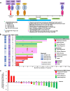
Fig. 1
Fig. 1. Trial design, Swimmer’s plot and Waterfall plot to evaluate the response of patients to vactosertib and pomalidomide.
a Vactosertib and pomalidomide dose administration schedule and vactosertib dose levels for the phase 1b dose escalation cohort. Also shown is the schedule for clinical and laboratory testing. b Swimmer’s plot indicates the individual trajectories and outcomes over the study duration in the 20 patients that received full-dose treatment. The magnitude of the Y-axis value for each patient indicates the months of study from the initiation of treatment. The color attributed to each patient’s lane in the Swimmer’s plot correlates with the response based upon IMWG criteria. c Waterfall plot indicates the maximal change in baseline of the biochemical marker, i.e., M-spike or LC, value for the same 20 patients as in (b). The magnitude of the vertical bar for each patient represented in the Waterfall represents the best change in the biochemical marker measured from the initiation of treatment. The color attributed to the vertical bar indicates with the patient response to treatment based upon IMWG criteria.
Fig. 2
Fig. 2. Effect of TGFβ1, vactosertib and pomalidomide on canonical and non-canonical TGFβ-signaling in MM cells.
a Representative western blots of lysates from MM.1S cells treated with vactosertib (1 μM) or pomalidomide (1 μM) for 18 h with or without TGFβ1 (10 ng/mL) included in the culture media. Whole cell lysates were prepared and probed with the following antibodies: phospho-SMAD2/3, total SMAD2/3, phospho-ERK1/2, total ERK1/2, cereblon, IKZF1, IKZF3 and GAPDH. The densitometric values for total SMAD2/3 and total ERK1/2 are relative that the values measured in the presence of TGFβ and in the absence of vactosertib and pomalidomide. Densitometric values for phospho-SMAD2/3 with each treatment represent the value determined in the presence of TGFβ relative to the total SMAD2/3 value with the same treatment. Similarly, the value for phospho-ERK1/2 with each treatment represent the value determined in the presence of TGFβ relative to the total SMAD2/3 value with the same treatment. b Representative western blot of MM.1S cells treated with vactosertib or pomalidomide individually and in combination at the indicated concentrations for 18 h in the presence of TGFβ1 (10 ng/mL). Experiments were independently performed twice. Whole cell lysates were collected and probed with the same antibodies as above. Source data are provided as a Source Data file.
Fig. 3
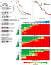
Fig. 3. Effectors upregulated in MM cells with acquired pomalidomide resistance.
a Representative western blots of MM.1S and MM.1S-pomalidomide-resistant cells cultured with or without TGFβ1 (10 ng/mL) treatment. Whole cell lysates were prepared and probed with the antibodies indicated in Fig. 2, along with Bcl-2 and cleaved caspase. Densitometric values for cereblon, IKZF1 and IKZF3 are relative to the value determined in WT (parental) cells in the absence of TGFβ. Densitometric values for total SMAD2/3 and total phospho-ERK1/2 are relative that the values measured in the absence of TGFβ in WT (parental) cells. Densitometric values for phospho-SMAD2/3 were determined relative to the total SMAD2/3 value determined with the same cells (either WT or pomalidomide-resistant) with or without TGFβ. Similarly, densitometric values for phospho-ERK1/2 were determined relative to the total ERK1/2 value determined with the same cells (WT or pomalidomide-resistant) with or without TGFβ. Experiments were independently performed twice. b Shown is the dose-dependent effect of pomalidomide and vactosertib on MM1.S parental and pomalidomide-resistant cells. Cells were treated with drugs at each indicated concentrations in triplicate and viability then determined using the XTT assay. Values shown represent the arithmetic mean of three independent experiments. c Concentration dependent combined effect of vactosertib and pomalidomide on MM.1S parental, pomalidomide-resistant and patient CD138+ cells. Cells were treated with drugs at each indicated concentrations in triplicate and viability then determined using the XTT assay. Values shown represent the arithmetic mean of three independent experiments. Green boxes indicate highest viability, yellow boxes intermediate viability and red boxes lowest viability. Source data are provided.
Fig. 4
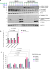
Fig. 4. Synergistic effect of vactosertib and pomalidomide on apoptosis in MM cells.
a Western blot analysis of MM1.S parental and pomalidomide-resistant cells treated with vactosertib and pomalidomide alone or in combination. Cells were treated with drugs as indicated, lysates prepared and probed with antibodies to phospho-ERK1/2, total ERK1/2, Bcl-2 and full-length (FL), cleaved caspase-3 and GADPH. Experiments were independently performed twice. b Combined effect of vactosertib and pomalidomide on MM1.S and MM1.S- pomalidomide-resistant cells. Cells (1 × 106/assay) were incubated with drugs for 20 h, washed with PBS, lysates prepared, probed using antibodies specific to annexin-V (BD Biosciences, 556419) and propidium iodide (PI) (Invitrogen, P21493) and analyzed by flow cytometry (Attune NxT). Relative % of annexin-V+/PI+ cells represents the percent of drug treated MM cells relative to untreated cells. c Combined effect of vactosertib and pomalidomide on patient BM CD138+ cells. Cells (~1 × 105/assay) were incubated with drugs for 20 h, washed with PBS, probed using antibodies specific to annexin-V (BD Biosciences, 556419) and PI and analyzed by flow cytometry. Annexin-V+/PI+ cells represent the percent of CD138+ cells positive for both annexin-V and PI relative to untreated cells. In (b) and (c), data are representative of three independent experiments. Error bars represent the SD of the mean (SEM). Two-way ANOVA was conducted to investigate potential interactions between more than two variables in (b) and (c).
Fig. 5
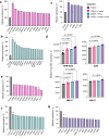
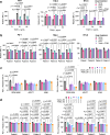
Fig. 5. Effect of TGFβ1, vactosertib and pomalidomide on myeloma immunophenotype.
High-throughput cell surface marker screening to determine the effect of vactosertib, pomalidomide and TGFβ1 on MM surface antigens using the human cell surface marker screening panel (BD Biosciences, #560747). The screening panel contained 242 purified monoclonal antibodies to detect individual human cell surface markers. RPMI-8226 cells were treated with either TGFβ1 (10 ng/mL), TGFβ1 + vactosertib (1 μM), TGFβ1 + pomalidomide (1 μM), or TGFβ1, vactosertib and pomalidomide for 48 h. Following treatment, cells were washed and probed with the antibody array panel for 30 min. Cells were probed with Alexa Fluor 647-conjugated goat anti-mouse Ig or goat anti-rat Ig secondary antibodies. Isotype controls were included to determine the isotype-specific background. Cells were analyzed using a high-throughput flow cytometry (BD LSR Fortessa). Shown are surface markers: a increased by vactosertib + TGFβ1, b increased by pomalidomide + TGFβ1 and c increased by vactosertib, pomalidomide, and TGFβ1. Green asterisks (*) in (ac) measure the effect of vactosertib, pomalidomide and both combined on the expression of HLA-ABC on RPMI-8226 cells. d Indicates the effect of TGFβ1 alone (grey), TGFβ1 + vactosertib (pink), TGFβ1 + pomalidomide (green) and TGFβ1 + vactosertib + pomalidomide (blue). In panel (e) are shown surface markers decreased by treatment with TGFβ1 and vactosertib, f surface markers decreased by treatment with TGFβ1 and pomalidomide and g surface markers decreased by treatment with TGFβ1, vactosertib and pomalidomide. Red asterisks (*) in (eg) indicate the immunoinhibitory receptor PD-L1. Values in ac and eg were determined relative to the value measured for cells incubated with TGFβ alone (red bar). Following treatment and antibody staining, fluorescent intensity was measured by flow cytometry acquired using ≥10,000 events. Data were analyzed using FlowJo v10.8.1 software (Ashland, OR). Baseline values for isotype controls were subtracted from each reading. Values in (d) represent the average of five replicates for RPMI-8226 cells and three replicates for other cell lines. Source data are provided as a Source Data file.
Fig. 6
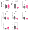
Fig. 6. Effect of vactosertib and pomalidomide in vivo treatment on patient BM cytokine and cellular components.
a Effect of vactosertib and pomalidomide on cytokine levels in patient BM samples. At the indicated times, latent TGFβ1, free (active) TGFβ1 and IL-6 levels were measured in patient BM samples (n = 8 samples at C3D1 and 6 samples at EOT). b Effect on vactosertib and pomalidomide on relative surface expression of the immunosuppressive markers PD-1, TIM-3, 2B4, BTLA and CTLA-4 on patient CD8+ T-cells present within the BMMC fraction. The percent of PD-1, TIM-3, 2B4, BTLA and CTLA-4-positive staining cells were quantitated at indicated times. The relative percent of marker-positive cells was determined compared to cells obtained at C3D1. Data represent three independent experiments. In (a, b), differences between two groups were analyzed using the unpaired T-test. Error bars represent the SD of the mean (SEM).
Fig. 7
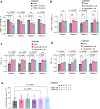
Fig. 7. Direct effect of vactosertib to CD8+ T-cell immunophenotype.
a Shown is the relative MFI value for the expression of each immunosuppressive marker (PD-1, TIM-3, BTLA, and LAG-3) on CD8+ T-cells isolated from healthy peripheral blood. T-cells were cultured in the presence of TGFβ1 at indicated concentrations. CTLA-4 was not detected by immunostaining of healthy human T-cells. Error bars represent the SD of the mean. b Shown are relative MFI values after staining for PD-1, TIM-3 and BTLA on MM patient CD8+ T-cells. Error bars represent the SD of the mean. c Effect of vactosertib on PD-L1 and PD-L2 expression on MMCLs. MMCLs were treated with vactosertib and pomalidomide as indicated for 72 h. Cells were then stained with PD-L1 and PD-L2 antibodies for 20 min and analyzed by flow cytometry. d Effect of vactosertib on PD-L1 and PD-L2 expression on MM patient BM-derived CD138+ cells. Patient CD138+ cells were treated with vactosertib and/or pomalidomide at various concentrations for 48 h. Cells were then stained with PD-L1 and PD-L2 specific antibodies simultaneously for 20 min and analyzed by flow cytometry. Statistical comparisons were made by comparing drug treated to untreated cells. In (ad), values represent the average of triplicate measurements. Two-way ANOVA was conducted to investigate potential interactions between more than two variables in (ad). Source data are provided as a Source Data file.
Fig. 8
Fig. 8. Direct effect of vactosertib on CD8+ T-cell cytolytic activity.
a Effect of TGFβ1 on the viability of patient CD8+ T-cells. Cells (5000/well) were treated as indicated for 24 h. Activated-XTT assay solution was added and plates were incubated in the dark for 3 h. Relative cell viability was quantified by measuring the absorbance at 450 nm on a SpectraMax i3x multi-mode microplate reader. b Effect of vactosertib on the viability of patient CD8+ T-cells in the presence of TGFβ1. Cells (5000/well) were treated as indicated for 24 h. Activated-XTT assay solution was added and plates were incubated in the dark for 3 h. Relative cell viability was quantified by measuring the absorbance at 450 nm on a SpectraMax i3x multi-mode microplate reader. c Effect of vactosertib on TNF-α production by MM patient CD8+ T-cells. Cells (5000/well) were incubated with vactosertib at indicated concentrations for 24 h. Culture supernatant was centrifuged at 1000 × g for 5 min and cytokine level quantitated using the human TNF-α Quantikine ELISA kit. d Effect of vactosertib on IFN-γ production by patient CD8+ T-cells. Cells (5000/well) were incubated with vactosertib at indicated concentrations for 24 h. Culture supernatants were centrifuged at 3000 rpm × 5 min. Cytokine levels were quantitated using the human IFN-γ Quantikine duoSet ELISA kit. e Effect of vactosertib on autologous CD8+ T-cell cytotoxic activity. Shown is the effect of vactosertib and pomalidomide on autologous CD8+ T-cell activity against patient CD138+ cells. CD138+ cells (20,000/well) were treated as indicated for 8 h and then co-cultured with CD8+ T-cells (50,000/well) for 18 h. Cells were then stained to detect CD138+ cells followed by annexin-V and PI staining. Cells that gated positively for CD138+, annexin-V and PI were quantitated by flow cytometry using FlowJo_10.8.1 software. In (ae), values represent the average of triplicate measurements (n = 3). Two-way ANOVA was conducted to investigate potential interactions between more than two variables (ae). In panel (a), two asterisks (**) indicate a p-value ≤ 0.01. Source data are provided.
Fig. 9
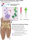
Fig. 9. The TGFβ type I receptor kinase inhibitor vactosertib in RRMM: a phase 1b trial.
Relapsed and/or refractory multiple myeloma patients previously treated with ≥2 lines of therapy were enrolled on a phase 1b study to assess safety and recommend a phase two dose of vactosertib in combination with the immunomodulatory drug pomalidomide. Shown are the putative combined in vivo effects of vactosertib and pomalidomide therapy in RRMM patients.

Update of

Similar articles

References

    1. van de Donk, N. W. C. J., Pawlyn, C. & Yong, K. L. Multiple myeloma. Lancet397, 410–427 (2021). 10.1016/S0140-6736(21)00135-5 - DOI - PubMed
    1. Cowan, A. J. et al. Diagnosis and management of multiple myeloma: a review. JAMA327, 464–477 (2022). 10.1001/jama.2022.0003 - DOI - PubMed
    1. Mateos, M. V., Nooka, A. K. & Larson, S. M. Moving toward a cure for myeloma. Am. Soc. Clin. Oncol. Educ. Book42, 1–12 (2022). - PubMed
    1. Pawlyn, C. & Morgan, G. J. Evolutionary biology of high-risk multiple myeloma. Nat. Rev. Cancer17, 543–556 (2017). 10.1038/nrc.2017.63 - DOI - PubMed
    1. Kastritis, E., Terpos, E. & Dimopoulos, M. A. How I treat relapsed multiple myeloma. Blood139, 2904–2917 (2022). 10.1182/blood.2020008734 - DOI - PubMed

Publication types

MeSH terms

Associated data