. 2024 Aug 27;15(1):7370.
doi: 10.1038/s41467-024-50976-9.

Simulation-driven design of stabilized SARS-CoV-2 spike S2 immunogens

Affiliations

Simulation-driven design of stabilized SARS-CoV-2 spike S2 immunogens

Xandra Nuqui et al. Nat Commun. .

Abstract

The full-length prefusion-stabilized SARS-CoV-2 spike (S) is the principal antigen of COVID-19 vaccines. Vaccine efficacy has been impacted by emerging variants of concern that accumulate most of the sequence modifications in the immunodominant S1 subunit. S2, in contrast, is the most evolutionarily conserved region of the spike and can elicit broadly neutralizing and protective antibodies. Yet, S2's usage as an alternative vaccine strategy is hampered by its general instability. Here, we use a simulation-driven approach to design S2-only immunogens stabilized in a closed prefusion conformation. Molecular simulations provide a mechanistic characterization of the S2 trimer's opening, informing the design of tryptophan substitutions that impart kinetic and thermodynamic stabilization. Structural characterization via cryo-EM shows the molecular basis of S2 stabilization in the closed prefusion conformation. Informed by molecular simulations and corroborated by experiments, we report an engineered S2 immunogen that exhibits increased protein expression, superior thermostability, and preserved immunogenicity against sarbecoviruses.


Conflict of interest statement

J.S.M., L.Z., R.E.A., X.N., L.C., and M.S. are inventors on a U.S. patent application describing the use of stabilized SARS-CoV-2 S proteins as vaccine antigens (Stabilized SARS-CoV-2 S Antigens, 63/583,090). K.C. is a member of the scientific advisory board of Integrum Scientific LLC and has consulted for Axon Advisors, LLC. K.C. owns shares in Integrum Scientific and Eitr Biologics, Inc. The other authors declare no competing interests.

Figures

Fig. 1
Fig. 1. HexaPro-SS-Δstalk S2-only trimer and context within the SARS-CoV-2 spike protein.
The model of the HexaPro-SS-Δstalk S2-only trimer in the closed prefusion conformation is depicted with a cartoon representation where the protomers are highlighted with different shades of purple. We note that the spike’s S1 subunit, here shown with a gray transparent surface only to provide context, is not present in this construct. N-glycans linked to S2 are shown as dark gray sticks. Glycans linked to S1 are omitted for clarity. HexaPro-SS-Δstalk is an S2-only construct derived from the prefusion-stabilized SARS-CoV-2 HexaPro spike. The distinctive substitutions (i.e., S704C, K790C, Q957E) incorporated in the HexaPro-SS-Δstalk are highlighted in the panels on the right. They stabilize S2 in a prefusion trimer assembly, preventing protomer dissociation via the formation of interprotomer salt bridges and disulfide bonds.
Fig. 2
Fig. 2. HexaPro-SS-Δstalk S2 trimer opening.
a Progress coordinates used in the WE simulation of HexaPro-SS-Δstalk, namely P987 triangle area and RMSDCH, are highlighted with yellow shapes (triangle and cylinders, respectively) drawn on top of the molecular structures. The CHs as in the closed conformation of the simulated construct are shown with shades of purple and are overlayed on the open conformation as in the crystal structure (colored in gray, PDB ID: 8U1G). b Distribution of the conformations sampled in the opening pathways obtained from the WE simulation. Each black point represents a conformation sampled along an opening pathway. The RMSD of the CH to CHOpen-crystal (y-axis) is plotted against the area of the triangle formed by the P987Cα at the CH apex (x-axis). The hexagonal bin color is scaled to the mean interprotomer distance of the black data points within the respective bin. A trace of one of the successful pathways is shown as a black line with white points corresponding to the closed, partially open, and open conformations. ce Molecular representation of closed (c), partially open (d), and open (e) conformations as highlighted in (b). Chain-A is depicted in light purple, chain-B in purple, and chain-C in dark purple. Residues 900–1030 encompassing HR1 and CH are illustrated with a cartoon representation, whereas the rest of the chain is shown as an opaque surface. Glycans are shown as gray sticks.
Fig. 3
Fig. 3. Contacts between residues in the central helices during S2 opening.
ac HexaPro-SS-Δstalk CHs in the closed conformation from a top-down view (a), side view in the context of the S2 trimer (gray cartoons) (b), and side view (c). CHs are highlighted with purple cartoons. The sidechains of the residue pointing toward the interior of the CHs are shown as sticks with the C atoms colored by the corresponding residue contact score as in the closed conformation of the HexaPro-SS-Δstalk. d, e Contact score heatmaps for the HexaPro-SS-Δstalk base construct (d) and HexaPro-SS-Δstalk tryptophan substitutions (e) in the closed, partially open, and open states. The scale ranges from 0 (weak or transient contacts) to 1 (persistent or extensive contacts, corresponding to the 95th percentile value of the HexaPro-SS-Δstalk contact scores). The star indicates the substituted residue.
Fig. 4
Fig. 4. V991W and T998W stabilize HexaPro-SS-Δstalk in the closed conformation.
a Thermodynamic stabilization of the closed prefusion conformation as imparted by each tryptophan variant relative to the base system (HexaPro-SS-Δstalk). For each variant, the value of ∆∆Gmutation-folding relative to the base construct is plotted with a colored bar indicating the free energy difference estimate (in kcal/mol) ± standard error as calculated from 1000 (forward and backward) non-equilibrium alchemical simulations. The standard errors, denoted by thin black lines, were calculated via bootstrapping with 100 drawn samples. b Distribution of the number of interprotomer contacts established per frame by residues at positions 991 + 998 from the ensemble of closed conformations extracted from respective WE simulations. Distributions are shown as kernel densities. c Relative frequency of occurrence (%) of the top six residue–residue interprotomer contacts occurring in the closed conformations observed in HexaPro-SS-Δstalk and HexaPro-SS-2W WE simulations. Relative frequencies are calculated with respect to the total number of closed conformations extracted from the respective WE simulation. d, e Molecular representation of the most important interprotomer contacts in HexaPro-SS-2W in (d) the closed conformation and (e) the partially open conformation. Chain-A is depicted in light purple, chain-B in purple, chain-C in dark purple, and interacting residues are highlighted with sticks. Solid lines connect residues that form hydrophobic (π–π) interactions, whereas dashed lines indicate electrostatic (cation–π and salt bridge) interactions. A representative closed conformation of HexaPro-SS-2W was selected for this purpose upon clustering of closed conformations retrieved from the respective WE simulation (Supplementary Fig. 18).
Fig. 5
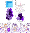
Fig. 5. Cellular expression, thermostability, and structural characterization of HexaPro-SS-2W.
a SDS-PAGE of purified S2 constructs (Base, HexaPro-SS-V991W, HexaPro-SS-T998W, and HexaPro-SS-2W). The ‘Base’ construct corresponds to HexaPro-SS-∆stalk. No statistical analysis was performed on the SDS-PAGE gel. The uncropped gel image is included in the Source Data file. b Size-exclusion chromatography (SEC) of purified S2 constructs. Both tryptophan substitutions increase protein expression yield, with V991W + T998W and T998W resulting in a greater increase. c Differential scanning fluorimetry of S2 constructs, including the original HexaPro (S1 + S2). HexaPro-SS-2W exhibits superior thermal stability to all HexaPro constructs, with a ~16 °C increase in Tm relative to HexaPro. d Cryo-EM map (Resolution: 2.8 Å) of closed prefusion state HexaPro-SS-2W (∆stalk) from side and top-down perspectives. eg Top-down perspective of HexaPro-SS-2W structure highlighting V991W (orange) and T998W (red) packing within the S2 interior; the tryptophan sidechains self-associate between chains in offset edge-to-edge and edge-to-face π–π stacking orientations. The EM map is shown as a transparent gray volume.
Fig. 6
Fig. 6. Immunogenicity of HexaPro-SS and HexaPro-SS-2W constructs.
a, b Sera from mice immunized with HexaPro-SS-2W can neutralize rVSV-CoV-2 variants. a 6–8-week-old female C57BL/6J mice (n = 6) were primed (week 0) and boosted (week 3) with 10 µg of immunogens HexaPro-SS, HexaPro-SS-2W, or PBS. b Sera was isolated at week 7 and evaluated for neutralization capacity against rVSVs bearing the spike proteins of SARS-CoV-2 Wuhan-1 or SARS-CoV-2 Omicron BA.1. Lower area under the curve (AUC) values correspond to better sera neutralizing capability. AUCs were compared across groups by ordinary one-way ANOVA with Tukey’s multiple comparisons test (** indicates a p-value < 0.01; n.s. indicates not significant). c Biolayer interferometry sensorgrams showing binding responses (colored lines indicate 1:1 binding fit, black lines indicate reference subtracted response) of previously reported neutralizing or non-neutralizing antibodies to epitopes on the S2 subunit in HexaPro-SS (top) and HexaPro-SS-2W (bottom) constructs.

Similar articles

  • Safety and immunogenicity of a modified mRNA-lipid nanoparticle vaccine candidate against COVID-19: Results from a phase 1, dose-escalation study.
    Essink BJ, Shapiro C, Isidro MGD, Bradley P, Pragalos A, Bloch M, Santiaguel J, Frias MV, Miyakis S, Alves de Mesquita M, Berrè S, Servais C, Waugh N, Hoffmann C, Baba E, Schönborn-Kellenberger O, Wolz OO, Koch SD, Ganyani T, Boutet P, Mann P, Mueller SO, Ramanathan R, Gaudinski MR, Vanhoutte N. Essink BJ, et al. Hum Vaccin Immunother. 2024 Dec 31;20(1):2408863. doi: 10.1080/21645515.2024.2408863. Epub 2024 Oct 18. Hum Vaccin Immunother. 2024. PMID: 39422261 Free PMC article. Clinical Trial.
  • SARS-CoV-2 BA.4/5 infection triggers more cross-reactive FcγRIIIa signaling and neutralization than BA.1, in the context of hybrid immunity.
    Richardson SI, Mzindle N, Motlou T, Manamela NP, van der Mescht MA, Lambson BE, Everatt J, Amoako DG, Balla S, von Gottberg A, Wolter N, de Beer Z, de Villiers TR, Bodenstein A, van den Berg G, Abdullah F, Rossouw TM, Boswell MT, Ueckermann V, Bhiman JN, Moore PL. Richardson SI, et al. J Virol. 2024 Jul 23;98(7):e0067824. doi: 10.1128/jvi.00678-24. Epub 2024 Jul 2. J Virol. 2024. PMID: 38953380
  • Altered TMPRSS2 usage by SARS-CoV-2 Omicron impacts infectivity and fusogenicity.
    Meng B, Abdullahi A, Ferreira IATM, Goonawardane N, Saito A, Kimura I, Yamasoba D, Gerber PP, Fatihi S, Rathore S, Zepeda SK, Papa G, Kemp SA, Ikeda T, Toyoda M, Tan TS, Kuramochi J, Mitsunaga S, Ueno T, Shirakawa K, Takaori-Kondo A, Brevini T, Mallery DL, Charles OJ; CITIID-NIHR BioResource COVID-19 Collaboration; Genotype to Phenotype Japan (G2P-Japan) Consortium; Ecuador-COVID19 Consortium; Bowen JE, Joshi A, Walls AC, Jackson L, Martin D, Smith KGC, Bradley J, Briggs JAG, Choi J, Madissoon E, Meyer KB, Mlcochova P, Ceron-Gutierrez L, Doffinger R, Teichmann SA, Fisher AJ, Pizzuto MS, de Marco A, Corti D, Hosmillo M, Lee JH, James LC, Thukral L, Veesler D, Sigal A, Sampaziotis F, Goodfellow IG, Matheson NJ, Sato K, Gupta RK. Meng B, et al. Nature. 2022 Mar;603(7902):706-714. doi: 10.1038/s41586-022-04474-x. Epub 2022 Feb 1. Nature. 2022. PMID: 35104837 Free PMC article.
  • The Key to Increase Immunogenicity of Next-Generation COVID-19 Vaccines Lies in the Inclusion of the SARS-CoV-2 Nucleocapsid Protein.
    Mendoza-Ramírez NJ, García-Cordero J, Shrivastava G, Cedillo-Barrón L. Mendoza-Ramírez NJ, et al. J Immunol Res. 2024 May 29;2024:9313267. doi: 10.1155/2024/9313267. eCollection 2024. J Immunol Res. 2024. PMID: 38939745 Free PMC article. Review.
  • Depressing time: Waiting, melancholia, and the psychoanalytic practice of care.
    Salisbury L, Baraitser L. Salisbury L, et al. In: Kirtsoglou E, Simpson B, editors. The Time of Anthropology: Studies of Contemporary Chronopolitics. Abingdon: Routledge; 2020. Chapter 5. In: Kirtsoglou E, Simpson B, editors. The Time of Anthropology: Studies of Contemporary Chronopolitics. Abingdon: Routledge; 2020. Chapter 5. PMID: 36137063 Free Books & Documents. Review.

Cited by

References

    1. Li, F. Structure, function, and evolution of coronavirus spike proteins. Annu. Rev. Virol.3, 237–261 (2016). 10.1146/annurev-virology-110615-042301 - DOI - PMC - PubMed
    1. Hsieh, C.-L. et al. Stabilized coronavirus spike stem elicits a broadly protective antibody. Cell Rep.37, 109929 (2021). 10.1016/j.celrep.2021.109929 - DOI - PMC - PubMed
    1. Pormohammad, A. et al. Comparison of confirmed COVID-19 with SARS and MERS cases—clinical characteristics, laboratory findings, radiographic signs and outcomes: a systematic review and meta-analysis. Rev. Med. Virol.30, e2112 (2020). 10.1002/rmv.2112 - DOI - PMC - PubMed
    1. World Health Organization. WHO COVID-19 Dashboard. https://covid19.who.int/ (2023).
    1. Barouch, D. H. Covid-19 vaccines—immunity, variants, boosters. N. Engl. J. Med.387, 1011–1020 (2022). 10.1056/NEJMra2206573 - DOI - PMC - PubMed

MeSH terms