Review
. 2024 Mar 7;3(2):e20230042.
doi: 10.1002/SMMD.20230042. eCollection 2024 Jun.

Emerging approaches for the development of artificial islets

Affiliations
Review

Emerging approaches for the development of artificial islets

Jingbo Li et al. Smart Med. .

Abstract

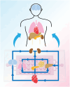
The islet of Langerhans, functioning as a "mini organ", plays a vital role in regulating endocrine activities due to its intricate structure. Dysfunction in these islets is closely associated with the development of diabetes mellitus (DM). To offer valuable insights for DM research and treatment, various approaches have been proposed to create artificial islets or islet organoids with high similarity to natural islets, under the collaborative effort of biologists, clinical physicians, and biomedical engineers. This review investigates the design and fabrication of artificial islets considering both biological and tissue engineering aspects. It begins by examining the natural structures and functions of native islets and proceeds to analyze the protocols for generating islets from stem cells. The review also outlines various techniques used in crafting artificial islets, with a specific focus on hydrogel-based ones. Additionally, it provides a concise overview of the materials and devices employed in the clinical applications of artificial islets. Throughout, the primary goal is to develop artificial islets, thereby bridging the realms of developmental biology, clinical medicine, and tissue engineering.

Keywords: artificial islet; diabetes; hydrogel; microfluidics; regenerative medicine.


Conflict of interest statement

The authors declare no competing financial interests.

Figures

FIGURE 1
FIGURE 1
Scheme of pancreas and islet cells. The islet consists of α, β, δ, and pp cells, which are perfused by microvessels and surrounded by the exocrine pancreas. The α cells secrete glucagon that elevates the blood glucose. The β cells secrete insulin, which is a unique hormone for reducing hyperglycemia. The δ cells secrete somatostatin, a hormone with various effects on countering other hormones, such as growth hormone, thyroid‐stimulating hormone, insulin, and glucagon. The pp cells secrete pancreatic polypeptide, regulating the metabolic behaviors of human bodies via inhibiting the release of cholecystokinin and pancreatic enzymes. Microvessels penetrate and accompany the islets, transporting the necessary nutrients and secreted hormones. Around the endocrine pancreas, the islets, spread the exocrine pancreas consisting of the acinar and ducts. The acinar cells secrete digestive enzymes including α‐amylase, lipase and proteases, which are responsible for the hydrolysis of carbohydrates, fats and proteins, respectively.
FIGURE 2
FIGURE 2
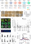
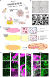
Differentiation of stem cells toward islet organoids. (A) Biomarkers for each stage are shown. (B) Representative images of the induced islet organoids from various hPSC cell lines. (C) Insulin contents of islet organoids derived from various hPSC cell lines. (D) Immunofluorescent images of islet organoids for C‐peptide, NKX6.1, glucagon (GCG), and somatostatin (SST). (E) Glucose responsiveness of islet organoids derived from hPSC under different genetic backgrounds. Source: Reproduced with permission. Copyright 2021, The Authors, published by Springer Nature. (F) Immunofluorescent images of islet organoids for insulin (Ins), GCG, SST, and polypeptide (PPY). Source: Reproduced with permission. Copyright 2022, Springer Nature. (G) Relative mRNA expression of islet genes in hPSC‐derived islet organoids and primary human islets. (H) Insulin secretion of islet organoids on dynamic GSIS assay. Source: Reproduced with permission. Copyright 2021, The Authors, published by Springer Nature.
FIGURE 3
FIGURE 3
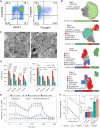
Identification of SC‐β and islet organoids. (A) Representative flow cytometric dot plots of dispersed stage 6 SC‐β cells immune‐stained for the indicated markers. Source: Reproduced with permission. Copyright 2021, The Authors, published by American Association for the Advancement of Science. (B) Single‐cell RNA sequencing of in vitro β‐cell differentiation. Source: Reproduced with permission. Copyright 2019, The Authors, published by Springer Nature. (C) Insulin granules observed via TEM. Source: Reproduced with permission. Copyright 2021, the American Diabetes Association. (D) The mitochondrial membrane potential evaluated by TMRE. (E) Typical OCR curve of islets. Source: Reproduced under terms of the CC‐BY license. Copyright 2022, The Authors, published by Springer Nature. (F) Glucose tolerance evaluated by IPGTT. Source: Reproduced under terms of the CC‐BY license. Copyright 2022, The Authors, published by Springer Nature.
FIGURE 4
FIGURE 4
Physiochemical and biological properties of hydrogels for the fabrication of artificial islets.
FIGURE 5
FIGURE 5
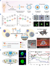
Alginate hydrogels for developing artificial islets. (A) Alginate hydrogel composite RGD motifs for pancreatic islet encapsulation. Source: Reproduced with permission. Copyright 2020, Elsevier B.V. (B) Design of zwitterionically modified alginates and their effects on reducing fibrosis. Source: Reproduced under terms of the CC‐BY license. Copyright 2019, The Authors, published by Springer Nature. (C) Core‐shell structure islet cell encapsulation based on microfluidic electrospray technology. Source: Reproduced under terms of the CC‐BY license. Copyright 2022, The Authors, published by Springer Nature.
FIGURE 6
FIGURE 6
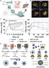
Chitosan for developing artificial islets. (A) All‐in‐water microfluidic system for fabricating binary capsules based on alginate and chitosan. (B) Relationship between hydrogel concentration and diameters of capsules. (C) Bright field and live/dead images of cells encapsulated in the capsules during the culture. Source: Reproduced under terms of the CC‐BY license. Copyright 2020, The Authors, published by John Wiley and Sons. (D) The enzymatic cross‐linking‐based hydrogel nanofilm caging system on pancreatic β cell spheroid prevented the infiltration of NK cells. (E) Illustrative and immunofluorescence images of the cells with/without the infiltration of NK cells. (F) Blood glucose levels of diabetic mice after pancreatic β cell transplantation. Source: Reproduced with permission. Copyright 2021, The Authors, published by American Association for the Advancement of Science.
FIGURE 7
FIGURE 7
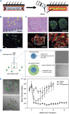
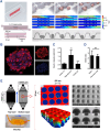
Agarose and hyaluronic acid for developing artificial islets. (A) SEK‐1005 with agarose‐SEK rods to pre‐vascularize a subcutaneous site for allogeneic islet transplantation without immunosuppression. (B) Histological images indicate microvessels at the graft site. (C) Immunofluorescence images of transplanted islets. Source: Reproduced with permission. Copyright 2018, Wolters Kluwer Health, Inc. (D) Schematic of core‐shell spherification method based on hyaluronic acid and polyethylene glycol diacrylate. (E) Live/dead images of the fabricated microspheres. (F) Blood glucose levels of mice transplanted with the microspheres. Source: Reproduced under terms of the CC‐BY license. Copyright 2021, The Authors, published by Mary Ann Liebert, Inc.
FIGURE 8
FIGURE 8
GelMA and PEG for developing artificial islets. (A) Porous microgels designed for the transplantation of β cells. (B) Confocal images of the porous microgels. (C) Blood glucose observation and IPGTT results in diabetic mice treated with microgels. Source: Reproduced with permission. Copyright 2023, John Wiley and Sons. (D) Synthesis scheme and structures of cell‐mimic polymersome (PSome)‐shielded islets for long‐term immune protection of neonatal porcine islet‐like cell clusters (NPCCs). Source: Reproduced with permission. Copyright 2020, Royal Society of Chemistry.
FIGURE 9
FIGURE 9
PEG for developing artificial islets. (A) Porous hydrogel microcarriers loading with pancreatic β cell aggregates using a microfluidic double emulsion strategy. (B) Bright field and SEM images of the porous microcarriers. Source: Reproduced with permission. Copyright 2022, Elsevier B.V. (C) The vascularized synthetic PEG macro‐encapsulation device for islet transplantation. (D) Confocal images of the hydrogel with and without VEGF. Source: Reproduced with permission. Copyright 2018, Elsevier B.V.
FIGURE 10
FIGURE 10
Islet‐on‐a‐chip systems. (A) Non‐invasive marker‐independent high content analysis of a micro‐physiological human pancreas‐on‐a‐chip model. (B) Immunofluorescence images of insulin staining (red). C–D, Insulin secretion (C) and stimulation index (D) under GSIS. Source: Reproduced under terms of the CC‐BY license. Copyright 2020, The Authors, published by Elsevier B.V. (E) Microwell‐based pancreas‐on‐chip model enhances gene expression and functionality of rat islets of Langerhans. Source: Reproduced with permission. Copyright 2020, Elsevier B.V.
FIGURE 11
FIGURE 11
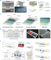
Multifunctional islet‐on‐a‐chip systems. (A) Acry‐Chip and Oxy‐Chip for constructing micro‐physiological systems. Source: Reproduced with permission. Copyright 2021, The Authors, published by American Association for the Advancement of Science. (B) Construction of the islet‐on‐a‐chip. (C) Perfused islet‐on‐a‐chip derived from stem cells. Source: Reproduced with permission. Copyright 2019, Royal Society of Chemistry. (D) Recapitulate liver‐islet axis on a chip. (E) The two‐organ‐on‐a‐chip and the enlarged view. Source: Reproduced under terms of the CC‐BY license. Copyright 2022, The Authors, published by John Wiley and Sons. (F) A pneumatically actuated micro‐physiological device that enables efficient crosstalk between 3D primary human liver cultures and intact human pancreatic islets on a chip. Source: Reproduced under terms of the CC‐BY license. Copyright 2022, The Authors, published by John Wiley and Sons.
FIGURE 12
FIGURE 12
Multi‐organs‐on‐a‐chip system based on the microfluidic chip.
FIGURE 13
FIGURE 13
Neovascularized Implantable Cell Homing and Encapsulation (NICHE) for islet allotransplantation. (A) Optical image of NICHE and annotated rendering of NICHE and scanning electron microscopy (SEM) images of the two‐layer mesh and nanoporous membrane. (B) Resin and PA devices implanted in rats for 6 weeks. (C) Quantification of fibrotic capsule thickness around medical grade titanium, resin, and PA. (D) Resin and g PA NICHE implanted subQ for 6 weeks. Dashed lines indicate cell reservoir. (E) BG measurements of diabetic rats transplanted with islets in NICHE cell reservoir receiving local (NICHE) or systemic (IP) immunosuppression, no immunosuppression (No IS), and healthy controls, iTx = islet transplant. Only NICHE and IP rats that achieved euglycemia are plotted. (F) Imaging mass cytometry of cell reservoir tissues from NICHE, IP, and rats with rejected grafts. Source: Reproduced under terms of the CC‐BY license. Copyright 2022, The Authors, published by Springer Nature.

Similar articles

Cited by

References

    1. Walker J. T., Saunders D. C., Brissova M., Powers A. C., Endocr. Rev. 2021, 42, 605. - PMC - PubMed
    1. Zhang X., Ma Z., Song E., Xu T., Protein Cell 2022, 13, 239. - PMC - PubMed
    1. Hogrebe N. J., Ishahak M., Millman J. R., Cell Stem Cell 2023, 30, 530. - PMC - PubMed
    1. Jain C., Ansarullah, Bilekova S., Lickert H., Nat. Metab. 2022, 4, 1097. - PubMed
    1. Xourafa G., Korbmacher M., Roden M., Nat. Rev. Endocrinol. 2023, 20, 27. - PubMed

LinkOut - more resources