[Preprint]. 2024 Aug 17:2024.08.16.608280.
doi: 10.1101/2024.08.16.608280.

Protein Arginine Methylation of the Translation Initiation Factor eIF1A Increases Usage of a Near-cognate Start Codon

Affiliations

Protein Arginine Methylation of the Translation Initiation Factor eIF1A Increases Usage of a Near-cognate Start Codon

Rebecca Wegman et al. bioRxiv. .

Abstract

Protein arginine methylation has emerged as a key post-translational modification responsible for many facets of eukaryotic gene expression. To better understand the extent of this modification in cellular pathways, we carried out bioorthogonal methylation profiling in Saccharomyces cerevisiae to comprehensively identify the in vivo substrates of the major yeast protein arginine methyltransferase Hmt1. Gene ontology analysis of candidate substrates revealed an enrichment of proteins involved in the process of translation. We verified one such factor, eIF1A, by in vitro methylation. Three sites on eIF1A were found to be responsible for its methylation: R13, R14, and R62, with varied capacity by which each site contributed to the overall methylation capacity in vitro. To determine the role of methylation in eIF1A function, we used a battery of arginine-to-alanine substitution mutants to evaluate translation fidelity in these mutants. Our data show that substitution mutants at R13 and R14 in the N-terminal tail improved the fidelity of start codon recognition in an initiation fidelity assay. Overall, our data suggest that Hmt1-mediated methylation of eIF1A fine-tunes the fidelity of start codon recognition for proper translation initiation.


Figures
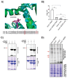
Figure 1.
Figure 1.. Tailored engineered yeast major protein arginine methyltransferase Hmt1-M36G preferentially utilizes S-adenosyl-L-methionine analogues
a) Sequence and structural alignment of S. cerevisae Hmt1 with S. pombe, R. norvegicus, and H. sapian PRMT1. The boxed amino acid at position 48 represents a highly conserved methionine residue in PRMT1, which corresponds to residue at position 36 in Hmt1 in both the three-dimensional structure of the two proteins (ribbon structure on the top panel) and the primary amino acid sequence (sequence alignment on the lower panel). b) Labeling of Npl3 by Hmt1 or Hmt1-M36G. Recombinantly purified Hmt1 or Hmt1-M36G was incubated with recombinantly purified Npl3 overnight in the presence of tritiated SAM followed by activity measurement by scintillation assay. The result plotted represents technical triplicates of three reads from a single methylation reaction. P-values were calculated using a two-tailed t-test, with *<=0.05, **<=0.001, and ***<=0.001. c) Specific in vitro labeling of Npl3 by Hmt1-M36G and Hey-SAM. The ability of Npl3 being labeled by Hmt1 or Hmt1-M36G using either Hey-SAM or Pob-SAM as a cofactor was measured by an in-gel fluorescence assay. The arrow denotes the band corresponding to labeled Npl3 by Hmt1 or Hmt1-M36G. d) In-gel fluorescence labeling assay was carried out using the following yeast lysates harvested in log phase: Hmt1-M36G, wild-type (parental), hmt1Δ, and Hmt1-G68R. The lysates were subjected to CuAAC in the presence of Hey-SAM and TAMRA-azide, followed by SDS-PAGE. The gels were subjected to analysis for fluorescence labeling (top panel) and coomassie staining (bottom) for loading control. Red arrows indicate bands that appear only in the lysates from Hmt1-M36G.
Figure 2.
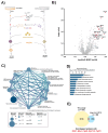
Figure 2.. Bioorthogonal profiling of Hmt1-M36G substrates.
a) Workflow schematic for bioorthogonal profiling of Hmt1 substrates. A yeast strain expressing Hmt1-M36G (MYY 2894) or lacking Hmt1 (MYY 432) was lysed and incubated with Hey-SAM. Following incubation, click chemistry was performed with cleavable biotin azide, and pulled-down peptides were labeled with tandem mass tag (TMT) technology to allow for quantitative mass spectrometry analysis. b) Volcano plot of proteins identified via Hmt1-M36G TMT mass spectrometry analysis. Any previously identified Hmt1 substrates with a p-value ≤ 0.05 and surpass the median enrichment threshold of 0.115 are labeled with red circles (e.g. Rps2). c) Connectivity map of major biological pathways enriched in the TMT mass spectrometry data from Hmt1-M36G substrates. d) GO term enrichment analysis of the Hmt1-M36G substrates with calculated score using Benjamini-Hochberg method. Shown are the top ten categories ranked by their scores. [ISP] e) Comparison of Hmt1 candidate substrates identified from this study to those identified by Low et al., 2015. The Venn diagram indicates the number of candidate substrates identified from this study or Low et al., 2015. Of the total number of candidates identified in this study, only those with a mean ratio of 1.1 or above was used to compare with the data from Low et al., 2015. The number of substrates identified from both studies is indicated at the intersection of both circles and listed.
Figure 3.
Figure 3.. Yeast eIF1A/Tif11 is able to act as an in vitro substrate of Hmt1.
In vitro methylation of intein-tagged wild-type eIF1A/Tif11, following purification from E. coli, was carried out using recombinant Hmt1 and [methyl-3H]-SAM. The full protein complement in each reaction was resolved on a 4-12% SDS-PAGE and transferred to a PVDF membrane. Methylated eIF1A/Tif11 was visualized by fluorography (arrow), after Ponceau S staining of the membrane to demonstrate protein loading levels (arrow). Recombinant GST-tagged Rps2 served as a positive control (highlighted by asterisk).
Figure 4.
Figure 4.. R13, R14, and R62 differentially contribute to the overall methylation capacity of eIF1A/Tif11.
a) The amino acid sequence of yeast eIF1A/Tif11, with potential methylated arginine sites at positions 13, 14, and 62 denoted by arrow. b) Intein-tagged wild-type eIF1A/Tif11 or eIF1A/Tif11 harboring R to A substitution(s) at amino acid positions 13, 14, and 62 were purified from E. coli and subjected to an in vitro methylation assay using recombinant Hmt1 and [methyl-3H] SAM as described in Figure 3. Methylated eIF1A/Tif11 was visualized by autoradiography (top panel), following Ponceau S staining of the membrane to demonstrate protein loading levels (bottom panel).
Figure 5.
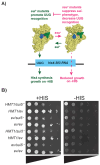
Figure 5.. Yeast strain lacking Hmt1 displays partial increase in the usage of proper start codon.
a) Overview of the his4-303 reporter assay used to test for sui and ssu phenotypes. The sui mutations promote a Pin/closed state of the preinitiation complex, decreasing translation fidelity, and allowing translation of his4-303 using a near-cognate UUG start codon. This allows histidine biosynthesis and growth on media lacking sufficient concentrations of histidine (-HIS). In contrast, ssu substitutions oppose the effects of sui mutations to promote the open/Pout conformation of the PIC, which prevents translation of the his4-303 mRNA and prevents growth on -HIS media. Structures of the 43S preinitiation complex were generated in pymol from structures 3J80 and 3J81 from the PDB (38). b) Translation initiation fidelity was assessed in wild-type (HMT1) or hmt1Δ yeast strains expressing a plasmid harboring indicated mutations in eIF5G31R (MYY 3290 and MYY 3255; indicated by sui5) or an empty vector (MYY 3292 and MYY 3257; indicated by ev). A spot assay with ten-fold serial dilution (triangles) was performed using SD medium containing either 0.3 mM histidine (indicated as +HIS) or 0.003 mM histidine (indicated as −HIS). Two biological replicates are shown from the same plate.
Figure 6.
Figure 6.. Substitution mutations of eIF1A/Tif11’s R13 and R14, but not R62, increase proper start codon usage when compared to the wild-type.
Translation initiation fidelity was assessed in wild-type eIF1A/TIF11 or eIF1A/tif11 R-to-A substitution mutant yeast strains expressing a plasmid with expression of eIF5G31R (indicated by sui5) or an empty vector (indicated by ev). Spot assays with ten-fold serial dilution (triangles) were performed using SD medium containing either 0.3 mM histidine (indicated as +HIS) or 0.003 mM histidine (indicated as −HIS). A) spot assay showing single substitution mutants of tif11R13A, tif11R14A, and tif11R62A; B) spot assay showing double substitution mutants of tif11R13,14A, tif11R13,62A, and tif11R14,62A; and C) spot assay showing triple substitution mutant tif11R13,14,62A. Two biological replicates are shown from the same plate in each spot assay.

Similar articles

References

    1. Bedford M. T., and Clarke S. G. (2009) Protein arginine methylation in mammals: who, what, and why. Molecular cell 33, 1–13 - PMC - PubMed
    1. Xu J., and Richard S. (2021) Cellular pathways influenced by protein arginine methylation: Implications for cancer. Mol Cell 81, 4357–4368 - PMC - PubMed
    1. Lorton B. M., and Shechter D. (2019) Cellular consequences of arginine methylation. Cell Mol Life Sci 76, 2933–2956 - PMC - PubMed
    1. Gary J. D., Lin W.-J., Yang M. C., Herschman H. R., and Clarke S. (1996) The predominant protein-arginine methyltransferase from Saccharomyces cerevisiae. Journal of Biological Chemistry 271, 12585–12594 - PubMed
    1. Tang J., Frankel A., Cook R. J., Kim S., Paik W. K., Williams K. R., Clarke S., and Herschman H. R. (2000) PRMT1 is the predominant type I protein arginine methyltransferase in mammalian cells. Journal of Biological Chemistry 275, 7723–7730 - PubMed

Publication types

LinkOut - more resources