. 2024 Sep 25;9(9):e0022224.
doi: 10.1128/msphere.00222-24. Epub 2024 Aug 21.

Genetic evidence for a regulated cysteine protease catalytic triad in LegA7, a Legionella pneumophila protein that impinges on a stress response pathway

Affiliations

Genetic evidence for a regulated cysteine protease catalytic triad in LegA7, a Legionella pneumophila protein that impinges on a stress response pathway

Dar Hershkovitz et al. mSphere. .

Abstract

Legionella pneumophila grows within membrane-bound vacuoles in phylogenetically diverse hosts. Intracellular growth requires the function of the Icm/Dot type-IVb secretion system, which translocates more than 300 proteins into host cells. A screen was performed to identify L. pneumophila proteins that stimulate mitogen-activated protein kinase (MAPK) activation, using Icm/Dot translocated proteins ectopically expressed in mammalian cells. In parallel, a second screen was performed to identify L. pneumophila proteins expressed in yeast that cause growth inhibition in MAPK pathway-stimulatory high-osmolarity medium. LegA7 was shared in both screens, a protein predicted to be a member of the bacterial cysteine protease family that has five carboxyl-terminal ankyrin repeats. Three conserved residues in the predicted catalytic triad of LegA7 were mutated. These mutations abolished the ability of LegA7 to inhibit yeast growth. To identify other residues important for LegA7 function, a generalizable selection strategy in yeast was devised to isolate mutants that have lost function and no longer cause growth inhibition on a high-osmolarity medium. Mutations were isolated in the two carboxyl-terminal ankyrin repeats, as well as an inter-domain region located between the cysteine protease domain and the ankyrin repeats. These mutations were predicted by AlphaFold modeling to localize to the face opposite from the catalytic site, arguing that they interfere with the positive regulation of the catalytic activity. Based on our data, we present a model in which LegA7 harbors a cysteine protease domain with an inter-domain and two carboxyl-terminal ankyrin repeat regions that modulate the function of the catalytic domain.

Importance: Legionella pneumophila grows in a membrane-bound compartment in macrophages during disease. Construction of the compartment requires a dedicated secretion system that translocates virulence proteins into host cells. One of these proteins, LegA7, is shown to activate a stress response pathway in host cells called the mitogen-activated protein kinase (MAPK) pathway. The effects on the mammalian MAPK pathway were reconstructed in yeast, allowing the development of a strategy to identify the role of individual domains of LegA7. A domain similar to cysteine proteases is demonstrated to be critical for impinging on the MAPK pathway, and the catalytic activity of this domain is required for targeting this path. In addition, a conserved series of repeats, called ankyrin repeats, controls this activity. Data are provided that argue the interaction of the ankyrin repeats with unknown targets probably results in activation of the cysteine protease domain.

Keywords: HOG pathway; Legionella pneumophila; MAP kinases; Saccharomyces cerevisiae; ankyrin repeats; cysteine proteases.


Conflict of interest statement

The authors declare no conflict of interest.

Figures

Fig 1
Fig 1
Growth defects caused by L. pneumophila IDTS expressed in S. cerevisiae. The L. pneumophila effectors (indicated on the right) were cloned under the GAL1 promoter and grown on plates containing glucose (Glu), galactose (Gal, inducing conditions), or galactose supplemented with 1 M sorbitol (Gal. + Sorb.) at 30°C, in the wild-type S. cerevisiae BY4741. pGREG523 (vector) was used as a negative control. Tenfold serial dilutions were performed, and the growth inhibition effect was compared to the one of the vector pGREG523 control (vector).
Fig 2
Fig 2
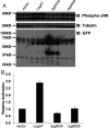
Ectopic expression of LegA7 results in elevated phosphorylation of SAPK/JNK in mammalian cells. (A) Quantitation of phospho-SAPK/JNK levels in the mammalian cell line HEK293T cells during ectopic expression of LegA7, Lpg0030, and Lpg0059 relative to empty vector control pDEST53 (vector). Forty hours after transfection, extracts were prepared, fractionated, and immunoprobed with anti-phospho-JNK (top panel), α-tubulin (middle panel), and green fluorescent protein (GFP) to identify hybrid proteins (bottom panel). Samples were run in triplicate, showing three independent transfections of plasmids into HEK293T. The arrow points to the LegA7 degradation product. Predicted sizes for GFP fusions are: empty vector, 27 kDa; LegA7, 84 kDa; Lpg0030, 62 kDa; Lpg0059 68 kDa. (B) Increased phosphorylation of LegA7 relative to other IDTS. Data were quantitated by determining phosphorylation levels relative to tubulin loading control (Materials and Methods). Data are the mean of three samples ± SE.
Fig 3
Fig 3
LegA7 affects MAPK pathways in yeast. (A) The L. pneumophila LegA7 effector cloned under the GAL1 promoter was grown on plates containing glucose (Glu), galactose (Gal, inducing conditions), galactose supplemented with 1 M sorbitol (Gal. Sorb.), galactose supplemented with 0.7 M NaCl (Gal. NaCl) at 30°C, and on SD plates containing galactose (Gal.) at 20°C and 37°C, in the wild-type S. cerevisiae BY4741. Tenfold serial dilutions were performed, and the growth inhibition effect was compared to the one of the vector pGREG523 control (vector). (B) Diagram of the yeast CWI MAPK pathway and the HOG MAPK pathway. The function of each protein is indicated on the left. T.F., transcription factor. (C) Examination of the inhibition of yeast growth mediated by the LegL7 effector in deletion mutants of the CWI and HOG MAPK pathways. LegA7 was overexpressed in the wild-type S. cerevisiae BY4741 (W.T.) and the hog1, pbs2, mpk1, and bck1 deletion mutants at 30°C and 37°C. Tenfold serial dilutions were performed, and the growth inhibition effect was compared to the one of the vector pGREG523 control (vector).
Fig 4
Fig 4
Similarity of LegA7 to bacterial cysteine protease family members. Sequence similarity alignment of LegA7 and several members of a bacterial cysteine protease family. Highlighted in red are the residues predicted to be part of the catalytic triad; highlighted in purple is the glycine residue that came out in the mutagenesis screen (see text). Accession numbers are as follows: lpg0403 (LegA7)—L. pneumophila, AAU26500; Lche_2717—L. cherrii, KTC80697; lpg2215 (LegA2)—L. pneumophila, AAU28280; Lboz_1599—L. bozemanae, KTC74159; LLO_1094—L. longbeachae, CBJ11449; HopN1—P. syringae, KPB86840; NopT—Sinorhizobium fredii, AAB91961; RipT—Ralstonia solanacearum, CBJ35895; and YopT—Yersinia pestis, WP_002213006.
Fig 5
Fig 5
Genetic evidence for a catalytic triad in LegA7. (A) Based on sequence similarity in Fig. 4, the residues C61, H206, N220, and N222 were selected as candidates for a catalytic triad. Point mutations were generated, and plating efficiency on galactose (Gal.), galactose and sorbitol (Gal. + Sorb.), and galactose and NaCl (Gal. + NaCl) plates of yeast strains harboring the mutant derivatives was determined. Tenfold serial dilutions were performed, and the growth inhibition effect was compared to the one of the vector pGREG523 control (vector). (B) LegA7 point mutations do not reduce steady-state levels of protein. To induce gene expression in yeast, yeast strains were grown on SD plates containing galactose. Lysates were analyzed by immunoblot with antibodies against the myc epitope, using PGK1 for loading control.
Fig 6
Fig 6
Ankyrin repeats are required for yeast growth inhibition. (A) Deletion of the Ank relieves LegA7-induced yeast growth inhibition. LegA7 (502 amino acids long) is predicted to have five Ank from residues 290–454 and an ID region upstream. To demonstrate that the Ank domain is important for LegA7 function, the entire Ank domain (residues 290–454), the four carboxy Ank (residues 331–454), the first two amino-terminal Ank (residues 290–361), or the first two amino-terminal Ank and the ID region (residues 264–361) were deleted from LegA7, and plating efficiency on galactose (Gal.), galactose and sorbitol (Gal. + Sorb.), and galactose and NaCl (Gal. + NaCl) plates of yeast strains harboring the mutant derivatives was determined. (B) Schematic of legA7 showing the cysteine peptidase domains (pink box), the ID region (green box), and the Ank domain-containing five repeats (blue boxes). (C) Western blot demonstrating expression of LegA7 and LegA7 deletions. To induce gene expression in yeast, overnight cultures were back-diluted into a medium containing 2% galactose for 5 hours. Lysates were analyzed by immunoblot with antibodies against the Xpress epitope and PGK1 for loading control. The ΔAnk (331–454) protein (partial ankyrin-repeat domain deletion) is predicted to migrate around 43 kDa. The ΔAnk (290–454) protein (full ankyrin-repeat domain deletion) is predicted to migrate around 39 kDa.
Fig 7
Fig 7
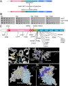
Identification of residues in the amino-terminal Ank that are essential for exerting yeast growth inhibition. (A) Selection and screening for LegA7 mutants that fail to cause hyperosmotic stress. Plasmid harboring LegA7-HIS3 protein fusion was mutagenized (Materials and Methods), transformed into yeast, and selected for HIS3+ in the presence of galactose. Surviving colonies were streaked onto sorbitol-containing plates on histidine dropout medium. Gal: galactose; srl: sorbitol. (B) Demonstration that legA7-HIS3 causes growth inhibition when expressed in yeast. The panel shows the plate assay of the growth of LegA7-HIS3 fusion as it compares to LegA7 alone. The L. pneumophila LegA7 and LegA7::HIS3 were cloned under the GAL1 promoter and grown on plates containing glucose (Glu), galactose (Gal; inducing conditions), galactose supplemented with 1 M sorbitol (Gal. Sorb.), or galactose supplemented with 0.7 M NaCl (Gal. NaCl) at 30°C. Tenfold serial dilutions were performed, and the growth inhibition effect was compared to the one of the vector pGREG523 control (vector). (C) Clustering of mutations in the amino-terminal region of the ankyrin repeats that relieve yeast growth inhibition. Schematic of LegA7 showing the cysteine peptidase domains (pink box), the ID region (green box), and the Ank domain-containing five repeats (blue boxes). The residues marked with red circles are the residues that make up the putative catalytic triad. The mutations indicated below schematic were identified in the legA7-HIS3 mutagenesis. The mutations indicated above are the directed point mutations that were used to identify the putative triad in Fig. 5. It is likely that GenBank incorrectly annotated the translation start site as 13 codons upstream from the designated start site in this panel. The transcriptional start site is downstream from the GenBank annotation, so the residue numbering system in this panel begins with the first start codon available after the transcription start, where there is also a consensus ribosome binding site. (D) Predicted LegA7 catalytic triad (gold), including the site of His42 residue (blue) mutation isolated in selection strategy. (E) The ID region links the catalytic domain to Ank repeats. Noted are surface-exposed residues that were mutation sites isolated in panel A. (F) Space-filling model of LegA7 residues 1–465 showing the arrangement of Ank repeats and catalytic site. (G) Image of panel F, rotated as noted, showing back surface relative to the catalytic domain, with sites of mutations described in panel C.
Fig 8
Fig 8
The cysteine protease domain of Icm/Dot and type III effectors. (A) Domain architecture of Legionella IDTS proteins harboring the cysteine protease (CP) domain. The known domains of each IDTS that contain the cysteine protease domain are shown. The domains presented are Ank (ankyrin domain) and PI (PI3P-binding domain). (B) The catalytic triad of the cysteine protease domain. Amino acid sequence alignments of cysteine protease catalytic triad I to III of five IDTS and predicted IDTS proteins as well as type III secreted effectors. The numbers indicated the position of the amino acids present in each of the proteins. Accession numbers are as follows: LegA7, AAU26500; LegA2, AAU28280; Lboz_1599, KTC74159; Lboz_0174, KTC77221; HopN1, PB86840; AvrPphB, SPD82586; HopC1, AAO54131; NopT, AAB91961; and YopT, WP_002213006.

Update of

Similar articles

References

    1. Burillo A, Pedro-Botet ML, Bouza E. 2017. Microbiology and epidemiology of Legionnaire’s disease. Infect Dis Clin North Am 31:7–27. doi:10.1016/j.idc.2016.10.002 - DOI - PubMed
    1. Cunha BA, Burillo A, Bouza E. 2016. Legionnaires' disease. Lancet 387:376–385. doi:10.1016/S0140-6736(15)60078-2 - DOI - PubMed
    1. Den Boer JW, Nijhof J, Friesema I. 2006. Risk factors for sporadic community-acquired Legionnaires' disease. A 3-year national case-control study. Pub Health 120:566–571. doi:10.1016/j.puhe.2006.03.009 - DOI - PubMed
    1. McDade JE, Shepard CC, Fraser DW, Tsai TR, Redus MA, Dowdle WR. 1977. Legionnaires' disease: isolation of a bacterium and demonstration of its role in other respiratory disease. N Engl J Med 297:1197–1203. doi:10.1056/NEJM197712012972202 - DOI - PubMed
    1. Boamah DK, Zhou G, Ensminger AW, O’Connor TJ. 2017. From many hosts, one accidental pathogen: the diverse protozoan hosts of Legionella. Front Cell Infect Microbiol 7:477. doi:10.3389/fcimb.2017.00477 - DOI - PMC - PubMed

MeSH terms

LinkOut - more resources