. 2024 Sep 15;13(9):bio060531.
doi: 10.1242/bio.060531. Epub 2024 Sep 2.

An atlas of Brachypodium distachyon lateral root development

Affiliations

An atlas of Brachypodium distachyon lateral root development

Cristovāo de Jesus Vieira Teixeira et al. Biol Open. .

Abstract

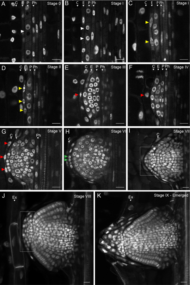
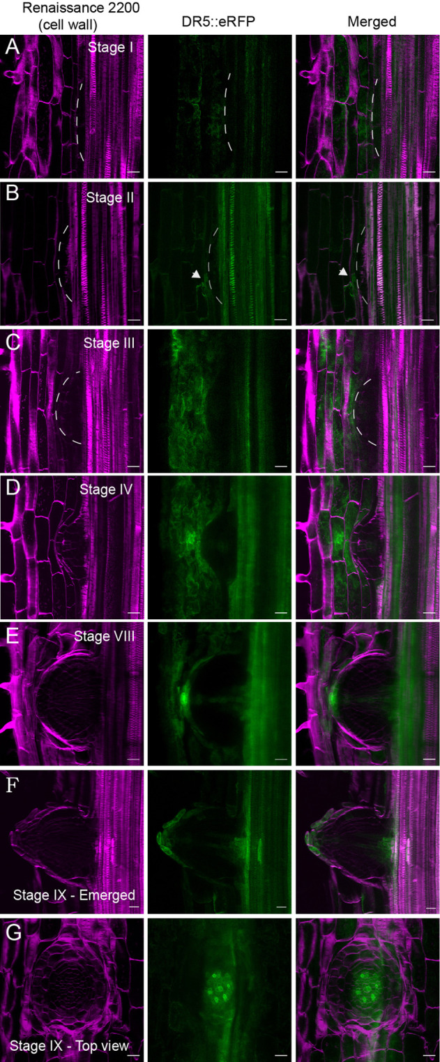
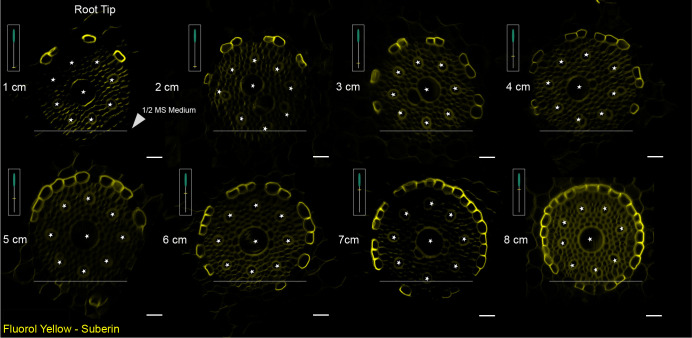
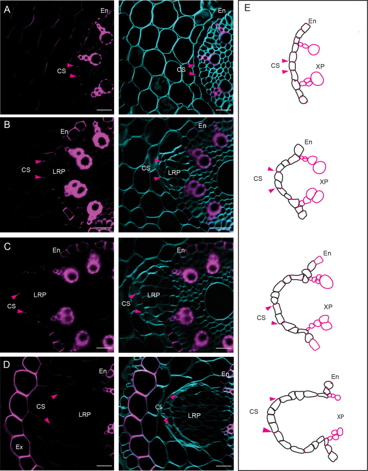
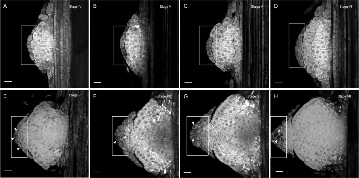
The root system of plants is a vital part for successful development and adaptation to different soil types and environments. A major determinant of the shape of a plant root system is the formation of lateral roots, allowing for expansion of the root system. Arabidopsis thaliana, with its simple root anatomy, has been extensively studied to reveal the genetic program underlying root branching. However, to get a more general understanding of lateral root development, comparative studies in species with a more complex root anatomy are required. Here, by combining optimized clearing methods and histology, we describe an atlas of lateral root development in Brachypodium distachyon, a wild, temperate grass species. We show that lateral roots initiate from enlarged phloem pole pericycle cells and that the overlying endodermis reactivates its cell cycle and eventually forms the root cap. In addition, auxin signaling reported by the DR5 reporter was not detected in the phloem pole pericycle cells or young primordia. In contrast, auxin signaling was activated in the overlying cortical cell layers, including the exodermis. Thus, Brachypodium is a valuable model to investigate how signaling pathways and cellular responses have been repurposed to facilitate lateral root organogenesis.

Keywords: Brachypodium distachyon; Endodermis; Exodermis; Lateral roots; Organogenesis.


Conflict of interest statement

Competing interests The authors declare no competing or financial interests.

Figures

Fig. 1.
Fig. 1.
Different stages of LRP development in Brachypodium. (A) Stage 0: No discernible cell divisions in the pericycle cells. (B) Stage I: White arrowheads indicate the first anticlinal cell division in the pericycle. (C) Stage I: Yellow arrowheads indicate the flattening of the endodermal cells preceding the cell divisions in the next stage. (D) Stage II: The endodermis starts to divide anticlinal (yellow arrowheads). (E) Stage III: Periclinal divisions take place at the center of the LRP resulting in three layers of cells. The red arrowhead indicates cell divisions in the overlying cortex. (F) Stage IV: The LRP undergoes radial expansion through constant anticlinal and periclinal cell divisions in the center of the LRP. Four cell layers can be observed. (G) Stage V: Five to six cell layers can still be distinguished. LRP boundaries are established, and the endodermis appears to become integrated in the LRP. Red arrowheads indicate more cell divisions in the cortex layer in the vicinity of the LRP. (H) Stage VI: The endodermal cells on the apex of the LRP start to divide again (green arrowheads). Cell layer counting is no longer used from this stage. (I) Stage VII: Formation of the root cap (white rectangular area. (J) Stage VIII: The LRP reaches the root exodermis. (K) Stage IX: Emerged: The LRP is fully formed and traverses the exodermis and epidermis. Ex, exodermis; C, cortex; E, endodermis; P, pericycle; Ph, phloem. Representative images were obtained from 45 seedling roots from three independent replicates each consisting of at least 15 plants of Bd21-3. Samples were cleared with DEEP-Clear and stained with 0.01% propidium iodide. Scale bars: 20 μm.
Fig. 2.
Fig. 2.
DR5pro::ER-mRFP activity during LR development in Brachypodium. (A) The DR5 signal is not evident in Stage I during the first pericycle cell divisions. (B) The DR5 signal could be observed in Stage II when the endodermis starts to divide (white arrowheads). (C,D) The DR5 signal is no longer observed in the endodermis but in the cortex cell layer in the vicinity of the LRP and in the central part of the LRP resembling vasculature. (E) The DR5 signal is intensified at the apex of the LRP, in the vasculature, and in the last cortex cell layer. (F,G) A fully emerged LR shows a similar DR5 pattern as usually observed in the primary root. Representative images were obtained from 45 seedlings from three independent replicates each consisting of at least 15 plants of Bd21-3. DR5pro::ER-mRFP (green) and cell walls stained with SCRI Renaissance (magenta) for cellulose. Scale bars: 50 μm.
Fig. 3.
Fig. 3.
Pattern of endodermal suberization along the Brachypodium root axis adjacent to the nutrient medium growth. Cross-sections of Brachypodium primary root showing asymmetric suberization. SL developed unilaterally on the side of the root exposed to the air from the root apex, but not on the side exposed to nutrient medium. Representative images were obtained from 30 seedlings of Bd21-3 from three independent replicates, each consisting of at least ten plants. Roots of similar length were positioned in parallel for consistency, and regions of interest of approximately 1 cm were sectioned. Scale bars: 20 μm.
Fig. 4.
Fig. 4.
Recently divided endodermal cells do not establish Casparian strips. (A-D) Cross-sections stained with BF (lignin) in magenta and Renaissance SR2200 (cellulose) in cyan. The images illustrate the progression of cell divisions in the endodermis and the distancing of the previously formed CS. (E) A graphical representation depicting the cell divisions in the endodermis and the separation of the CS is shown on the right. The arrows indicate the position of the CS. Roots of seedlings (six DAG) with similar length were positioned in parallel for consistency, and regions of interest of approximately 1 cm from the root tip were sectioned. Representative images were obtained from 30 seedlings of Bd21-3 from three independent replicates, each consisting of at least ten plants. Scale bars: 50 µm.
Fig. 5.
Fig. 5.
Endodermal cells give rise to the columella cells of the root cap. (A-H) Starch granules (dark structures in boxed area) were detected using Lugol staining. The boxed area shows cell divisions in the endodermis and its progression in differentiating into columella cells from Stage VI (E) to Stage VII (H) marked by the sediments of starch granules (arrowheads) in the first cell layer on the apex of a Stage VI LRP. Representative images were obtained from 30 seedlings from three independent replicates, each consisting of at least ten plants of Bd21-3. The root cortex was mechanically removed with forceps preserving the LRPs integrity. Samples were cleared with DEEP-Clear and stained with Lugol. Scale bars: 50 µm.

Similar articles

References

    1. Andersen, T. G., Molina, D., Kilian, J., Franke, R. B., Ragni, L. and Geldner, N. (2021). Tissue-autonomous phenylpropanoid production is essential for establishment of root barriers. Curr. Biol. 31, 965-977.e5. 10.1016/j.cub.2020.11.070 - DOI - PubMed
    1. Atkinson, J. A., Rasmussen, A., Traini, R., Voß, U., Sturrock, C., Mooney, S. J., Wells, D. M. and Bennett, M. J. (2014). Branching out in roots: uncovering form, function, and regulation. Plant Physiol. 166, 538-550. 10.1104/pp.114.245423 - DOI - PMC - PubMed
    1. Banda, J., Bellande, K., von Wangenheim, D., Goh, T., Guyomarcc'h, S., Laplaze, L. and Bennett, M. J. (2019). Lateral root formation in arabidopsis: a well-ordered LRexit. Trends Plant Sci. 24, 826-839. 10.1016/j.tplants.2019.06.015 - DOI - PubMed
    1. Bao, Y., Aggarwal, P., Robbins, N. E., Sturrock, C. J., Thompson, M. C., Tan, H. Q., Tham, C., Duan, L., Rodriguez, P. L., Vernoux, T.et al. (2014). Plant roots use a patterning mechanism to position lateral root branches toward available water. Proc. Natl Acad. Sci. USA 111, 9319-9324. 10.1073/pnas.1400966111 - DOI - PMC - PubMed
    1. Barberon, M., Vermeer, J. E. M., De Bellis, D., Wang, P., Naseer, S., Andersen, T. G., Humbel, B. M., Nawrath, C., Takano, J., Salt, D.et al. (2016). Adaptation of root function by nutrient-induced plasticity of endodermal differentiation. Cell 164, 447-459. 10.1016/j.cell.2015.12.021 - DOI - PubMed

MeSH terms

Substances

LinkOut - more resources