This is a preprint.
Physiological oxygen concentration during sympathetic primary neuron culture improves neuronal health and reduces HSV-1 reactivation
- PMID: 39149301
- PMCID: PMC11326244
- DOI: 10.1101/2024.08.09.607366
Physiological oxygen concentration during sympathetic primary neuron culture improves neuronal health and reduces HSV-1 reactivation
Update in
-
Physiological oxygen concentration during sympathetic primary neuron culture improves neuronal health and reduces HSV-1 reactivation.Microbiol Spectr. 2024 Nov 11;12(12):e0203124. doi: 10.1128/spectrum.02031-24. Online ahead of print. Microbiol Spectr. 2024. PMID: 39526754 Free PMC article.
Abstract
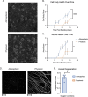
Herpes simplex virus-1 (HSV-1) establishes a latent infection in peripheral neurons and periodically reactivates in response to a stimulus to permit transmission. In vitro models using primary neurons are invaluable to studying latent infection because they use bona fide neurons that have undergone differentiation and maturation in vivo. However, culture conditions in vitro should remain as close to those in vivo as possible. This is especially important when considering minimizing cell stress, as it is a well-known trigger of HSV reactivation. We recently developed an HSV-1 model system that requires neurons to be cultured for extended lengths of time. Therefore, we sought to refine culture conditions to optimize neuronal health and minimize secondary effects on latency and reactivation. Here, we demonstrate that culturing primary neurons under conditions closer to physiological oxygen concentrations (5% oxygen) results in cultures with features consistent with reduced stress. Furthermore, culture in these lower oxygen conditions diminishes the progression to full HSV-1 reactivation despite minimal impacts on latency establishment and earlier stages of HSV-1 reactivation. We anticipate that our findings will be useful for the broader microbiology community as they highlight the importance of considering physiological oxygen concentration in studying host-pathogen interactions.
Figures


Similar articles
-
DLK-Dependent Biphasic Reactivation of Herpes Simplex Virus Latency Established in the Absence of Antivirals.J Virol. 2022 Jun 22;96(12):e0050822. doi: 10.1128/jvi.00508-22. Epub 2022 May 24. J Virol. 2022. PMID: 35608347 Free PMC article.
-
Herpes Simplex Virus 1 Strains 17syn+ and KOS(M) Differ Greatly in Their Ability To Reactivate from Human Neurons In Vitro.J Virol. 2020 Jul 16;94(15):e00796-20. doi: 10.1128/JVI.00796-20. Print 2020 Jul 16. J Virol. 2020. PMID: 32461310 Free PMC article.
-
A primary neuron culture system for the study of herpes simplex virus latency and reactivation.J Vis Exp. 2012 Apr 2;(62):3823. doi: 10.3791/3823. J Vis Exp. 2012. PMID: 22491318 Free PMC article.
-
A comparison of herpes simplex virus type 1 and varicella-zoster virus latency and reactivation.J Gen Virol. 2015 Jul;96(Pt 7):1581-602. doi: 10.1099/vir.0.000128. Epub 2015 Mar 20. J Gen Virol. 2015. PMID: 25794504 Free PMC article. Review.
-
[The mechanisms for latency and reactivation of alpha herpesviridae].Nihon Rinsho. 2000 Apr;58(4):807-14. Nihon Rinsho. 2000. PMID: 10774199 Review. Japanese.
References
Publication types
Grants and funding
LinkOut - more resources
Full Text Sources
