

Melatonin supplementation counteracts fiber loss in knee ligaments of diabetes-induced rats
- PMID: 39135805
- PMCID: PMC11317382
- DOI: 10.3389/fphar.2024.1399719
Melatonin supplementation counteracts fiber loss in knee ligaments of diabetes-induced rats
Abstract
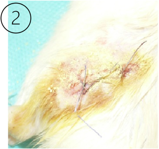
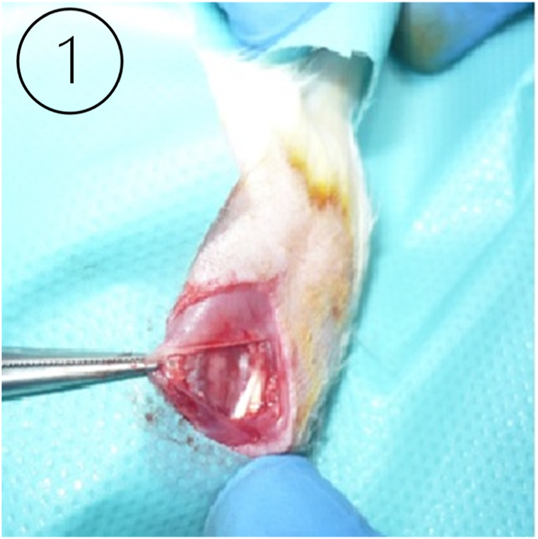
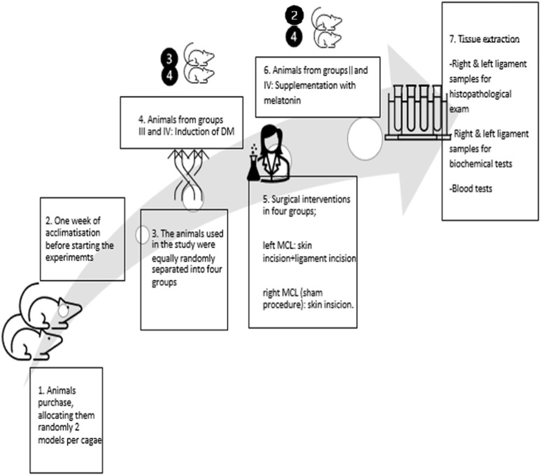
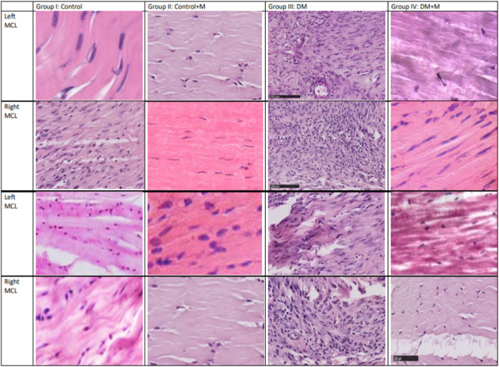
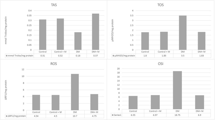
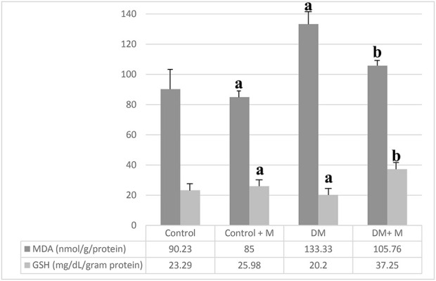
Diabetes mellitus (DM) is a prevalent metabolic disease. The clinical impact of sustained hyperglycemia on ligament healing has not been well characterized. Diabetes is a known cause of macro-, microvascular, and diabetic ulcer healing difficulties among tissues. Therefore, we aimed to investigate the healing potential occurring in injured and healthy ligaments among diabetic and healthy individuals using a rat model. We hypothesize that DM may contribute to altering the knee medial collateral ligament (MCL), thus its morphology, biochemical fitness, and functionality. The study cohort consisted of 40 rats. The animals were randomized into four equal groups. Groups I and II (20 rats) received saline subcutaneously and served as controls. Groups III and IV (20 rats) were injected with a single dose of streptozotocin (STZ). All animals underwent surgery to cut the left tibial collateral ligament in the hind limb and suture it. The access site was sutured to create inflammation and study the regenerative capacities of animals with normal carbohydrate metabolism and pharmacologically induced diabetes. Each animal then underwent sham surgery to access and suture the right tibial collateral ligament in the hind limb without ligament intervention. After the animals had undergone surgeries, groups II and IV were given melatonin supplementation for 4 weeks. Rats with DM presented with more fibrosis and calcification of the MCL and decreased healing potential. Treatment with melatonin in diabetic rats mitigated alterations and improved the antioxidant status of ligaments from the diabetic group.
Keywords: diabetes ligaments; diabetes mellitus; ligaments alteration; ligaments alteration in diabetes mellitus; orthopedic complications.
Copyright © 2024 Adamska, Wnuk, Kamińska, Poniatowska, Maciąg, Kamiński, Stolarczyk, Matin, Atanasov, Łapiński and Jóźwik.
Conflict of interest statement
The authors declare that the research was conducted in the absence of any commercial or financial relationships that could be construed as a potential conflict of interest.
Figures
Similar articles
-
Comparison between Operative and Non-Operative Treatment of the Medial Collateral Ligament: Histological and Ultrastructural Findings during Early Healing in the Epiligament Tissue in a Rat Knee Model.Cells Tissues Organs. 2018;206(3):165-182. doi: 10.1159/000496985. Epub 2019 Mar 15. Cells Tissues Organs. 2018. PMID: 30879007
-
Early expression of marker genes in the rabbit medial collateral and anterior cruciate ligaments: the use of different viral vectors and the effects of injury.J Orthop Res. 1999 Jan;17(1):37-42. doi: 10.1002/jor.1100170107. J Orthop Res. 1999. PMID: 10073645
-
Instrument-assisted cross fiber massage increases tissue perfusion and alters microvascular morphology in the vicinity of healing knee ligaments.BMC Complement Altern Med. 2013 Sep 28;13:240. doi: 10.1186/1472-6882-13-240. BMC Complement Altern Med. 2013. PMID: 24073942 Free PMC article.
-
Mesenchymal Stem Cells for Enhanced Healing of the Medial Collateral Ligament of the Knee Joint.Medicina (Kaunas). 2023 Apr 7;59(4):725. doi: 10.3390/medicina59040725. Medicina (Kaunas). 2023. PMID: 37109683 Free PMC article. Review.
-
Functional tissue engineering for ligament healing: potential of antisense gene therapy.Ann Biomed Eng. 2004 Mar;32(3):342-51. doi: 10.1023/b:abme.0000017551.93144.1a. Ann Biomed Eng. 2004. PMID: 15098538 Review.
References
Grants and funding
LinkOut - more resources
Full Text Sources