. 2024 Sep 20;5(3):103253.
doi: 10.1016/j.xpro.2024.103253. Epub 2024 Aug 10.

Protocol for the isolation and purification of endoplasmic reticulum-plasma membrane junctions from the mouse brain

Affiliations

Protocol for the isolation and purification of endoplasmic reticulum-plasma membrane junctions from the mouse brain

Jason A Weesner et al. STAR Protoc. .

Abstract

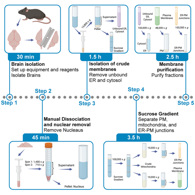
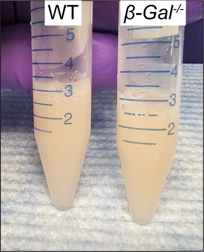
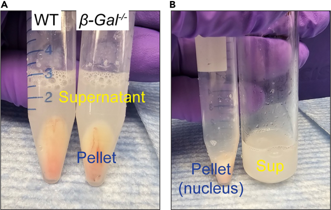
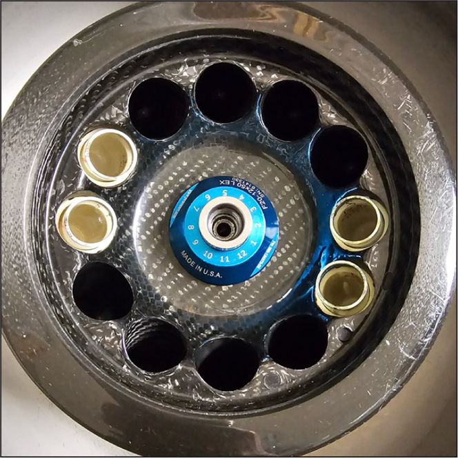
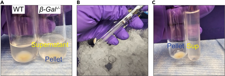
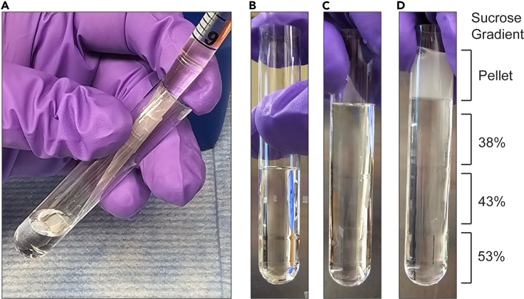
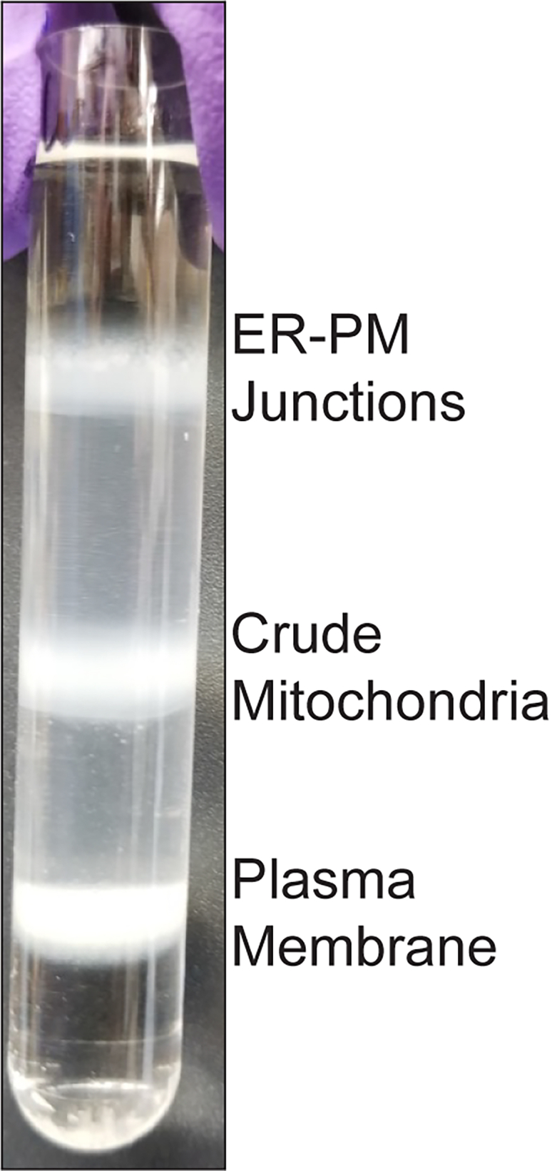
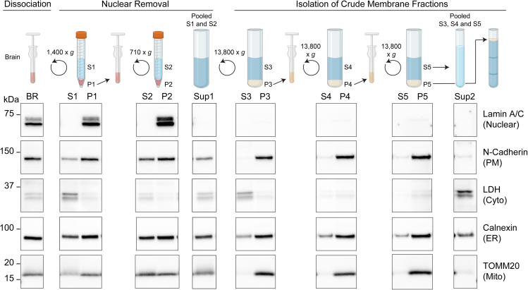
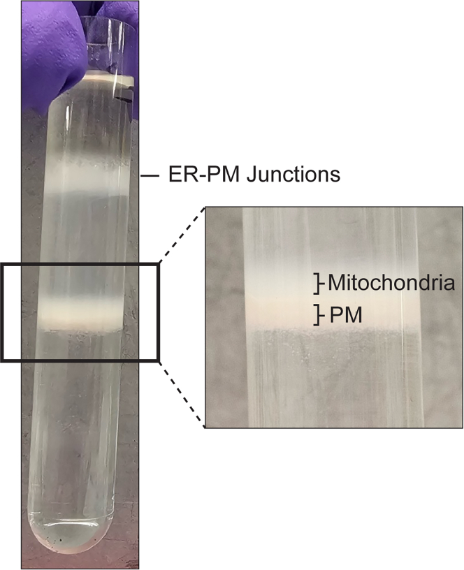
Dynamic communication between intracellular organelles often takes place at specialized membrane contact sites that form between their membranes. Here we detail a procedure for the purification of endoplasmic reticulum-plasma membrane (ER-PM) junctions from the mouse brain. We describe steps for homogenizing isolated brain hemispheres and sequential centrifugation to remove the nuclear fraction from the other membrane fractions. We then detail procedures for separating the resulting crude membrane fractions by sucrose density gradients and purifying into their respective pellets. For complete details on the use and execution of this protocol, please refer to Weesner et al.1.

Keywords: Cell Membrane; Cell separation/fractionation; Neuroscience.


Conflict of interest statement

Declaration of interests The authors declare no competing interests.

Figures

None
Graphical abstract
Figure 1
Figure 1
Isolate the mouse brain Isolate the brain in Step 1 a-f by (A) euthanizing the mouse, (B) removing the skull, (C and D) cutting open the skull and peeling the scalp from the brain, (E) removing the brain, and (F) cutting the brain in half.
Figure 2
Figure 2
Homogenize the mouse brain in Dounce Homogenizer After weighing brain, (A) place brain in a Dounce homogenizer and (B) homogenize with Pestle A. Step 3.
Figure 3
Figure 3
Remove nucleus and cell debris from lysate Representative image of pellet and supernatant obtained in Step 6 from WT and β-Gal−/− mice.
Figure 4
Figure 4
Resuspend the pellet in Solution A Add solution A to the pellet in Step 7 and pipette up and down.
Figure 5
Figure 5
Homogenize the pellet Representative image of pellet homogenate collected in Step 9 from WT and β-Gal−/− mice.
Figure 6
Figure 6
Nuclear removal (A) Representative image of pellet (containing the nucleus) and supernatant collected in Step 10 from WT and β-Gal−/− mice. (B) Collect supernatant and transfer to a 30 mL Corex glass tube in Step 11.
Figure 7
Figure 7
Pellet the supernatant containing membrane fractions Insert the Corex tubes from Step 12 and place them in a Sorvall Centrifuge.
Figure 8
Figure 8
Separate the unbound ER and cytosolic fraction from the crude membrane fraction (A) Representative image of pellet (containing the membranes) and supernatant (containing unbound ER and cytosol) collected in Step 14 from WT and β-Gal−/− mice. (B) Carefully remove the supernatant and (C) transfer the supernatant to a 13.2 mL ultra-clear Beckman centrifuge tube.
Figure 9
Figure 9
Layer the sucrose gradient (A) Add 2 mL of 53% sucrose in a 13.2 mL Beckman centrifuge tube. (B–D) Carefully layer (B) 43% and (C) 38% sucrose on top. (D) Layer the homogenized pellet obtained in Step 21 on top of the gradient. Related to Step 22a-d.
Figure 10
Figure 10
Separate the membrane fractions on a sucrose gradient After 2.5 h spin in Step 24, there are 3 bands containing the ER-PM junctions, crude mitochondria, and plasma membrane fractions.
Figure 11
Figure 11
Purify the ER-PM junctions Representative image of pellet containing purified ER-PM junctions obtained in Step 25g from WT and β-Gal−/− mice.
Figure 12
Figure 12
Purify the plasma membrane fraction Representative image of pellet containing purified plasma membrane fraction obtained in Step 26f from WT and β-Gal−/− mice.
Figure 13
Figure 13
Purify the plasma membrane fraction Representative image of pellet containing purified ER fraction obtained in Step 27d from WT and β-Gal−/ mice.
Figure 14
Figure 14
Expected purity marker for first 2 major steps Schematic representation of “Manual dissociation of mouse brain” and “Isolation of crude membrane fractions” major steps. Western blot analyses demonstrate purity markers for each fraction described in the expected outcomes. (Adapted from Weesner et al. 20241).
Figure 15
Figure 15
Improper separation of plasma membrane and mitochondria bands Example of improper separation of plasma membrane and mitochondria band from sucrose gradient described in problem 1.
Figure 16
Figure 16
Diffuse plasma membrane band Examples of improper separation of plasma membrane band from sucrose gradient described in problem 2.
Figure 17
Figure 17
Removal of mitochondria contamination from fractions Examples of (A) low mitochondria contamination and (B) too much mitochondria contamination as described in problem 3.
Figure 18
Figure 18
Low ER-PM junction yield Examples of low ER-PM junction recovery as described in problem 5.
Figure 19
Figure 19
Example of a high-performance thin-layer chromatography plate with sucrose contamination as described in problem 6
Figure 20
Figure 20
Example of a high-performance thin-layer chromatography with low sucrose contamination after wash with Washing (−) Buffer as described in Solution 6

Similar articles

References

    1. Weesner J.A., Annunziata I., van de Vlekkert D., Robinson C.G., Campos Y., Mishra A., Fremuth L.E., Gomero E., Hu H., d'Azzo A. Altered GM1 catabolism affects NMDAR-mediated Ca(2+) signaling at ER-PM junctions and increases synaptic spine formation in a GM1-gangliosidosis model. Cell Rep. 2024;43 doi: 10.1016/j.celrep.2024.114117. - DOI - PMC - PubMed
    1. Weesner J.A., Annunziata I., van de Vlekkert D., d'Azzo A. Glycosphingolipids within membrane contact sites influence their function as signaling hubs in neurodegenerative diseases. FEBS Open Bio. 2023;13:1587–1600. doi: 10.1002/2211-5463.13605. - DOI - PMC - PubMed
    1. Gray E.G., Whittaker V.P. The isolation of nerve endings from brain: an electron-microscopic study of cell fragments derived by homogenization and centrifugation. J. Anat. 1962;96:79–88. - PMC - PubMed
    1. Annunziata I., Weesner J.A., d'Azzo A. Isolation of Mitochondria-Associated ER Membranes (MAMs), Synaptic MAMs, and Glycosphingolipid Enriched Microdomains (GEMs) from Brain Tissues and Neuronal Cells. Methods Mol. Biol. 2021;2277:357–370. doi: 10.1007/978-1-0716-1270-5_22. - DOI - PubMed
    1. Hahn C.N., del Pilar Martin M., Schröder M., Vanier M.T., Hara Y., Suzuki K., Suzuki K., d'Azzo A. Generalized CNS disease and massive GM1-ganglioside accumulation in mice defective in lysosomal acid beta-galactosidase. Hum. Mol. Genet. 1997;6:205–211. doi: 10.1093/hmg/6.2.205. - DOI - PubMed

LinkOut - more resources