. 2024 Aug 1;25(15):8430.
doi: 10.3390/ijms25158430.

RiboScreenTM Technology Delivers a Ribosomal Target and a Small-Molecule Ligand for Ribosome Editing to Boost the Production Levels of Tropoelastin, the Monomeric Unit of Elastin

Affiliations

RiboScreenTM Technology Delivers a Ribosomal Target and a Small-Molecule Ligand for Ribosome Editing to Boost the Production Levels of Tropoelastin, the Monomeric Unit of Elastin

Bjoern Wimmer et al. Int J Mol Sci. .

Abstract

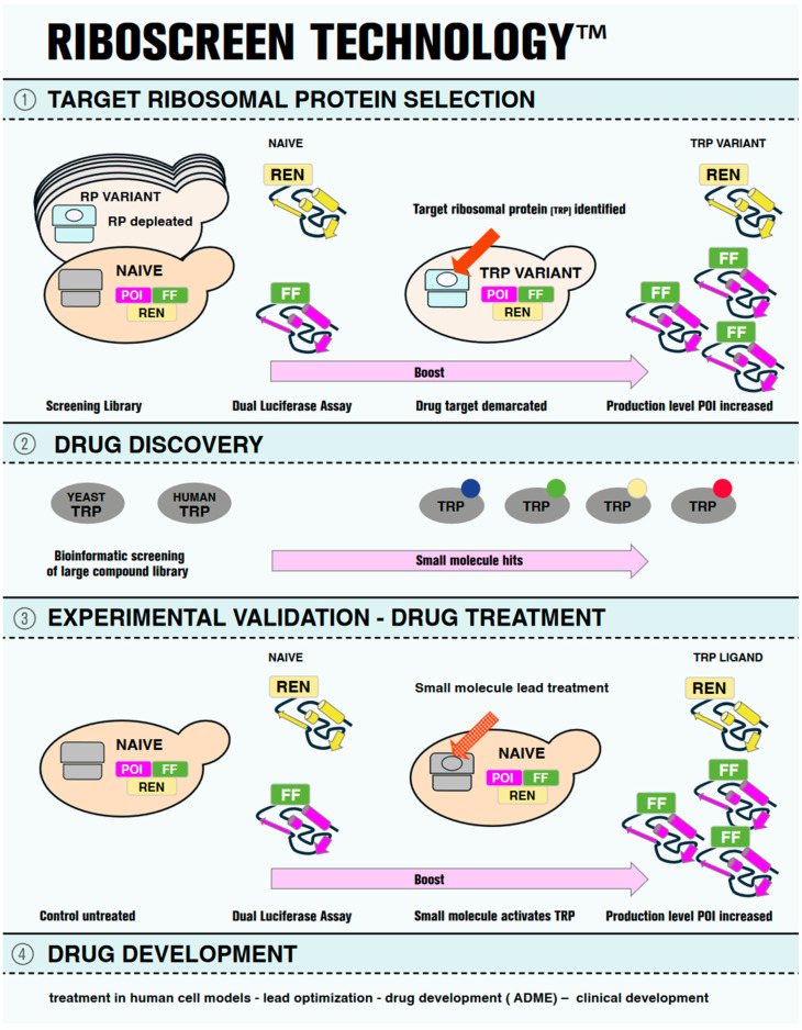
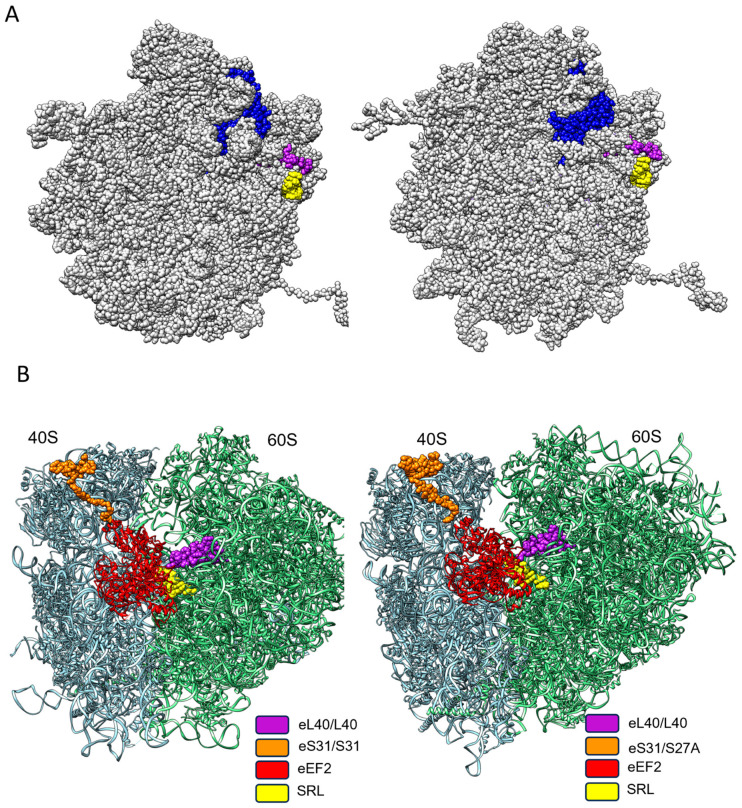
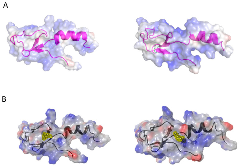
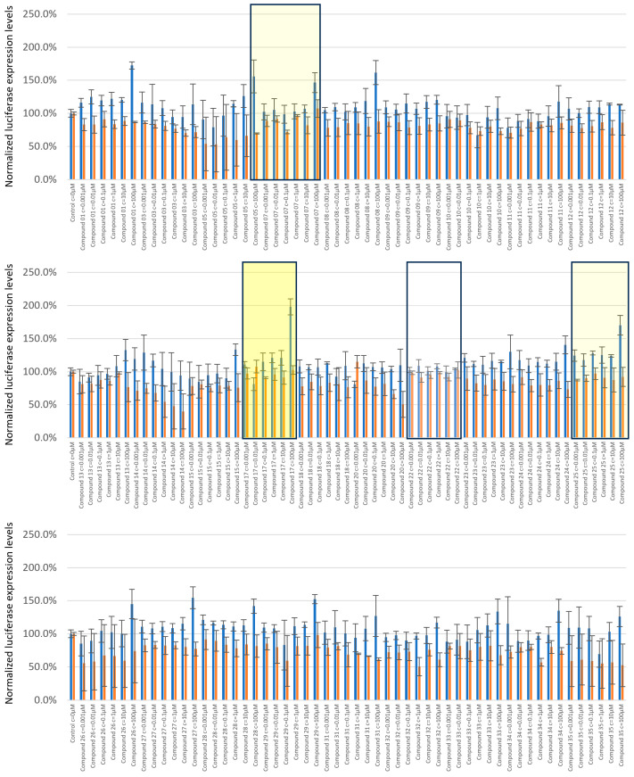
Elastin, a key structural protein essential for the elasticity of the skin and elastogenic tissues, degrades with age. Replenishing elastin holds promise for anti-aging cosmetics and the supplementation of elastic activities of the cardiovascular system. We employed RiboScreenTM, a technology for identifying molecules that enhance the production of specific proteins, to target the production of tropoelastin. We make use of RiboScreenTM in two crucial steps: first, to pinpoint a target ribosomal protein (TRP), which acts as a switch to increase the production of the protein of interest (POI), and second, to identify small molecules that activate this ribosomal protein switch. Using RiboScreenTM, we identified ribosomal protein L40, henceforth eL40, as a TRP switch to boost tropoelastin production. Drug discovery identified a small-molecule hit that binds to eL40. In-cell treatment demonstrated activity of the eL40 ligand and delivered increased tropoelastin production levels in a dose-dependent manner. Thus, we demonstrate that RiboScreenTM can successfully identify a small-molecule hit capable of selectively enhancing tropoelastin production. This compound has the potential to be developed for topical or systemic applications to promote skin rejuvenation and to supplement elastic functionality within the cardiovascular system.

Keywords: RiboScreenTM Technology; ageing of elastic tissues; customized protein expression; elastin; ribosomal protein eL40; small-molecule hit; tropoelastin expression.


Conflict of interest statement

H.B.-K., J.W.B., H.H. and J.K. are shareholders of KBHB Consult GmbH, Salzburg, Austria. J.V.H. and H.B.-K. are named as inventors in the patent family represented by WO24099952 A1 based on EP4368729, which is filed in the name of Merck Patent GmbH. The author Christin Rakers was employed by the company Merck Healthcare KGaA, Darmstadt, Germany. The author Daniel Kuhn was employed by the company Merck Healthcare KGaA, Darmstadt, Germany. The remaining authors declare that the research was conducted in the absence of any commercial or financial relationships that could be construed as a potential conflict of interest.

Figures

Figure 1
Figure 1
Pictographic representation of RiboScreenTM Technology. On the left-hand side, the first step of the RiboScreenTM technology is shown in Panel (1). Two tools are used. First is a screening library of yeast vehicles, each of which is depleted for one of the eighty eukaryotic ribosomal proteins (RPs), the ribosomal variant strain (RVS) screening library. In cyan, a depleted ribosome for a ribosomal protein (white oval) is shown. In grey, a wild-type ribosome is shown in a naïve vehicle. The second tool is a dual luciferase assay to monitor protein expression levels of the protein of interest (POI, magenta), the cellular target. The reporter of the POI carries a C-terminally tagged Firefly luciferase (FF, green), and a Renilla (REN, yellow) luciferase reporter serves as internal control. In the middle section, panel (1) presents the identification of the actual drug target, the target ribosomal protein (TRP) (red arrow), demarcated by its altered functional availability (white oval), which leads to an increase in the production level of the POI, (panel (1), right). Panel (2) shows orthologous yeast and human TRPs (grey ovals) on the left, which serve as protein baits for structure-based virtual screening to identify small-molecule binders. The compounds identified in this way from the screening of a library of small molecules are then computationally docked into the binding sites of the TRP and scored for their binding affinities to identify potential ligands. The virtual screening results are post-processed to select the representative hits (small colored circles) for further analysis (panel (2), left). Panel (3) shows the experimental validation of the hit molecules using RiboScreenTM technology. Naïve vehicles, equipped with identical protein reporters as employed in the initial screening (POI-FF and REN), are used (Panel (3), left), but here treated with TRP ligands (red dotted arrow). Hit compounds are identified based on their demonstrated activity to boost POI production levels. In Panel (4), further steps of drug development are listed for completeness.
Figure 2
Figure 2
Exon structure of ELN mRNA encoding Tropoelastin. On top, the ELN exon structure is depicted, with the individual exons numbered as present in the human mRNA (modified from [47]). The bottom shows isoform 6 splice variant, lacking several exons, among them exon 26A.
Figure 3
Figure 3
Reporter protein production levels in the ribosomal variant strain (RVS) screening library. This plot correlates, in a single data point for each ribosomal variant strain vehicle, the mean protein production level of the internal control REN reporter, normalized to the mean wild-type signal on the y-axis and the mean protein production level of the TE-FF reporter normalized to the mean wild type signal on the x-axis. A circle centered at a 100% expression level (i.e., 1) of both reporter proteins in the wild-type vehicle (marked in yellow) and with a radius of 50% difference in expression level, corresponding to the 2-fold standard deviation of the overall mean of measurements in all RVS contains the TE-FF and REN reporter expression profiles from the majority of screening vehicles. The data point obtained for the RVS, carrying a depletion for eL40, as encoded by RPL40A (marked in green), but not as encoded by RPL40B (marked in grey), signals that the modification of this functional availability of eL40 boosts TE-FF production 2.8-fold while leaving REN expression levels unaltered. This identifies eL40 as a potential target ribosomal protein (TRP) for customized boost of protein production levels of tropoelastin. Minor TRP species, where REN expression levels are also affected, are represented by the reporter protein expression levels observed in RVS depleted for ribosomal proteins uS15 and uL2 as encoded by RPS13 and by RPL2B (marked in grey). Note that the gene nomenclature and protein nomenclature are different for ribosomal proteins in yeast [40].
Figure 4
Figure 4
Ribosomal protein eL40 is conserved between yeast and humans in sequence, structure, and topological position on the ribosome. (A) Sequence comparison between the two yeast paralogous eL40 proteins, as well as that to their human orthologue, is shown, with the N-terminal ubiquitin tag included. (B) eL40 resides in the 60S subunit of eukaryotic ribosomes, yeast shown to the left (PDB code 7B7D) and human shown to the right (PDB code 6QZP), and ribbon models of yeast and human eL40 show their integration into the 60S subunit, with a close-up in (C). In (D), a visualization of the domain architecture of eL40 depicts the non-globular extension of the protein, which anchors the protein within the rRNA scaffold of the ribosome (grey box). The central domain, as well as the N-terminus and C-terminus, remains accessible for other intermolecular interactions.
Figure 5
Figure 5
The spatial–functional conservation of eL40 on yeast and human ribosome. (A) During the generation of the translation-competent ribosomal subunits, eL40 (blue) is the penultimate ribosomal protein to arrive at the 60S subunit, as shown from the inter-subunit side for yeast on the left (PDB code 7B7D) and for human on the right (PDB code 6QZB). eL40 is positioned atop the Sarcin–Ricin loop (yellow). Upon the arrival of the last ribosomal protein to be incorporated into the 60S subunit, uL16 (magenta), the 60 S subunit, attains its final, translation-competent configuration, which is ready to form the translation-competent ribosome by joining the mRNA-associated 40S subunit. (B) On the translation-competent ribosome, yeast to the left (PDB code 5JUU) and human to the right (PDB code 6Z6N) eL40, in close proximity to the small subunit protein eS31 and the Sarcin–Ricin loop of the 60S subunit, respectively, form the landing platform (factor binding site), for elongation factors (EF), which drive the rate of protein synthesis. eEF2, in exemplary form, is shown binding to the factor binding site.
Figure 6
Figure 6
Ligand binding sites of eL40 and the visualization of binding pockets. (A) Coulombic surface representation of yeast (left, PDB code 3J77) and human (right, PDB code 6XA1) eL40 proteins, overlaid on their secondary structures. (B) Potential small-molecule binding pockets on yeast eL40, to the left, (yellow, near Ser94, PDB code 3J77) and human eL40, to the right (yellow, near Asp92, PDB code 6EK0), were identified by druggability analysis (Sitemap [72]) using the amino acid sequence of the eL40 proteins from the ribosome X-ray structures (minus rRNA). These pockets are adjacent (yellow patches) and, depending on protein dynamics, could potentially form a larger, common binding cavity.
Figure 7
Figure 7
Validation of small-molecule eL40 ligands for customized boost of tropoelastin production levels. Candidate small-molecule ligands were tested in a range of concentrations from 1 nM to 100 µM. Firefly tagged tropoelastin luciferase signals (blue) and Renilla signals (orange) obtained upon treatments are shown normalized to the untreated control. The compounds are listed per increasing number, and C17 showed a dose-dependent and significant response at 100 μM, with a 1.7-fold boost in tropoelastin reporter expression, and Renilla expression was unaltered (middle panel, yellow rectangle). Examples of minor candidate eL40 activators are C7 and C25 (mute yellow). A representative of non-active eL40 ligands is C22 (lower panel, white).
Figure 8
Figure 8
Molecular structures of rpL40 ligands. UPAC nomenclature, molecular structure and space filling models of compounds C17, C7, C25, and C22 are shown.
Figure 9
Figure 9
Molecular interactions of C17 with yeast and human rpL40. (A) Predicted docking poses for C17 with yeast (PDB code 3J77) and human (PDB code 6EK0) eL40 structures. This figure illustrates the predicted binding modes of compound C17 to both yeast and human eL40 orthologues. On the left, the complex structure with the yeast eL40 is shown, where the carbon atoms of C17 are highlighted in green. On the right, the complex structure with the human eL40 is displayed, with C17’s carbon atoms in cyan. The molecular surfaces of both structures are depicted in panel (A). Panel (B) highlights the key interactions: the charged interactions between the piperidine group of C17 and the aspartate 92 residue, and the hydrogen bond between the phenolic group of C17 and the backbone of alanine 107.

Similar articles

References

    1. Ozsvar J., Yang C., Cain S.A., Baldock C., Tarakanova A., Weiss A.S. Tropoelastin and Elastin Assembly. Front. Bioeng. Biotechnol. 2021;9:643110. doi: 10.3389/fbioe.2021.643110. - DOI - PMC - PubMed
    1. Weihermann A.C., Lorencini M., Brohem C.A., de Carvalho C.M. Elastin structure and its involvement in skin photoageing. Int. J. Cosmet. Sci. 2017;39:241–247. doi: 10.1111/ics.12372. - DOI - PubMed
    1. Baumann L., Bernstein E.F., Weiss A.S., Bates D., Humphrey S., Silberberg M., Daniels R. Clinical Relevance of Elastin in the Structure and Function of Skin. Aesthetic Surg. J. Open Forum. 2021;3:ojab019. doi: 10.1093/asjof/ojab019. - DOI - PMC - PubMed
    1. Wise S.G., Weiss A.S. Tropoelastin. Int. J. Biochem. Cell Biol. 2009;41:494–497. doi: 10.1016/j.biocel.2008.03.017. - DOI - PubMed
    1. Vindin H., Mithieux S.M., Weiss A.S. Elastin architecture. Matrix Biol. 2019;84:4–16. doi: 10.1016/j.matbio.2019.07.005. - DOI - PubMed

Grants and funding

For the further development of RiboScreenTM Technology, we gratefully acknowledge financial support from Land Salzburg WISS 2025 Research Initiative Grant (number P_147200_30). The authors declare that this study received funding from Merck Healthcare KGaA, Darmstadt, Germany. This funder had the following involvement with the study: Providing small molecules to be assayed in this report. The funder was not involved in the study design, collection, analysis, interpretation of data, the writing of this article, or the decision to submit it for publication.

LinkOut - more resources