. 2024 Jul 23;25(15):8012.
doi: 10.3390/ijms25158012.

Potential Transcriptional Enhancers in Coronaviruses: From Infectious Bronchitis Virus to SARS-CoV-2

Affiliations

Potential Transcriptional Enhancers in Coronaviruses: From Infectious Bronchitis Virus to SARS-CoV-2

Roberto Patarca et al. Int J Mol Sci. .

Abstract

Coronaviruses constitute a global threat to human and animal health. It is essential to investigate the long-distance RNA-RNA interactions that approximate remote regulatory elements in strategies, including genome circularization, discontinuous transcription, and transcriptional enhancers, aimed at the rapid replication of their large genomes, pathogenicity, and immune evasion. Based on the primary sequences and modeled RNA-RNA interactions of two experimentally defined coronaviral enhancers, we detected via an in silico primary and secondary structural analysis potential enhancers in various coronaviruses, from the phylogenetically ancient avian infectious bronchitis virus (IBV) to the recently emerged SARS-CoV-2. These potential enhancers possess a core duplex-forming region that could transition between closed and open states, as molecular switches directed by viral or host factors. The duplex open state would pair with remote sequences in the viral genome and modulate the expression of downstream crucial genes involved in viral replication and host immune evasion. Consistently, variations in the predicted IBV enhancer region or its distant targets coincide with cases of viral attenuation, possibly driven by decreased open reading frame (ORF)3a immune evasion protein expression. If validated experimentally, the annotated enhancer sequences could inform structural prediction tools and antiviral interventions.

Keywords: IBV; SARS-CoV-2; coronavirus; enhancer; host immune evasion; long-range RNA interactions; viral attenuation.


Conflict of interest statement

The authors declare no conflicts of interest.

Figures

Figure 1
Figure 1
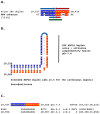
Nonanucleotide enhancer in transmissible gastroenteritis virus (TGEV, Alphacoronavirus, subgenus Tegacovirus, GenBank Accession: NC_038861). The TGEV enhancer upregulates the expression of the subgenomic RNA encoding the nucleocapsid (N) protein, possibly by approximating the TRS-L and TRS-B of N via duplex formation between the distal (close to the middle of the membrane [M] gene) and proximal (7 nucleotides upstream of the N TRS-B) enhancer elements. Despite its sequence similarity to the distal element, the intermediate element shown does not contribute to enhancer activity consistently with its high minimum free energy.
Figure 2
Figure 2
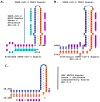
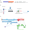
(A). TGEV enhancer-based model for mechanism underlying MHV (Betacoronavirus, subgenus Embecovirus, GenBank Accession: NC_048217.1 GenBank Accession: NC_048217.1) enhancer function. Located immediately after the leader sequence, the MHV enhancer (green) could pair with a sequence towards the end of the genomic region encoding the ORF1b polyprotein. Because ORF1ab covers approximately two-thirds of the genome, the pairing would bring the TRS-L (red) closer to the first genomic TRS-B upstream of ORF2, potentially enhancing the transcription of its subgenomic RNA in a sequence-specific manner. (B). Bovine CoV (Betacoronavirus, subgenus Embecovirus) has an MHV-like enhancer also immediately after the 5′-UTR leader sequence and similar sequences at three distal positions in the genome, which could pair with the 5′-UTR sequence. The minimum free energies (kcal/mol) are shown relative to the 5′-UTR sequence and the one located at position 4434; the latter pairings are predicted to be more stable. Similarities with the leader, TRS-L, enhancer, and beyond are highlighted in purple, red, green, and blue, respectively (C). The enhancer sequence shared between MHV and bovine CoV includes an octanucleotide reading the same in the sense and antisense strands (green arrows) and with complementary halves (blue and red arrows).
Figure 3
Figure 3
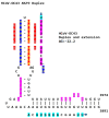
(A). Similarity between the NSP16 duplex in avian infectious bronchitis virus (IBV; Gammacoronavirus, subgenus Igacovirus) and the MHV enhancer (green). Red and blue arrows indicate complementary halves that can form a duplex (B). NSP16 duplex (with same sense and antisense sequences) and extended duplex. ΔG is the minimum free energy in kcal/mol. (C). Similar distal sequences potentially pairing with the NSP16 duplex. One sequence is proximal (region encoding the first protein in ORF1a) and the other distal (region encoding spike [S]) to the duplex.
Figure 4
Figure 4
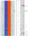
Mutations (highlighted in light blue) in avian IBV NSP16 extended duplex (complementary halves in blue and red), duplex minimum free energy (ΔG), number of GenBank strains with each mutation combination (mutations in light blue), translated amino sequence (conservative substitutions in green, nonconservative ones in magenta), and avian IBV origin other than chicken.
Figure 5
Figure 5
Examples of changes in the NSP16 duplex, the distal binding sequence, or both, which may underlie published cases of viral attenuation (Massachusetts and China strains) or its reversal (Taiwan strains). The NSP16 duplex is shown vertically in an open configuration, with complementary halves in blue and red. Mutations are highlighted in light blue.
Figure 6
Figure 6
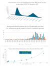
Analyses of possible association between extended NSP16 duplex variation and viral attenuation or its reversal.
Figure 7
Figure 7
Relationship between extended NSP16 duplex minimum free energy and frequency of viral attenuated/vaccine-derived/vaccine revertant strains.
Figure 8
Figure 8
Similarities of the IBV NSP16 extended duplex with Rousettus bat betacoronaviruses (Nobecoviruses), closely related to SARS-CoV-1 and -2, which can utilize the human ACE2 receptor in vitro. 1. Extended duplex in IBV (NC_001451). 2. OQ175246.1 (Bat CoV RlYN17 [Rousettus leschenaultia], China/Yunnan, 2016, isolate BtR1-BetaCoV/YN2016-Q319, toward end of ORF1ab); OQ175248.1 (Bat CoV RlYN17 [Rousettus leschenaultia], China/Yunnan, 2016, isolate BtR1-BetaCoV/YN2016-Q320, toward end of ORF1ab); OQ175341.1 (Bat CoV RlYN17 [Rousettus leschenaultia], China/Yunnan, 2017, isolate BtR1-BetaCoV/YN2017-Q321, toward end of ORF1ab starting at position 20,136). 3. MK492263.1 (Rousettus Bat CoV strain BtCoV92, Cynopterus brachyotis, Singapore, 2015). 4. OM219649.1 (Bat CoV GCCDC1, Eonycteris spelaea, Cambodia, 12/18,19/2010, isolate RK091); KU762332.1 (Rousettus leschneaulti Bat CoV isolate GCCDC1 356, China, 05/28/2014); NC_030886.1 (Rousettus leschneaulti Bat CoV isolate GCCDC1 356, China, 05/28/2014); KU762337.1 (Rousettus leschneaulti Bat CoV isolate GCCDC1 346, China, 05/28/2014); MT350598.1 (Rousettus bat CoV GCCDC1, Eonycteris spelaea, Singapore, 10/2016, betaCoV, Nobecovirus); OQ175331.1 (Bat CoV EsYN16, Eonycteris spelaea, China/Yunnan, 2016, BtEs-13BetaCoV/YN2016-Q311); OQ175332.1 (Bat CoV EsYN17, Eonycteris spelaea, China/Yunnan, 2017, BtEs-13BetaCoV/YN2017-Q312); OQ175333.1 (Bat CoV EsYN17, Eonycteris spelaea, China/Yunnan, 2017, BtEs-13BetaCoV/YN2017-Q313); OQ175242.1 (Bat CoV EsYN17, Eonycteris spelaea, China/Yunnan, 2017, BtEs-13BetaCoV/YN2017-Q309). Duplex complementary halves are highlighted in blue and red. Differences in bat sequences relative to IBV extended duplex are highlighted in light blue. Nucleotides shared among sequences are highlighted in gray. Minimum free energy (ΔG) is shown for each duplex, as is degree of similarity of duplexes relative to IBV reference duplex expressed as expect (e).
Figure 9
Figure 9
Duplex-forming sequences reading the same in the sense and antisense directions in the NSP3 gene of SARS-CoV-2 (A) and SARS-CoV-1 (B) and in the NSP16 of IBV (C). The complementary halves are highlighted in blue and red. Extended duplex regions are also shown. Regions of similarity within the SARS-CoV-2 extended duplex are highlighted in light blue, with arrows indicating that they are in inverted orientations. Similar sequences within and among SARS-CoV-2 and SARS-CoV-1 and in IBV are highlighted in pink. Minimum free energy is shown for all duplexes.
Figure 10
Figure 10
(A). Region in the SARS-CoV-2 5′-UTR with an MHV-like enhancer and similarity to the NSP3 duplex, with which it can pair, leaving the duplex free to pair with three other distal genomic regions. The pairings involving the S gene (the first gene after ORF1ab; e = 2.4 for sequence similarity to the NSP3 duplex) would approximate the TRS-L to the TRS-B of the gene encoding the viroporin ORF3a. The pairings involving NSP4 and NSP10 (e = 0.6 for both sequence comparisons to the NSP3 duplex) would also decrease the distance between the TRS sequences. (B). Sequence of the SARS-CoV-2 leader (blue box) that precedes the MHV-like enhancer (yellow letters with a dark green background) and that is added via discontinuous transcription to all accessory and structural genes. (C). Similarities among SARS-CoV-2 5′-UTR sequence after leader, NSP3 duplex, and TGEV proximal enhancer element.
Figure 11
Figure 11
(A). NSP3 duplex and complementary sequence in the SARS-CoV-2 genome. The pair can form a duplex with a minimum free energy (ΔG) similar to the NSP3 duplex. (B). Switch model for the opening of the NSP3 duplex and interaction with a 17-nucleotide complementary sequence in ORF3a. Initially, the duplex could be stabilized by RNA-RNA, RNA–protein, and protein–protein interactions involving undetermined factors, here termed X. Upon removal of said factors by epigenetic modification or interaction with other proteins or regulatory RNAs, the duplex could open and interact with other genomic sequences. In the illustration, the duplex and complementary sequences are 20,252 nucleotides apart, and their interaction is more stable in terms of ΔG and the length of the sequences involved than those between TRS-L (6 nucleotides long) and TRS-Bs during the discontinuous transcription of accessory and structural genes. A protein (named here Y or a combination of factors) could stabilize the new duplex between distant complementary sequences. (C). Positions (depicted with stars) in the SARS-CoV-2 genome of the NSP3 duplex (purple) and complementary sequences in Panel A and Figure 10. Numbers next to positions correspond from lowest to highest ΔG; e ranged from 0.6 to 2.4.
Figure 12
Figure 12
(A). The SARS-CoV-2 NSP3 duplex-forming 36-nucleotide sequence is present only in closely related bat Sarbecoviruses and SARS-CoV-1 among all Viridae. Nucleotide changes among all isolates are highlighted in light blue and those unique to SARS-CoV-1 are in light green. Sequences in SARS-CoV-2-related bat coronaviruses could be divided into four groups: With an identical 36–nucleotide segment (ΔG = −22.5): BetaCoV_Yunnan_Rp_JCC9_2020 (OK287355.1); BANAL-20-236/Laos/2020 (MZ937003.2); BANAL-20-247/Laos/2020 (MZ937004.1); BANAL-20-116/Laos/2020 (MZ937002.1); BANAL-20-103/Laos/2020 (MZ937001.1); BANAL-20-52/Laos/2020 (MZ937000.1); RpYN06 strain bat/Yunnan/RpYN06/2020 (MZ081381.1); isolate PrC31 (MW703458.1). With identical nucleotides 1–33 except nucleotide 3 (ΔG = −17.3): isolates RsHB20 BtRs-BetaCoV/HB2020-Q329 (OQ175349.1), Jingmen Rhinolophus sinicus betacoronavirus 1 (MZ328294.1), SC2018B (OK017846.1), and BM48-31/BGR/2008 (NC_014470.1). With identical nucleotides 4–33 (ΔG = −17.1): Horseshoe bat Sarbecovirus isolates Rt22QT53 (OR233321.1), Rt22QT48 (OR233320.1), Rt22QT46 (OR233319.1), Rt22QT36 (OR233318.1), Rt22QT178 (OR233317.1), Rt22QT161 (OR233316.1), Rt22QT124 (OR233300.1), Rt22QB8 (OR233299.1), and Rt22QB78 (OR233298.1); and isolates BtSY1 (OP963575.1), HN2021F (OK017835.1), and HN2021E (OK017834.1). With identical nucleotides 1–36 except nucleotides 3 and 7 (ΔG = −15.1): isolates GD2019E (OK017828.1), GD2019D (OK017827.1), GD2019B (OK017826.1), GD2019A (OK017825.1), GD2017W (OK017824.1), GD2017P (OK017822.1). SARS-CoV-1 sequence in Tor2 (NC_004718) and Urbani (MT308984) strains. (B). A complementary segment (11 nucleotides, e = 0.3) to the SARS-CoV-1 Tor2 (NC_004718.3) and Urbani (MT308984) strains NSP3 duplex is similar to that in SARS-CoV-2. However, the minimum free energy is higher for the pairing between the switch duplex and the complementary sequence, rendering the pairing less stable. (C). SARS-CoV-1 ORF3a duplex and complementary sequence in SARS-CoV-1 genome with similar minimum free energy. In this case, the duplex switch structure is distal to the complementary sequence with which it can pair with a similar ΔG. However, the effect of reducing the distance between TRS-L and the TRS-B of the gene distal to the viroporin ORF3a, namely the viroporin E, is achieved. (D). Comparison between the SARS-CoV-2 NSP3 duplex (highlighted in green) and the equivalent region in SARS-CoV-1, and between the SARS-CoV-1 ORF3a duplex (highlighted in blue) and the equivalent region in SARS-CoV-2. The SARS-CoV-2 NSP3 duplex is relatively well conserved in SARS-CoV-1, while the SARS-CoV-1 ORF3a duplex is not conserved in SARS-CoV-2 (nucleotides differing between SARS-CoV-2 and -1 are shown in red). However, the encoded amino acid sequences (highlighted in yellow) are relatively well conserved..
Figure 13
Figure 13
Similarity between the enhancer elements of the TGEV and MERS-CoV sequences. The intermediate sequence is the one with the highest similarity, yet in TGEV, it does not contribute to the enhancer activity. The TGEV enhancer distal and proximal sequences are partly present. The minimum free energy of the distal–proximal element pairing is higher than that in TGEV, rendering this potential enhancer unlikely to be active in MERS-CoV.
Figure 14
Figure 14
Comparison between the NSP3 duplexes and extended duplexes of the Betacoronaviruses (subgenus Sarbecovirus) SARS-CoV-2 (A) and SARS-CoV-1 (B) with the NSP16 duplex and extended duplex in the Betacorinavirus (subgenus Merbecovirus) MERS-CoV (C). Regions of similarity are highlighted in light blue and pink, and arms of the duplex reading similarly in the sense and antisense directions are highlighted in dark blue and red.
Figure 15
Figure 15
NSP3 extended duplex in hCoV-OC43 (Betacoronavirus, subgenus Embecovirus). Regions of similarity with the NSP3 duplexes in SARS-CoV-2 and -1 and MERS-CoV are highlighted in light blue and pink, and arms of the duplex reading the same in the sense and antisense directions are highlighted in dark blue and red. Sequences in green letters highlighted in red correspond to repeated sequences in the duplexes described in this paper and shown in Figure 16.
Figure 16
Figure 16
Extended duplexes of the Betacoronaviruses infecting humans. (A). Multiple alignments of the duplexes with similarity percentages. The highest duplex similarity was between SARS-CoV-2 and -1, while the MERS-CoV extended duplex sequence was closest to that of IBV (61% similarity), both in NSP16 but also to the NSP3 duplex of SARS-CoV-2 and -1 (67% and 58%). Asterisks denote nucleotide positions conserved among all duplexes, highlighted in either blue or green. (B). Repeated sequences within duplexes and similarity among them. Repeated similar sequences within and among duplexes are highlighted in red, fuchsia (reverse of those in red), light blue (shared between SARS-CoV-2 and MERS-CoV), and green (shared between MERS-CoV and IBV).
Figure 17
Figure 17
Annotated coronaviral duplex-forming sequences that read similarly in the sense and antisense directions, with complementary halves. Minimum free energies (ΔG) are shown next to each hairpin. GenBank accession numbers are shown in parentheses. Positions that do not read the same in the sense and antisense directions are underlined. The MERS-CoV NSP16 duplex is present in bat Merbecoviruses and pangolin CoV HKU4 (GenBank OM009282.1). Most of the MHV M duplex is in the bovine coronavirus M gene (OP866729.1), and the one in the bovine CoV NSP13 helicase is in the canine respiratory coronavirus (ON133844.1), also an Embecovirus. The sequences annotated here do not comprise an exhaustive list, and other coronaviruses that share some of these duplexes are mentioned in the text.

Similar articles

References

    1. Gorbalenya A.E., Enjuanes L., Ziebuhr J., Snijder E.J. Nidovirales: Evolving the largest RNA virus genome. Virus Res. 2006;117:17–37. doi: 10.1016/j.virusres.2006.01.017. - DOI - PMC - PubMed
    1. Lauber C., Goeman J.J., Parquet Mdel C., Nga P.T., Snijder E.J., Morita K., Gorbalenya A.E. The footprint of genome architecture in the largest genome expansion in RNA viruses. PLoS Pathog. 2013;9:e1003500. doi: 10.1371/journal.ppat.1003500. - DOI - PMC - PubMed
    1. Masters P.S. The molecular biology of coronaviruses. Adv. Virus Res. 2006;66:193–292. doi: 10.1016/S0065-3527(06)66005-3. - DOI - PMC - PubMed
    1. Forni D., Cagliani R., Clerici M., Sironi M. Molecular Evolution of Human Coronavirus Genomes. Trends Microbiol. 2017;25:35–48. doi: 10.1016/j.tim.2016.09.001. - DOI - PMC - PubMed
    1. Ziv O., Gabryelska M.M., Lun A.T.L., Gebert L.F.R., Sheu-Gruttadauria J., Meredith L.W., Liu Z.Y., Kwok C.K., Qin C.F., MacRae I.J., et al. COMRADES determines in vivo RNA structures and interactions. Nat. Methods. 2018;15:785–788. doi: 10.1038/s41592-018-0121-0. - DOI - PMC - PubMed

Grants and funding

This research was funded by ACCESS Health International, a nonprofit organization with which both authors are affiliated.

LinkOut - more resources