. 2024 Aug 9;14(1):19.
doi: 10.1186/s13395-024-00350-6.

Pilot investigations into the mechanistic basis for adverse effects of glucocorticoids in dysferlinopathy

Affiliations

Pilot investigations into the mechanistic basis for adverse effects of glucocorticoids in dysferlinopathy

Erin M Lloyd et al. Skelet Muscle. .

Abstract

Background: Dysferlinopathies are a clinically heterogeneous group of muscular dystrophies caused by gene mutations resulting in deficiency of the membrane-associated protein dysferlin. They manifest post-growth and are characterised by muscle wasting (primarily in the limb and limb-gridle muscles), inflammation, and replacement of myofibres with adipose tissue. The precise pathomechanism for dysferlinopathy is currently unclear; as such there are no treatments currently available. Glucocorticoids (GCs) are widely used to reduce inflammation and treat muscular dystrophies, but when administered to patients with dysferlinopathy, they have unexpected adverse effects, with accelerated loss of muscle strength.

Methods: To investigate the mechanistic basis for the adverse effects of GCs in dysferlinopathy, the potent GC dexamethasone (Dex) was administered for 4-5 weeks (0.5-0.75 µg/mL in drinking water) to dysferlin-deficient BLA/J and normal wild-type (WT) male mice, sampled at 5 (Study 1) or 10 months (Study 2) of age. A wide range of analyses were conducted. Metabolism- and immune-related gene expression was assessed in psoas muscles at both ages and in quadriceps at 10 months of age. For the 10-month-old mice, quadriceps and psoas muscle histology was assessed. Additionally, we investigated the impact of Dex on the predominantly slow and fast-twitch soleus and extensor digitorum longus (EDL) muscles (respectively) in terms of contractile function, myofibre-type composition, and levels of proteins related to contractile function and metabolism, plus glycogen.

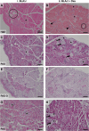
Results: At both ages, many complement-related genes were highly expressed in BLA/J muscles, and WT mice were generally more responsive to Dex than BLA/J. The effects of Dex on BLA/J mice included (i) increased expression of inflammasome-related genes in muscles (at 5 months) and (ii) exacerbated histopathology of quadriceps and psoas muscles at 10 months. A novel observation was pronounced staining for glycogen in many myofibres of the damaged quadriceps muscles, with large pale vacuolated myofibres, suggesting possible myofibre death by oncosis.

Conclusion: These pilot studies provide a new focus for further investigation into the adverse effects of GCs on dysferlinopathic muscles.

Keywords: Adipocytes; Complement; Dexamethasone; Dysferlin; Dysferlinopathy; Glucocorticoids; Glycogen; Inflammasome; Limb-girdle muscular dystrophy; Skeletal muscle.


Conflict of interest statement

The authors declare no competing interests.

Figures

Fig. 1
Fig. 1
Whole body mass of wild-type (WT) and dysferlin-deficient BLA/J mice aged 5 months, without and with dexamethasone (Dex) treatment. Data were analysed by two-way ANOVA: * BLA/J (± Dex) vs. WT (± Dex) (p < 0.05, strain effect). Data are presented as mean ± SD (n = 8)
Fig. 2
Fig. 2
Body and muscle mass of WT and dysferlin-deficient BLA/J mice aged 10 months, without and with dexamethasone (Dex) treatment. A Body mass measured at sampling, and B quadriceps (quad), C soleus, and D extensor digitorum longus (EDL) muscle mass normalised to body mass (mg/gBM). Data were analysed by two-way ANOVA: **** BLA/J (± Dex) vs WT (± Dex) (p < 0.0001, strain effect); # Dex-treated vs. untreated (p < 0.05, treatment effect); ^^ BLA/J + Dex vs WT + Dex (p < 0.01, strain/treatment interaction effect). Data are presented as mean ± SD (n = 8–9)
Fig. 3
Fig. 3
Relative metabolism-associated gene expression in psoas muscle of wild-type (WT) C57Bl/6J and dysferlin-deficient BLA/J mice aged 5 months, without and with dexamethasone (Dex) treatment. A Glucose transporter type 4 (Glut4); B Lipoprotein lipase (Lpl); C Sterol regulatory element-binding transcription factor 1 (Srebf1); D Peroxisome proliferator-activated receptor gamma (Pparγ); E CCAAT/enhancer-binding protein delta (Cepbδ); F Fatty Acid Synthase (Fasn); G ATP citrate lyase (Acly); H Acetyl-CoA carboxylase 1 (Acaca). Skeletal actin (Acta1) was used as a reference gene to normalise gene expression values in the psoas muscle. Relative gene expression was calculated using the 2−ΔΔCT Method, and values were normalised to the untreated WT for each gene. Data were analysed by two-way ANOVA: **, ***, **** BLA/J (± Dex) vs WT (± Dex) (p < 0.01, 0.001, 0.0001, respectively, strain effect). Data are log10 transformed and presented on a log10 y-axis scale as mean ± SD (n = 5–8)
Fig. 4
Fig. 4
Relative metabolism-associated gene expression in quadriceps and psoas muscles of wild-type (WT) C57Bl/6J and dysferlin-deficient BLA/J mice aged 10 months, without and with dexamethasone (Dex) treatment. A, K Glucose transporter type 4 (Glut4); B, L Lipoprotein lipase (Lpl); C, M Sterol regulatory element-binding transcription factor 1 (Srebf1); D Peroxisome proliferator-activated receptor gamma (Pparγ); E, O Carbohydrate-responsive element-binding protein (Chrebp); F, P CCAAT enhancer binding protein alpha (Cebpα); G, Q CCAAT/enhancer-binding protein delta (Cepbδ); H, R Fatty Acid Synthase (Fasn); I, S ATP citrate lyase (Acly); J, T Acetyl-CoA carboxylase 1 (Acaca). All mRNA expression values were standardised against the reference genes peptidylprolyl isomerase A (Ppia) and TATA-box binding protein (Tbp1) using the GeNorm algorithm [64]. Data were analysed by two-way ANOVA: **, ***, **** BLA/J (± Dex) vs WT (± Dex) (p < 0.01, 0.001, 0.0001, respectively, strain effect); #, ## Dex-treated vs untreated (p < 0.05, 0.01, respectively, treatment effect); ^, ^^, ^^^, ^^^^ significant difference between groups (p < 0.05, 0.01, 0.001, 0.0001, respectively, strain/treatment interaction effect). Data are log10 transformed and presented on a log10 y-axis scale as mean ± SD (n = 7)
Fig. 5
Fig. 5
Relative immune-associated gene expression in psoas muscle of wild-type (WT) C57Bl/6J and dysferlin-deficient BLA/J mice aged 5 months, without and with dexamethasone (Dex) treatment. A Complement C1q B Chain (C1qb); B Complement component 3 (C3); C Complement C3a Receptor 1 (C3ar1); D Complement component 4 (C4); E Complement component 5 (C5); F Complement C5a Receptor 2 (C5ar2); G Decay-accelerating factor 1 for complement (Daf1 or CD55); H Decay-accelerating factor 2 for complement B (Daf2 or CD55b); I Caspase 1 (Casp1); J NLR family pyrin domain containing 3 (Nlrp3). Skeletal actin (Acta1) was used as a reference gene to normalise gene expression values in the psoas muscle. Relative gene expression was calculated using the 2−ΔΔCT Method, and values were normalised to the untreated WT for each gene. Data were analysed by two-way ANOVA: **** BLA/J (± Dex) vs WT (± Dex) (p < 0.0001, strain effect); ## Dex-treated vs untreated (p < 0.01, treatment effect); ^, ^^, ^^^, ^^^^ significant difference between groups (p < 0.05, 0.01, 0.001, 0.0001, respectively, strain/treatment interaction effect). Data are log10 transformed and presented on a log10 y-axis scale as mean ± SD (n = 5–8)
Fig. 6
Fig. 6
Relative immune-related gene expression in quadriceps and psoas muscles of wild-type (WT) C57Bl/6J and dysferlin-deficient BLA/J mice aged 10 months, without and with dexamethasone (Dex) treatment. A, H Tumour necrosis factor (Tnf); B, I Complement C1q B Chain (C1qb); C, J Complement component 3 (C3); D, K Complement component 4 (C4); E, L Complement C5a Receptor 1 (C5ar1); F, M Caspase 1 (Casp1); G, N NLR family pyrin domain containing 3 (Nlrp3). All mRNA expression values were standardised against the reference genes peptidylprolyl isomerase A (Ppia) and TATA-box binding protein (Tbp1) using the GeNorm algorithm [64]. Data were analysed by two-way ANOVA: *, **, **** BLA/J (± Dex) vs WT (± Dex) (p < 0.05, 0.01, 0.0001, respectively, strain effect); # Dex-treated vs untreated (p < 0.05, treatment effect); ^, ^^^, ^^^^ significant difference between groups (p < 0.05, 0.001, 0.0001, respectively, strain/treatment interaction effect). Data are log10 transformed and presented on a log10 y-axis scale as mean ± SD (n = 4–10)
Fig. 7
Fig. 7
Effect of dexamethasone (Dex) treatment on histopathology of quadriceps muscles of BLA/J mice (aged 10 months). Representative images of consecutive paraffin sections showing highly variable histopathology of (i) BLA/J and (ii) BLA/J + Dex quadriceps stained by (A, B) haematoxylin and eosin (H&E), indicating the location of some adipocytes (circled), foci of inflammatory cells (arrowheads), and large pale myofibres (asterisks, *). C, D The presence of glycogen is shown by periodic acid-Schiff (PAS) staining, with (E, F) the absence of glycogen following diastase-induced breakdown (as a control to verify glycogen PAS staining; PAS-D). More (G, H) PAS-stained sections are shown from additional BLA/J mice. Dex treatment (in B, D, and H) appeared to increase the number of large pale myofibres (some marked by *), and some with conspicuous vacuoles/fragmentation (arrows) evident with PAS staining. In contrast, such pale myofibres did not appear as large (nor vacuolated) in untreated BLA/J muscle. Shown for four 10-month-old male mice: (i) BLA/J (ID for panels A, C, E. 16/21, and G. 16/23) and (ii) BLA/J + Dex (ID for panels B, D, F. 16/32, and H. 16/31). Scale bar = 100 μm
Fig. 8
Fig. 8
Glycogen content of (A) soleus and (B) EDL muscles of WT and dysferlin-deficient BLA/J mice aged 10 months, without and with dexamethasone (Dex) treatment. Whole muscle homogenates analysed for glycogen content. Data were analysed by two-way ANOVA: #### Dex-treated vs untreated (p < 0.0001, treatment effect). ^^ significant difference between groups (p < 0.01, strain/treatment interaction effect). Data are presented as mean ± SD (n = 6)

Similar articles

References

    1. Cárdenas AM, González-Jamett AM, Cea LA, Bevilacqua JA, Caviedes P. Dysferlin function in skeletal muscle: possible pathological mechanisms and therapeutical targets in dysferlinopathies. Exp Neurol. 2016;283(Pt A):246–54. 10.1016/j.expneurol.2016.06.026 - DOI - PubMed
    1. Anwar S, Yokota T. The dysferlinopathies conundrum: clinical spectra, disease mechanism and genetic approaches for treatments. Biomolecules. 2024;14(3):256. 10.3390/biom14030256 - DOI - PMC - PubMed
    1. Lek A, Evesson FJ, Sutton RB, North KN, Cooper ST. Ferlins: regulators of vesicle fusion for auditory neurotransmission, receptor trafficking and membrane repair. Traffic. 2012;13(2):185–94. 10.1111/j.1600-0854.2011.01267.x - DOI - PubMed
    1. Bulankina AV, Thoms S. Functions of vertebrate ferlins. Cells. 2020;9(3):534. 10.3390/cells9030534 - DOI - PMC - PubMed
    1. Klinge L, Harris J, Sewry C, Charlton R, Anderson L, Laval S, Chiu YH, Hornsey M, Straub V, Barresi R, Lochmüller H, Bushby K. Dysferlin associates with the developing T-tubule system in rodent and human skeletal muscle. Muscle Nerve. 2010;41(2):166–73. 10.1002/mus.21166 - DOI - PubMed

MeSH terms

Supplementary concepts

LinkOut - more resources