[Preprint]. 2024 Jul 30:2024.07.30.605768.
doi: 10.1101/2024.07.30.605768.

Passive infusion of an S2-Stem broadly neutralizing antibody protects against SARS-CoV-2 infection and lower airway inflammation in rhesus macaques

Affiliations

Passive infusion of an S2-Stem broadly neutralizing antibody protects against SARS-CoV-2 infection and lower airway inflammation in rhesus macaques

Christopher T Edwards et al. bioRxiv. .

Abstract

The continued evolution of SARS-CoV-2 variants capable of subverting vaccine and infection-induced immunity suggests the advantage of a broadly protective vaccine against betacoronaviruses (β-CoVs). Recent studies have isolated monoclonal antibodies (mAbs) from SARS-CoV-2 recovered-vaccinated donors capable of neutralizing many variants of SARS-CoV-2 and other β-CoVs. Many of these mAbs target the conserved S2 stem region of the SARS-CoV-2 spike protein, rather the receptor binding domain contained within S1 primarily targeted by current SARS-CoV-2 vaccines. One of these S2-directed mAbs, CC40.8, has demonstrated protective efficacy in small animal models against SARS-CoV-2 challenge. As the next step in the pre-clinical testing of S2-directed antibodies as a strategy to protect from SARS-CoV-2 infection, we evaluated the in vivo efficacy of CC40.8 in a clinically relevant non-human primate model by conducting passive antibody transfer to rhesus macaques (RM) followed by SARS-CoV-2 challenge. CC40.8 mAb was intravenously infused at 10mg/kg, 1mg/kg, or 0.1 mg/kg into groups (n=6) of RM, alongside one group that received a control antibody (PGT121). Viral loads in the lower airway were significantly reduced in animals receiving higher doses of CC40.8. We observed a significant reduction in inflammatory cytokines and macrophages within the lower airway of animals infused with 10mg/kg and 1mg/kg doses of CC40.8. Viral genome sequencing demonstrated a lack of escape mutations in the CC40.8 epitope. Collectively, these data demonstrate the protective efficiency of broadly neutralizing S2-targeting antibodies against SARS-CoV-2 infection within the lower airway while providing critical preclinical work necessary for the development of pan-β-CoV vaccines.


Conflict of interest statement

Conflicting Interests: RA, TFR, and DRB are listed as inventors on pending patent applications describing the SARS-CoV-2 and HCoV-HKU1 S cross-reactive antibodies. DRB and RA are listed as inventors on a pending patent application describing the S2 stem epitope immunogens identified in this study. DRB is a consultant for IAVI. All other authors declare that they have no competing interests.

Figures
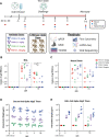
Fig. 1.
Fig. 1.. Preinfusion of S2-targeting bnAb CC40.8 reduced viral loads in RMs on SARS CoV-2 challenge
(A) 24 RMs (6 females and 18 males; mean age of 5 years and 11 months old; range 5–6 years old) were infused intravenously 5 days pre infection with either a 0.1 mg/kg, 1 mg/kg, or 10mg/kg concentration of SARS-CoV-2 bnAb CC40.8 or with control mAb PGT121, with each experimental group consisting of 6 RMs (1 female and 5 males). RMs were screened for preexisting, SARS-CoV-2 spike specific antibodies prior to CC40.8 administration RMs were euthanized at 7 dpi (n = 3 RMs per treatment arm) or 8 dpi (n = 3 RMs per treatment arm). Levels of SARS-CoV-2 sgRNA N in BAL (B) and nasopharyngeal swabs (C). (D) Anti-Spike hIgG titers in the serum (D) and BAL (E) measured via ELISA. Control PGT121-treated RMs are depicted with red squares, CC40.8 0.1mg/kg-treated RMs depicted with green upward pointing triangles, CC40.8 1mg/kg-treated RMs depicted with purple downward pointing triangles, CC40.8 10mg/kg-treated RMs depicted with blue circles. Black lines represent the median viral loads for each treatment group at each time point. Statistical analyses were performed using nonparametric Mann-Whitney tests. *P < 0.05.
Fig. 2.
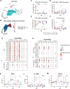
Fig. 2.. CC40.8-treated RMs had lower frequencies of inflammatory CD163+ MRC1- macrophages compared with PGT121-treated RMs.
(A) Representative staining of macrophages for CD163 and MRC1 in the BAL at −4, 2, and 7/8 dpi with frequency as a percentage of total CD163+ cells. Macrophages were gated on singlets, CD45+, FSC and SSC characteristic of granulocytes and alveolar macrophages, live cells, and CD14+ populations. (B to D) Frequency as a percentage of total CD163+ cells for (B) MRC1-, (C) MRC1 intermediate and (D) MRC1++ cells. (E to F) Fold change from −4 dpi baseline as a percentage of total CD163+ cells for (E) MRC1-, (F) MRC1 intermediate, and (G) MRC1++ cells. Control PGT121-treated RMs are depicted with red squares, CC40.8 0.1mg/kg-treated RMs depicted with green upward pointing triangles, CC40.8 1mg/kg-treated RMs depicted with purple downward pointing triangles, CC40.8 10mg/kg-treated RMs depicted with blue circles. Black lines represent the median frequency or fold change in RMs from each respective treatment group. Statistical analyses were performed using nonparametric Mann-Whitney tests. *P < 0.05, **P < 0.01
Fig. 3.
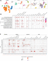
Fig. 3.. Effect of CC40.8 treatment on gene expression of BAL single cells during SARS-CoV-2 infection using 10X.
A) Uniform Manifold Approximation and Projection (UMAP) of BAL samples (107830 cells) integrated using reciprocal principal components analysis (PCA) showing cell type annotations. Captures were performed on BAL cells from all RMs at 2 and 7/8 dpi. (B) Mapping of macrophage/monocyte cells in the BAL of SARS-CoV-2-infected PGT121- and CC40.8-treated RMs to different lung macrophage/monocyte subsets from healthy RMs (67). (C to F) Percentage of different macrophage/monocyte subsets of all the macrophage/monocytes in BAL at 2 and 7/8 dpi from PGT121- and CC40.8-treated RMs. Frequency as a percentage of total CD163+ cells for (C) MRC1-, (D) MRC1+ TREM2+, (E) MRC1++, and (F) CD16+ monocytes. (G to J) Dot plots showing the expression of selected (G) ISGs, (I) inflammatory genes, (H) chemokines, and (J) inflammasome genes in CD163+ MRC1- macrophages. The size of the dot indicates the percentage of cells that express a given gene, and the color indicates the level of expression. (K to M) Fold change of cytokines and chemokines in BAL fluid relative to −4 dpi measured by MSD immunoassay. Control PGT121-treated RMs are depicted with red squares, CC40.8 0.1mg/kg-treated RMs depicted with green upward pointing triangles, CC40.8 1mg/kg-treated RMs depicted with purple downward pointing triangles, CC40.8 10mg/kg-treated RMs depicted with blue circles. Black lines represent the median frequency or fold change in RMs from each respective treatment group. Statistical analyses were performed using two-sided nonparametric Mann-Whitney tests. *P < 0.05.
Fig. 4.
Fig. 4.. Effect of CC40.8 treatment on lung cells during SARS-CoV-2 infection.
(A) UMAP based on reciprocal PCA of lung single cells (101,766 cells) collected at 7/8 dpi (n = 3 PGT121, 3 CC40.8 0.1mg/kg, and 3 CC40.8 10mg/kg). The cells were classified into four broad categories—epithelial, lymphoid, myeloid, and other, followed by subsetting and separate clustering within each category. UMAPs for each category with cell type annotations are also shown. (B) Selected gene sets that were found to be enriched (P-adjusted value < 0.05) in lung cells from PGT121-treated RMs compared to CC40.8 10mg/kg-treated RMs at 7/8 dpi based on overrepresentation analysis using Hallmark, Reactome, Kyoto Encyclopedia of Genes and Genomes, and BioCarta gene sets from MSigDB. The size of the dots represents the number of genes that were enriched in the gene set, and the color indicates the P-adjusted value. The gene set IDs in order are M983, M15913, M27255, M27253, M5902, M5890, M5921, M27250, M41804, M5897, M5932, M27698, M27251, M29666, M27436, M27895, M27897, and M1014. (C) Dot plots showing gene expression in lung cells present at higher frequencies from PGT121- and CC40.8-treated macaques at 7/8 dpi. Plot is organized by epithelial, myeloid, lymphoid and other subsets. The size of the dot represents the percentage of cells expressing a given gene, and the color indicates the average expression.
Fig. 5.
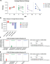
Fig. 5.. Effect of CC40.8 treatment on SARS-CoV-2 mutant frequency
(A) The average Shannon entropy calculated from intra-sample single nucleotide variant (iSNV) frequency in replicate SARS-CoV-2 ARTIC libraries generated from BAL supernatant at 2 dpi and NX (7/8 dpi). Black lines connect libraries from same animal at different timepoints (B) Mean change in Shannon entropy for each treatment group from 2 dpi to 7/8 dpi. (C to D) Frequency of intra-sample, single nucleotide variations at the CC40.8 S2 stem helix epitope at (C) 2 dpi and (D) 7/8 dpi.

Similar articles

References

    1. Tan W, Zhao X, Ma X, Wang W, Niu P, Xu W, et al. A Novel Coronavirus Genome Identified in a Cluster of Pneumonia Cases - Wuhan, China 2019–2020. China CDC Wkly. 2020;2(4):61–2. - PMC - PubMed
    1. Cui J, Li F, Shi Z-L. Origin and evolution of pathogenic coronaviruses. Nat Rev Microbiol. 2019;17(3):181–92. - PMC - PubMed
    1. Su S, Wong G, Shi W, Liu J, Lai ACK, Zhou J, et al. Epidemiology, Genetic Recombination, and Pathogenesis of Coronaviruses. Trends Microbiol. 2016;24(6):490–502. - PMC - PubMed
    1. Forni D, Cagliani R, Clerici M, Sironi M. Molecular Evolution of Human Coronavirus Genomes. Trends Microbiol. 2017;25(1):35–48. - PMC - PubMed
    1. Mackay IM, Arden KE. MERS coronavirus: diagnostics, epidemiology and transmission. Virol J. 2015;12(1):222. - PMC - PubMed

Publication types