. 2024 Jul 22:15:1397761.
doi: 10.3389/fphar.2024.1397761. eCollection 2024.

The mechanism of low molecular weight fucoidan-incorporated nanofiber scaffolds inhibiting oral leukoplakia via SR-A/Wnt signal axis

Affiliations

The mechanism of low molecular weight fucoidan-incorporated nanofiber scaffolds inhibiting oral leukoplakia via SR-A/Wnt signal axis

Ming Xu et al. Front Pharmacol. .

Abstract

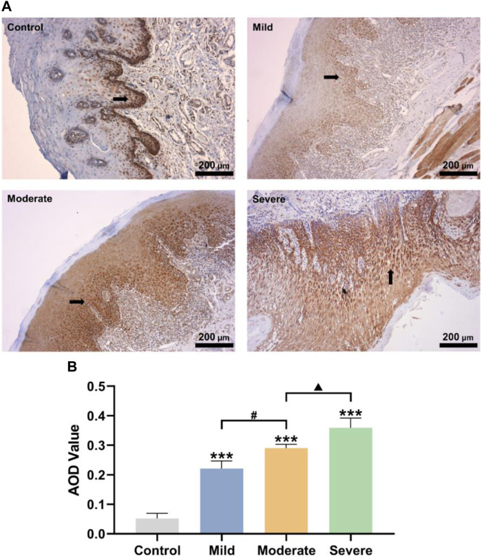
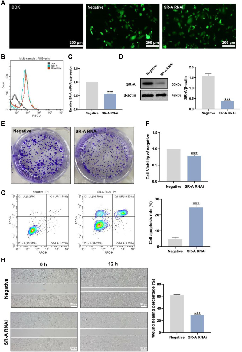
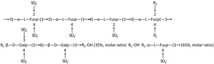
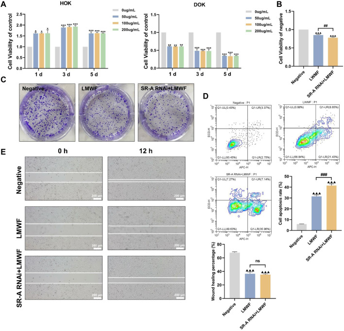
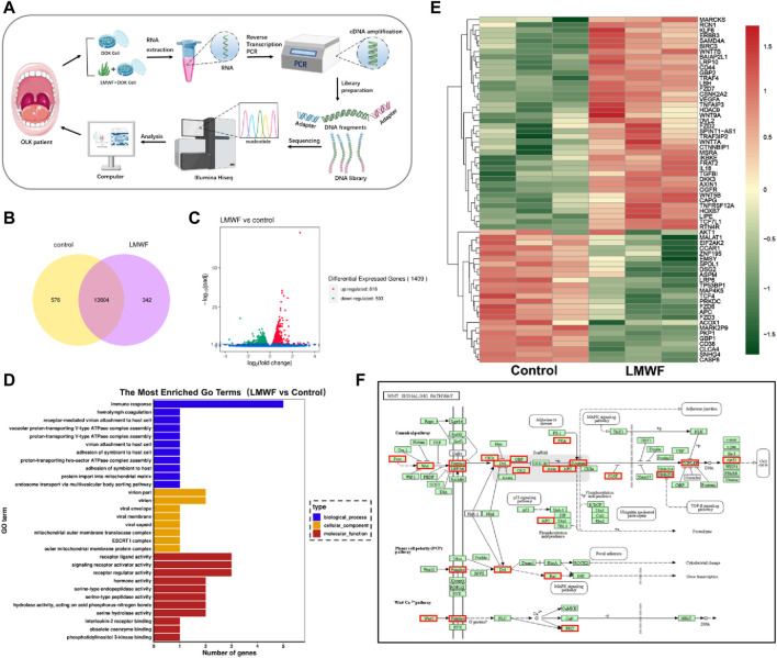
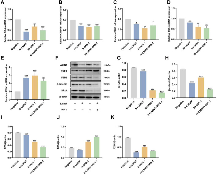
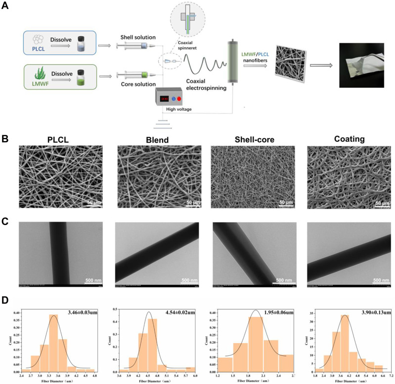
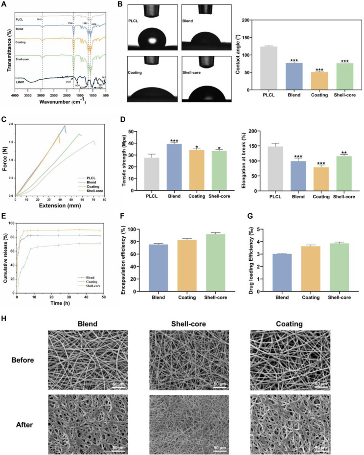
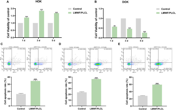
Oral leukoplakia (OLK) is the most common oral precancerous lesion, and 3%-17% of OLK patients progress to oral squamous cell carcinoma. OLK is susceptible to recurrence and has no effective treatment. However, conventional drugs have significant side effects and limitations. Therefore, it is important to identify drugs that target OLK. In this study, scavenger receptor A (SR-A) was found to be abnormally highly expressed in the oral mucosal epithelial cells of OLK patients, whereas molecular biology studies revealed that low molecular weight fucoidan (LMWF) promoted apoptosis of dysplastic oral keratinocytes (DOK) and inhibited the growth and migration of DOK, and the inhibitory effect of LMWF on OLK was achieved by regulating the SR-A/Wnt signaling axis and related genes. Based on the above results and the special situation of the oral environment, we constructed LMWF/poly(caprolactone-co-lactide) nanofiber membranes with different structures for the in-situ treatment of OLK using electrospinning technology. The results showed that the nanofiber membranes with a shell-core structure had the best physicochemical properties, biocompatibility, and therapeutic effect, which optimized the LMWF drug delivery and ensured the effective concentration of the drug at the target point, thus achieving precise treatment of local lesions in the oral cavity. This has potential application value in inhibiting the development of OLK.

Keywords: Wnt/β-catenin; electrospun nanofiber; fucoidan; oral leukoplakia; scavenger receptor A.


Conflict of interest statement

The authors declare that the research was conducted in the absence of any commercial or financial relationships that could be construed as a potential conflict of interest.

Figures

FIGURE 1
FIGURE 1
Impact of SR-A in the development of OLK occurrence. (A) Control: Representative pictures of oral epithelial immunohistochemistry in healthy people; mild, moderate, and severe: representative images of the oral epithelium of OLK patients with different degrees of abnormal hyperplasia. Brownish-yellow particles represented SR-A positive expressions. Scale bar = 200 μm. (B) AOD values of SR-A positive substances in control, mild, moderate, and severe groups. Data were expressed as the means ± SD. ***p < 0.001 vs. Control group; p < 0.05 vs. Moderate group; # p < 0.05 vs. Mild group.
FIGURE 2
FIGURE 2
Establishment of the SR-A RNAi model and the effect of SR-A on DOK cells. (A) Representative images of lentiviral transfection efficiency. Green fluorescence indicates lentiviral vector particles. Scale bar = 200 μm. (B) Lentiviral transfection efficiency by flow cytometry assay. (C) and (D) The mRNA and protein expression levels of SR-A. (E) Colony-forming ability of DOK negative cells and SR-A RNAi groups (F) Cell viability in DOK negative cells and SR-A RNAi groups (G) Apoptosis rates in DOK negative cells and SR-A RNAi groups (H) Migration ability in DOK negative cells and SR-A RNAi groups. Scale bar = 200 μm. Data were expressed as the means ± SD. ***p < 0.001 vs. DOK negative group.
FIGURE 3
FIGURE 3
Speculation on the structure of fucoidan sulfate in Laminaria japonica (Wang et al., 2010).
FIGURE 4
FIGURE 4
Effect of LMWF on DOK cells. (A) Effect of different concentrations of LMWF on the viability of DOK and HOK cells (B) Proliferative capacity of cells (C) Colony-forming ability of different groups (D) Apoptosis rates of cells (E) Migration ability of cells. Scale bar = 200 μm. Data were expressed as the means ± SD. ***p < 0.001, **p < 0.01 and *p < 0.05 vs. 0 μg/mL group; ▲▲▲ p < 0.001 vs. DOK negative group; ### p < 0.001 and ## p < 0.01 vs. LMWF group.
FIGURE 5
FIGURE 5
Effect of LMWF on related signaling pathways and genes. (A) Schematic diagram of the high-throughput sequencing process. (B) Venn diagram of differentially expressed genes (FPKM > 1). (C) Gene differential volcano plot. Red dots (upregulated) and green dots (downregulated) indicated genes with significant differential expression. The horizontal coordinates represented gene expression fold changes in different samples, and the vertical coordinates represented the statistical significance of the differences in gene expression changes. Blue dots denoted genes with negligible differential expression. (D) The enriched GO term was the vertical coordinate, and the number of differentially expressed genes in that word was the horizontal coordinate. Different colors denote various cellular components, molecular activities, and biological processes. (E) Map of the differential gene clustering. Each row represented one gene, and each column represented one sample. The hue changes from red to green, indicating that lg (FPKM+1) went from large to tiny. (F) Diagram of the metabolic pathway considerably enriched in the KEGG database; red boxes indicated differential genes.
FIGURE 6
FIGURE 6
Effect of LMWF on the SR-A/Wnt signaling axis and related gene expression. (A–E) After different treatments, the expression of mRNA for SR-A, CTNNB1, FZD6, TCF4, and AXIN1. (F) Representative protein expression bands for SR-A, CTNNB1, FZD6, TCF4, AXIN1, and β-actin. (G–K) Relative expression levels of SR-A, CTNNB1, FZD6, TCF4, and AXIN1 proteins compared to β-actin. Data were expressed as the means ± SD. ***p < 0.001, **p < 0.01 and *p < 0.05 vs. DOK negative group.
FIGURE 7
FIGURE 7
Construction and surface morphology of LMWF/PLCL nanofiber membranes. (A) Schematic diagram of nanofiber membrane spinning process. (B) SEM images of different nanofiber membranes. Scale bar = 50 μm. (C) TEM images of different nanofiber membranes. Scale bar = 500 nm. (D) Diameter distribution of different nanofibers.
FIGURE 8
FIGURE 8
Physicochemical characterization of LMWF/PLCL nanofiber membranes. (A) FTIR spectra of LMWF and nanofiber membranes. (B) WCA images and analytical plots of different nanofiber membranes at 10 s. (C) Schematic diagram of the mechanical properties of nanofiber membranes. (D) Tensile strength and elongation at break of nanofiber membranes. (E) Cumulative drug release curves of nanofiber membranes. (F) Encapsulation rate of nanofiber membranes. (G) The drug loading rate of nanofiber membranes. (H) The change of fiber morphology after LMWF/PLCL nanofiber membranes were soaked in artificial saliva. Scale bar = 50 μm *** p < 0.001, **p < 0.01 and *p < 0.05 vs. PLCL group.
FIGURE 9
FIGURE 9
Evaluation of biocompatibility and treatment efficacy of LMWF/PLCL nanofiber membranes. (A) Effect of nanofiber membranes on HOK cell viability. (B) Effect of nanofiber membranes on DOK cell viability. (C–E) The apoptosis rate of DOK cells co-cultured with nanofibrous membrane for 1, 3, and 5 days. ***p < 0.001, **p < 0.01 vs. Control group.

Similar articles

References

    1. Aguirre-Urizar J. M., Lafuente-Ibáñez de Mendoza I., Warnakulasuriya S. (2021). Malignant transformation of oral leukoplakia: systematic review and meta-analysis of the last 5 years. Oral Dis. 27 (8), 1881–1895. 10.1111/odi.13810 - DOI - PubMed
    1. Al-Dhahebi A. M., Gopinath S. C. B., Saheed M. S. M. (2020). Graphene impregnated electrospun nanofiber sensing materials: a comprehensive overview on bridging laboratory set-up to industry. Nano Converg. 7 (1), 27. 10.1186/s40580-020-00237-4 - DOI - PMC - PubMed
    1. Amin M. L., Mawad D., Dokos S., Koshy P., Martens P. J., Sorrell C. C. (2021). Fucoidan- and carrageenan-based biosynthetic poly(vinyl alcohol) hydrogels for controlled permeation. Mater Sci. Eng. C Mater Biol. Appl. 121, 111821. 10.1016/j.msec.2020.111821 - DOI - PubMed
    1. Bhattacharyya S., Ray S., Saha D., Mustafi S. M., Alam N., Sarkar A., et al. (2021). Chewing tobacco may act as a risk factor for dysplastic transformation of squamous cells in Oral leukoplakia- A cytochemistry based approach. Pathology, Res. Pract. 218, 153287. 10.1016/j.prp.2020.153287 - DOI - PubMed
    1. Bi D., Yu B., Han Q., Lu J., White W. L., Lai Q., et al. (2018). Immune activation of RAW264.7 macrophages by low molecular weight fucoidan extracted from New Zealand Undaria pinnatifida. J. Agric. food Chem. 66 (41), 10721–10728. 10.1021/acs.jafc.8b03698 - DOI - PubMed

Grants and funding

The author(s) declare that financial support was received for the research, authorship, and/or publication of this article. The authors declare no competing financial interest. This work was supported by the National Natural Science Foundation of China (82301083), Natural Science Foundation of Shandong Province (ZR2021MH388), Qingdao Traditional Chinese Medicine Science and Technology Project of 2021 (2021-zyyq15), Qingdao Medical Research Guidance Project of 2020 (2020-WJZD142), National Natural Science Foundation of China (32201049), Qingdao Postdoctoral Funding Program (QDBSH20220202192), Qingdao Key Health Discipline Development Fund (2022-2024), Qingdao Clinical Research Center for Oral Diseases (22-3-7-lczx-7-nsh) and Shandong Provincial Key Medical and Health Discipline of Oral Medicine (2024-2026).

LinkOut - more resources