. 2024 Aug 6;14(1):18160.
doi: 10.1038/s41598-024-68268-z.

Antox targeting AGE/RAGE cascades to restore submandibular gland viability in rat model of type 1 diabetes

Affiliations

Antox targeting AGE/RAGE cascades to restore submandibular gland viability in rat model of type 1 diabetes

Marwa M Ahmad et al. Sci Rep. .

Abstract

Diabetes mellitus (DM) is a chronic disorder of glucose metabolism that threatens several organs, including the submandibular (SMG) salivary glands. Antox (ANX) is a strong multivitamin with significant antioxidant benefits. The goal of this study was to demonstrate the beneficial roles of ANX supplementation in combination with insulin in alleviating diabetic SMG changes. For four weeks, 30 rats were divided into equal five groups (n = 6): (1) control group; (2) diabetic group (DM), with DM induced by streptozotocin (STZ) injection (50 mg/kg i.p.); (3) DM + ANX group: ANX was administrated (10 mg/kg/day/once daily/orally); (4) DM + insulin group: insulin was administrated 1U once/day/s.c.; and (5) DM + insulin + ANX group: co-administrated insulin. The addition of ANX to insulin in diabetic rats alleviated hyposalivation and histopathological alterations associated with diabetic rats. Remarkably, combined ANX and insulin exerted significant antioxidant effects, suppressing inflammatory and apoptotic pathways associated with increased salivary advanced glycation end-product (AGE) production and receptor for advanced glycation end-product expression (RAGE) activation in diabetic SMG tissues. Combined ANX and insulin administration in diabetic rats was more effective in alleviating SMG changes (functions and structures) than administration of insulin alone, exerting suppressive effects on AGE production and frustrating RAGE downstream pathways.


Conflict of interest statement

The authors declare no competing interests.

Figures
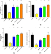
Figure 1
Figure 1
Effect of ANX plus insulin on SMG weights (A), total saliva amount (B), saliva flow rate (C) and salivary alpha-amylase enzyme (B) in STZ-induced diabetic rats.Data were presented as the mean ± SE (n = 6). a p < 0.05 versus control group; b p < 0.05 versus DM group; c p < 0.05 versus DM + ANX group and d p < 0.05 versus DM + insulin group (STZ, streptozotocin; ANX, antox; DM, diabetes mellitus and SMG, submandibular gland).
Figure 2
Figure 2
Effect of ANX plus insulin on fasting blood glucose concentration (A) and glycated hemoglobin (B), in STZ-induced diabetic rats. Data were analyzed and presented as the mean ± SE (n = 6). a p < 0.05 versus control group; b p < 0.05 versus DM group; c p < 0.05 versus DM + ANX group and d p < 0.05 versus DM + insulin group. (STZ, streptozotocin; ANX, antox; DM, diabetes mellitus and SMG, submandibular gland).
Figure 3
Figure 3
Effect of ANX and insulin on the activities of SOD (A), GPX (B) and MDA concentration (C) IL-1β concentration (D) and AGEs concentration (E) in SMG tissues of STZ-induced diabetic rats. Data were presented as the mean ± SE (n = 6). a p < 0.05 versus control group; b p < 0.05 versus DM group; c p < 0.05 versus DM + ANX group and d p < 0.05 versus DM + insulin group. (STZ, streptozotocin; ANX, antox; SOD, superoxide dismutase; MDA, malondiahyde; GPX, IL-1β, interleukine-1 beta: AGEs, advanced glycation end products; DM, diabetes mellitus and SMG, submandibular gland).
Figure 4
Figure 4
Photomicrographs of sections of the SMG from the different studied groups; (A): The control group shows well-defined closely packed serous acini (S), granular convoluted tubules (G), and striated ducts (SD). The acinar pyramidal cells have darkly stained cytoplasm and spherical basophilic nuclei (bifid arrow). The striated duct is lined by columnar cells having characteristic basal striations (arrowhead) and oval vesicular nuclei (zigzag arrow). The granular convoluted tubules are lined by columnar cells having abundant apical dark-stained eosinophilic granules (arrow) and basal rounded nuclei (curved arrow). (B-D): The DM group shows parenchymal destruction with degenerative areas (*), apparent shrunken acini (angled arrow), and the acini losing their uniform typical architecture (rectangle). The granular convoluted tubules appear atrophied leaving remnants (circle) and their cells display darkly stained nuclei (curved arrow) and cytoplasmic vacuoles (arrow). The acinar cells appear with intracytoplasmic vacuoles of different sizes (tailed arrow). Some ducts show marked dilatation with stagnant secretion (SS), flattening of its epithelial lining cells (zigzag arrow) cytoplasmic vacuoles (arrowhead), and surrounded by extensive connective tissue stroma ( +). (E, F): The DM + ANX/The DM + insulin groups respectively show cytoplasmic vacuoles (V, V1 &V2) in cells of serous acini(S), granular convoluted tubules (G), and striated ducts (SD) respectively. Some acini still show disturbed architecture (oval red shape). Some cells of granular convoluted tubules exhibit diminished acidophilic content (arrow). (G): The DM + insulin + ANX group shows more or less normal histological appearance; closely packed acini (S) having pyramidal cells with rounded nuclei (bifid arrow). The granular convoluted tubules (G) appear to have abundant eosinophilic granules (arrow) and basal nuclei (curved arrow). The striated ducts (SD) exhibit basal striation (arrowhead) with round nuclei (zigzag arrow) (H & E, X 400).
Figure 5
Figure 5
Photomicrographs of submandibular gland sections from different studied groups; (A): control group shows scanty collagen fibers surrounding the acini (arrow), blood vessels (arrowhead), and striated ducts (curved arrow). (B): The DM group showing abundant deposition of collagen fibers around acini (arrow), ducts (curved arrow) and blood vessels (arrowhead). (C, D): The DM + ANX/The DM + insulin groups, respectively show mild deposition of collagen fibers around striated ducts (bifid arrow) and blood vessels (arrowhead). Notice some acini are still surrounded by a considerable amount of collagen fibers (arrow) (E): The DM + insulin + ANX group shows very little amount of collagen fiber surrounding the acini (arrow), blood vessels (arrowhead), and striated ducts (curved arrow). (Masson trichrome, X 400) (F): Effect of ANX plus insulin on area % of collage fibers in SMG streptozotocin-induced diabetic rats. a p < 0.05 versus control group; b p < 0.05 versus DM group; c p < 0.05 versus DM + ANX group; and d p < 0.05 versus insulin-treated group (ANX, antox; DM, diabetes mellitus).
Figure 6
Figure 6
photomicrographs of submandibular gland sections from different studied groups: (A): control group shows Negative cytoplasmic immunoreaction for 8-OHdG in the acini (arrow), granular convoluted tubules (curved arrow), and striated ducts (arrowhead). (B): The DM group showing intense positive immunoreaction for 8-OHdG in cytoplasm acini (arrow), granular convoluted tubules (curved arrow), and striated ducts (arrowhead). (C, D): The DM + ANX/The DM + insulin groups respectively show some acini with mild reaction (arrow) and some still have an intense reaction (tailed arrow). Also, some granular convoluted tubules appear with mild reaction (curved arrow) while others with intense reaction (angled arrow). The ducts show mild reaction (arrowhead). (E): The DM + insulin + ANX group shows faint or nearly negative immunoreaction to 8-OHdG in the acini (arrow), granular convoluted tubules (curved arrow), and striated ducts (arrowhead) (8-OHdG immunostaining, X 400). (F): Effect of ANX plus insulin on OD of 8 OHdG in SMG streptozotocin-induced diabetic rats a p < 0.05 versus control group; b p < 0.05 versus DM group; c p < 0.05 versus DM + ANX group; and d p < 0.05 versus insulin-treated group (ANX, antox; DM, diabetes mellitus).
Figure 7
Figure 7
photomicrographs of submandibular gland sections from different studied groups; (A): control group showing faint immunoreaction for α-SMA in the myoepithelial cells surrounding the acini (arrow) and negative reaction around granular tubules (curved arrow). (B): The DM group showing strong positive immunoreaction at the myoepithelial cells surrounding the acini (arrow) and positive reaction around granular tubules (curved arrow) notice negative reaction around the duct (bifid arrow). (C, D): The DM + ANX/The DM + insulin groups respectively show some acini with a faint immune reaction to α-SMA at the myoepithelial cells surrounding the acini (arrow), around granular tubules (curved arrow) and around the duct (bifid arrow) while some acini with a strong positive reaction to α-SMA (arrowhead). (E): The DM + insulin + ANX group shows a faint immune reaction to α-SMA surrounding the acini (arrow) with a negative reaction at the periphery of the granular tubules (curved arrow). (Immunostaining for α-SMA X 400) (F) Effect of ANX and insulin on area % of α SMA in SMG streptozotocin-induced diabetic rats. a p < 0.05 versus control group; b p < 0.05 versus DM group; c p < 0.05 versus DM + ANX group; and d p < 0.05 versus insulin-treated group (ANX, antox; DM, diabetes mellitus).
Figure 8
Figure 8
Photomicrographs of submandibular gland sections from different studied groups: (A): The control group shows negative cytoplasmic immunoreactivity for BAX in the acini (arrow), granular convoluted tubules (curved arrow), and striated ducts (arrowhead). (B): The DM group showing strong positive immunoreaction for BAX in cytoplasm acini (arrow), granular convoluted tubules (curved arrow), and striated ducts (arrowhead). (C, D): The DM + insulin/ANX groups showing mild reaction in the acini (arrow) and granular convoluted tubules (curved arrow) while the striated ducts show intense reaction (arrowhead). (E): The DM + insulin + ANX group shows faint immunoreactivity to BAX in the acini (arrow), granular convoluted tubules (arrowhead), and striated ducts (curved arrow). (BAX immunostaining, X 400) (F): Effect of ANX plus insulin on OD of BAX streptozotocin-induced diabetic rats. a p < 0.05 versus control group; b p < 0.05 versus DM group; c p < 0.05 versus DM + ANX group; and d p < 0.05 versus insulin-treated group (ANX, antox; DM, diabetes mellitus).

Similar articles

References

    1. Mata, A. D. et al. Effects of diabetes mellitus on salivary secretion and its composition in the human. Mol. Cell. Biochem.261(1–2), 137–142. 10.1023/b:mcbi.0000028748.40917.6f (2004). 10.1023/b:mcbi.0000028748.40917.6f - DOI - PubMed
    1. Fouani, M. et al. Salivary gland proteins alterations in the diabetic milieu. J. Mol. Histol.52(5), 893–904. 10.1007/s10735-021-09999-5 (2021). 10.1007/s10735-021-09999-5 - DOI - PMC - PubMed
    1. Asseri, S. M. et al. Glycyrrhizic acid ameliorates submandibular gland oxidative stress, autophagy and vascular dysfunction in rat model of type 1 diabetes. Sci. Rep.12(1), 725 (2022). 10.1038/s41598-021-04594-w - DOI - PMC - PubMed
    1. de Morais, H. et al. Increased oxidative stress in prefrontal cortex and hippocampus is related to depressive-like behavior in streptozotocin-diabetic rats. Behav. Brain Res.258, 52–64. 10.1016/j.bbr.2013.10.011 (2014). 10.1016/j.bbr.2013.10.011 - DOI - PubMed
    1. Nowotny, K., Jung, T., Höhn, A., Weber, D. & Grune, T. Advanced glycation end products and oxidative stress in type 2 diabetes mellitus. Biomolecules5(1), 194–222 (2015). 10.3390/biom5010194 - DOI - PMC - PubMed

MeSH terms

LinkOut - more resources