. 2024 Aug 1;65(10):8.
doi: 10.1167/iovs.65.10.8.

IL-23 Priming Enhances the Neuroprotective Effects of MSC-Derived Exosomes in Treating Retinal Degeneration

Affiliations

IL-23 Priming Enhances the Neuroprotective Effects of MSC-Derived Exosomes in Treating Retinal Degeneration

Hong Zhou et al. Invest Ophthalmol Vis Sci. .

Abstract

Purpose: Neuroinflammation is a characteristic feature of neurodegenerative diseases. Mesenchymal stem cell-derived exosomes (MSC-exo) have shown neuroprotective effects through immunoregulation, but the therapeutic efficacy remains unsatisfactory. This study aims to enhance the neuroprotective capacity of MSC-exo through IL-23 priming for treating retinal degeneration in mice.

Methods: MSC were primed with IL-23 stimulation in vitro, and subsequently, exosomes (MSC-exo and IL-23-MSC-exo) were isolated and characterized. Two retinal degenerative disease models (NaIO3-induced mice and rd10 mice) received intravitreal injections of these exosomes. The efficacy of exosomes was assessed by examining retinal structural and functional recovery. Furthermore, exosomal microRNA (miRNA) sequencing was conducted, and the effects of exosomes on the M1 and M2 microglial phenotype shift were evaluated.

Results: IL-23-primed MSC-derived exosomes (IL-23-MSC-exo) exhibited enhanced capability in protecting photoreceptor cells and retinal pigment epithelium (RPE) cells against degenerative damage and fostering the restoration of retinal neural function in both NaIO3-induced retinal degeneration mice and rd10 mice when compared with MSC-exo. The exosomal miRNA suppression via Drosha knockdown in IL-23-primed MSC would abolish the neuroprotective role of IL-23-MSC-exo, highlighting the miRNA-dependent mechanism. Bioinformatic analysis, along with further in vivo biological studies, revealed that IL-23 priming induced a set of anti-inflammatory miRNAs in MSC-exo, prompting the transition of M1 to M2 microglial polarization.

Conclusions: IL-23 priming presents as a potential avenue for amplifying the immunomodulatory and neuroprotective effects of MSC-exo in treating retinal degeneration.


Conflict of interest statement

Disclosure: H. Zhou, None; Y. Liu, None; T. Zhou, None; Z. Yang, None; B. Ni, None; Y. Zhou, None; H. Xu, None; X. Lin, None; S. Lin, None; C. He, None; X. Liu, None

Figures

Figure 1.
Figure 1.
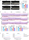
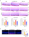
IL-23 priming promoted MSC-exo to preserve the neuroretinal structure and function in NaIO3-induced retinal degenerative mice. (A) Representative images of OCT scanning. Compared with C57 normal control (NC), the PBS-treated NaIO3 model displayed obvious RPE layer discontinuity with high-reflective deposits (red arrows) and decreased ONL thickness (as indicated by the distance between the yellow lines). Upon the administration of exosomes, the RPE layer became continuous and flat, and the thickness of ONL was preserved, particularly evident in the IL-23-MSC-exo group (Scale bar = 200 µm, n = 6 mice, 1-way ANOVA and Tukey’s post hoc test). (B) H&E staining images revealed a decrease in the thickness of ONL and the accumulation of melanin in the RPE layer (yellow arrows) in PBS-treated NaIO3-retina. Exosomes could significantly rescue the retina structure, especially the IL-23-MSC-exo. The ONL thickness of the central, mid-peripheral, and peripheral retinas was calculated and compared (Scale bar = 100 µm, n = 6 mice, 1-way ANOVA, and Tukey’s post hoc test). (C) ERG showed that intravitreal injection of IL-23-MSC-exo could significantly increase the amplitudes of a- and b- waves in comparison with the MSC-exo group (n = 6 mice, 1-way ANOVA, and Tukey’s post hoc test). GCL, ganglion cell layer; INL, inner nuclear layer; ONL, outer nuclear layer. All data are shown as mean ± SEM, *P < 0.05, **P < 0.01, ***P < 0.001, ns: no significance.
Figure 2.
Figure 2.
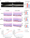
IL-23-MSC-exo exerted the neuroprotective effect in a miRNAs-dependent manner. (A) The OCT images of NaIO3 mice treated with IL-23-MSC-siDrosha-exo (Drosha siRNA group) showed more hyper-reflective foci (red arrows) along the RPE layer and thinner thickness of ONL (distance between the yellow lines) than those mice treated with IL-23-MSC-siNC-exo (negative control NC siRNA group) (Scale bar = 200 µm, n = 6 mice, unpaired Student's t-test). (B) H&E staining of retinal sections showed that Drosha siRNA group presented more melanin deposited (yellow arrows) along RPE layer and also less ONL thickness (Scale bar = 100 µm, n = 6 mice, unpaired Student's t-test). (C) ERG showed decreased amplitudes of a- and b- waves after IL-23-MSC-siDrosha-exo treatment compared with IL-23-MSC-siNC-exo treatment (n = 6 mice, unpaired Student's t-test). All data are shown as mean ± SEM, *P < 0.05, **P < 0.01, ***P < 0.001, ns: no significance.
Figure 3.
Figure 3.
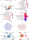
Exosomal miRNA profile and analysis. (A) miRNA sequencing was performed on IL-23-MSC-exo and MSC-exo (n = 3 for each group). Volcano plot [x-axis = log2 (fold change); y-axis = −log10 (P value)] showing 117 significantly differentially expressed miRNAs between IL-23-MSC-exo and MSC-exo. Red dots indicate the upregulated miRNAs in IL-23-MSC-exo; and the blue dots indicate the downregulated miRNAs in IL-23-MSC-exo with fold change >1. (B) Putative target genes of differentially expressed miRNAs were analyzed for GO biological process enrichment (P < 0.05). The enriched pathways included neuron death, regulation of immune system process and other biological processes. (C) Selected pathways from the GO biological process enrichment of the predicted target genes belong to immune process system (P < 0.05), which included macrophage/microglia cell activation. (D) The miRNA enrichment analysis was performed using the experimentally validated miRNA Enrichment Analysis and Annotation Tool (miEAA). The miRNAs enriched in IL-23-MSC-exo were related to microglia cell activation and inflammatory response. (E) Cytoscape and plug-in StringApp were used to construct the protein-protein interaction (PPI) network ground on the predicted genes of up-regulated miRNAs. The hub genes of this network included M1 markers CD86, IL-1β, and IFN-y. (F) The similar process was performed for the target genes of downregulated miRNAs to establish the other PPI network. The core genes of this network included M2 markers IL-4 and IL-10. (G) The molecular network was established to show the putative miRNAs targeting M1 microglial markers (IL-1β, TNF-α, CD86, and Nos2) among the upregulated miRNAs. (H) The molecular network showed the putative miRNAs targeting M2 microglial markers (Arg1, IL-4, and IL-10) among the downregulated miRNAs.
Figure 4.
Figure 4.
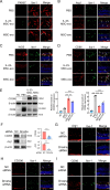
IL-23-MSC-exo promoted microglia polarization from M1 to M2 phenotype via miRNAs transport in the NaIO3-induced mice. (A) Representative confocal images showed that the PKH67-labeled IL-23-MSC-exo or MSC-exo (green) were co-located with Iba-1+ microglia (red), indicating the exosomes were internalized by microglia (Scale bar = 50 µm, n = 3 retinas). (BD) Intravitreal injection of exosomes increased the Arg1+Iba-1+ microglia and decreased the iNOS+/CD86+Iba-1+microglia, particularly evident in the IL-23-MSC-exo treatment (Scale bar = 50 µm, n = 3 eyeballs). (E) Application of exosome especially the IL-23-MSC-exo could significantly upregulate protein expression of CD206 and downregulate CD86 (n = 3 retinas, 1-way ANOVA and Tukey's post hoc test). (F) IL-23-MSC-siDrosha-exo suppressed the expressions of CD206 and Arg1 in the NaIO3-retinas (n = 3 retinas, unpaired Student's t test). All data are shown as mean ± SEM, **P < 0.01, ***P < 0.001. (GI) Intravitreal injection of IL-23-MSC-siDrosha-exo decreased Arg1+Iba-1+ as well as CD206+Iba-1+ microglia and increased CD86+ Iba-1+ microglia (n = 3 eyeballs, Scale bar = 50 µm).
Figure 5.
Figure 5.
IL-23-MSC-exo also protected rd10 mice against retinal degeneration. (A) The H&E staining images showed that intravitreal injection of IL-23-MSC-exo preserved the photoreceptors in the ONL layer and increased the thickness of ONL layer in the rd10 mice (Scale bar = 100 µm, n = 4 mice, 1-way ANOVA, and Tukey’s post hoc test). (B) The rhodopsin was highly preserved in the IL-23-MSC-exo-treated rd10 retinae, whereas the least Tunel+ cells in the ONL layer in comparison to PBS-treated or MSC-exo-treated rd10 mice (Scale bar = 50 µm, n = 6 eyeballs, 1-way ANOVA, and Tukey’s post hoc test). Data are shown as mean ± SEM, *P < 0.05, **P < 0.01, ns: no significance.
Figure 6.
Figure 6.
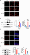
IL-23-MSC-exo induced microglia to adopt M2 phenotype via miRNAs transport in rd10 mice. (A) Representative confocal images depicted the co-localization of PKH67-labeled IL-23-MSC-exo or MSC-exo (green) with Iba-1+ microglia (red), suggesting the internalization of exosomes by microglia (n = 3 retinas, Scale bar = 50 µm). (B) Western blot shows that exosome treatment could significantly increase the protein levels of CD206 and Arg1 in rd10 mice, especially in the IL-23-MSC-exo group (n = 3 retinas, 1-way ANOVA and Tukey's post hoc test). (C) Intravitreal injection of exosomes promoted the CD206 expression on the Iba-1+ microglia, especially the IL-23-MSC-exo injection (n = 3 eyeballs, Scale bar = 50 µm). (D) Intravitreal injection of IL-23-MSC-siDrosha-exo decreased the protein levels of CD206 and Arg1 compared with IL-23-MSC-siNC-exo in rd10 mice (n = 3 retinas, unpaired Student's t test). Data are shown as mean ± SEM, *P < 0.05, **P < 0.01, ***P < 0.001.

Similar articles

References

    1. Jin ZB, Gao ML, Deng WL, et al. .. Stemming retinal regeneration with pluripotent stem cells. Prog Retin Eye Res. 2019; 69: 38–56. - PubMed
    1. Van Gelder RN, Chiang MF, Dyer MA, et al. .. Regenerative and restorative medicine for eye disease. Nat Med. 2022; 28: 1149–1156. - PMC - PubMed
    1. Dhodapkar RM, Martell D, Hafler BP.. Glial-mediated neuroinflammatory mechanisms in age-related macular degeneration. Semin Immunopathol. 2022; 44: 673–683. - PubMed
    1. Karlstetter M, Scholz R, Rutar M, Wong WT, Provis JM, Langmann T.. Retinal microglia: just bystander or target for therapy? Prog Retin Eye Res. 2015; 45: 30–57. - PubMed
    1. Arnhold S, Absenger Y, Klein H, Addicks K, Schraermeyer U.. Transplantation of bone marrow-derived mesenchymal stem cells rescue photoreceptor cells in the dystrophic retina of the rhodopsin knockout mouse. Graefes Arch Clin Exp Ophthalmol. 2007; 245: 414–422. - PubMed

MeSH terms

LinkOut - more resources