. 2024 Aug 1;15(8):552.
doi: 10.1038/s41419-024-06936-8.

Triggering of endoplasmic reticulum stress via ATF4-SPHK1 signaling promotes glioblastoma invasion and chemoresistance

Affiliations

Triggering of endoplasmic reticulum stress via ATF4-SPHK1 signaling promotes glioblastoma invasion and chemoresistance

Beiwu Lan et al. Cell Death Dis. .

Abstract

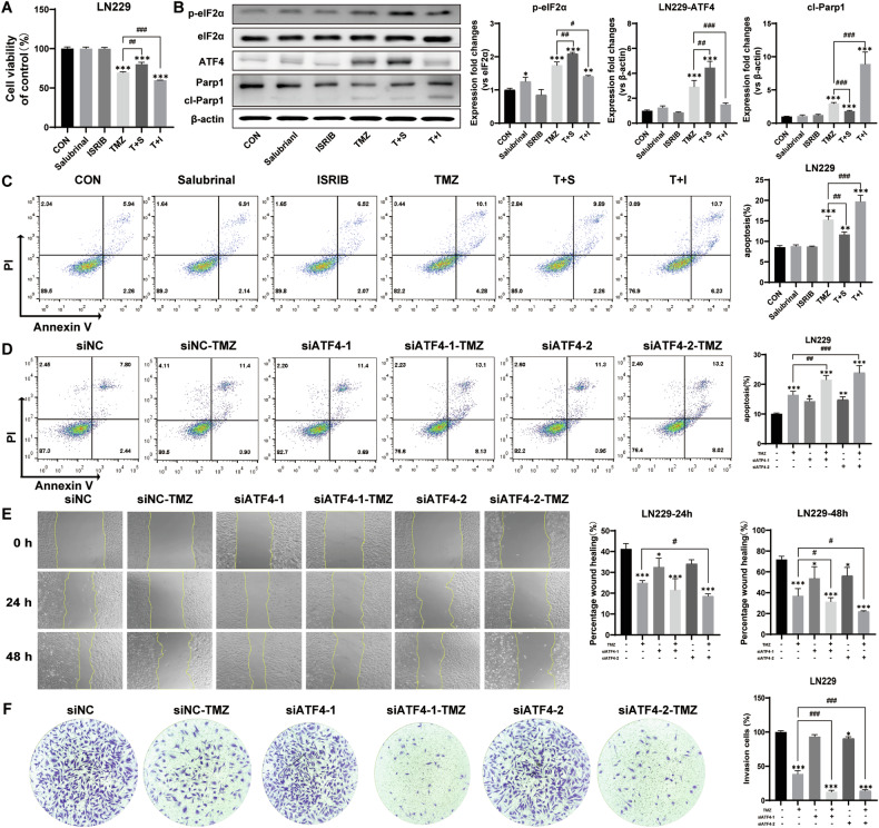
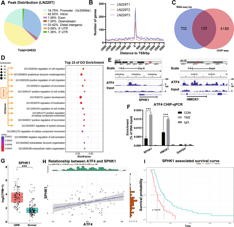
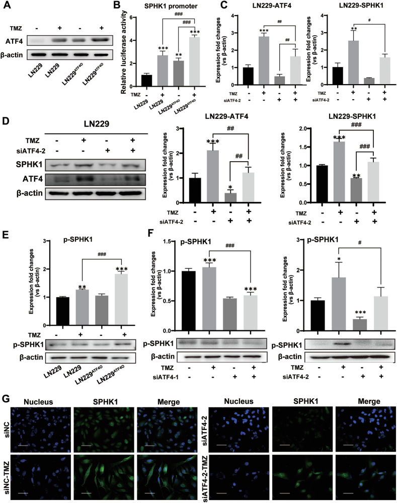
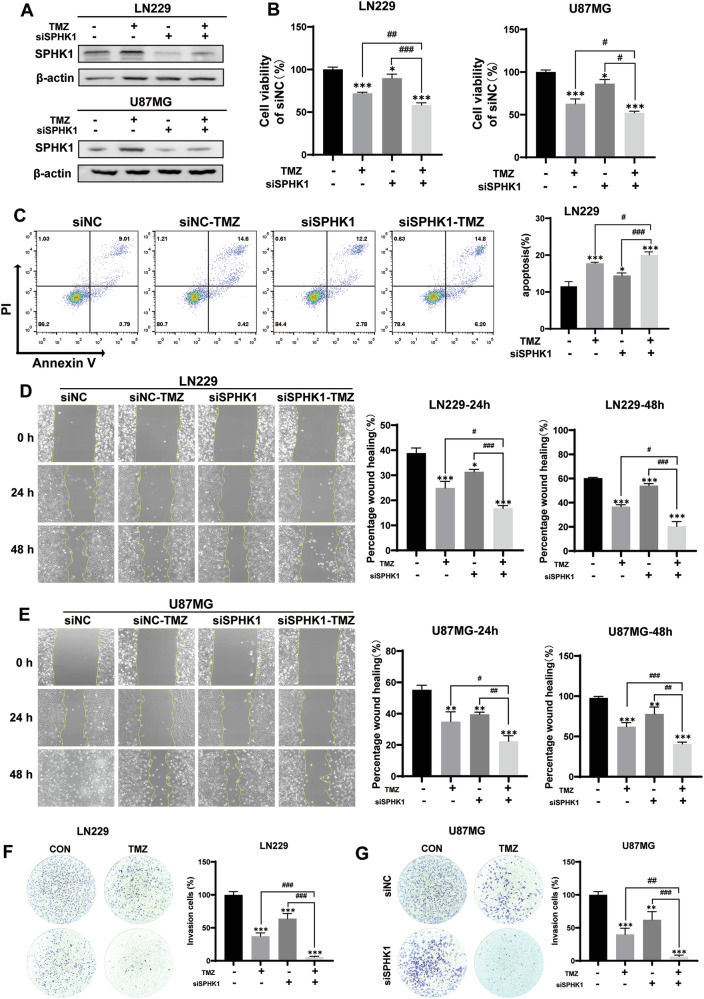
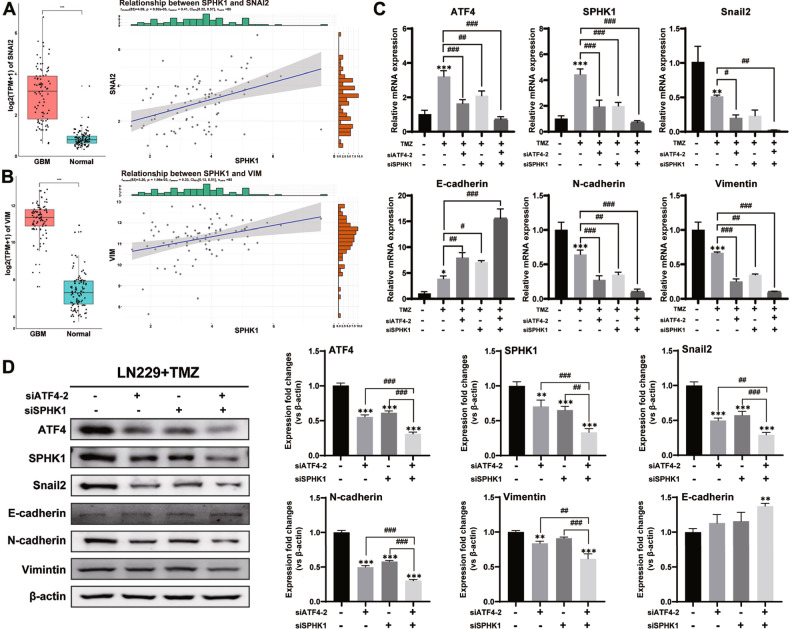
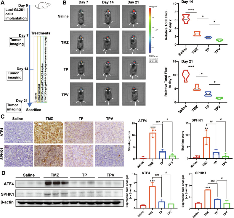
Despite advances in therapies, glioblastoma (GBM) recurrence is almost inevitable due to the aggressive growth behavior of GBM cells and drug resistance. Temozolomide (TMZ) is the preferred drug for GBM chemotherapy, however, development of TMZ resistance is over 50% cases in GBM patients. To investigate the mechanism of TMZ resistance and invasive characteristics of GBM, analysis of combined RNA-seq and ChIP-seq was performed in GBM cells in response to TMZ treatment. We found that the PERK/eIF2α/ATF4 signaling was significantly upregulated in the GBM cells with TMZ treatment, while blockage of ATF4 effectively inhibited cell migration and invasion. SPHK1 expression was transcriptionally upregulated by ATF4 in GBM cells in response to TMZ treatment. Blockage of ATF4-SPHK1 signaling attenuated the cellular and molecular events in terms of invasive characteristics and TMZ resistance. In conclusion, GBM cells acquired chemoresistance in response to TMZ treatment via constant ER stress. ATF4 transcriptionally upregulated SPHK1 expression to promote GBM cell aggression and TMZ resistance. The ATF4-SPHK1 signaling in the regulation of the transcription factors of EMT-related genes could be the underlying mechanism contributing to the invasion ability of GBM cells and TMZ resistance. ATF4-SPHK1-targeted therapy could be a potential strategy against TMZ resistance in GBM patients.


Conflict of interest statement

The authors declare no competing interests.

Figures

Fig. 1
Fig. 1. ER signaling pathways in GBM patients and GBM cells.
A Enriched genes for protein processing in the endoplasmic reticulum by GSEA. B HSPA5 mRNA expressions in GBM tissues and cerebral cortex tissues. C The survival probability analysis between the GBM patients with HSPA5-high expression and HSPA5-low expression based on the median number of GBM samples. D Western blotting analysis of the expression changes of ER stress-related proteins in LN229 cells treated with TMZ for 3 days. E Immunofluorescence detection of ATF4 expression and subcellular localization in LN229 cells treated with TMZ for 3 days (bar = 60 μm). F Cell viability by MTT in SHG44, U251, U87MG, T98G, LN229, and LN229ATF4O cells with TMZ treatment for 3 days. G qPCR to detected the mRNA levels of ATF4, SPHK1, and cleaved caspase-3 in LN229 and LN229ATF4O cells with TMZ treatment for 3 days. H Western blotting analysis of the expression changes of SPHK1, ATF4 and cleaved caspase-3 in LN229 and LN229ATF4O cells with TMZ treatment for 3 days. I Heatmap of the top 30 upregulated genes (based on Z-score) by RNA-seq analysis of LN229 cells treated with TMZ for 3 days. J GSEA analysis of protein processing in endoplasmic reticulum pathway. CON control, D day, cl-casp-3 cleaved caspase-3. (*p < 0.05, **p < 0.01, ***p < 0.001).
Fig. 2
Fig. 2. PERK/eIF2α/ATF4 signaling promotes GBM cell aggression and TMZ resistance.
A Cell viability by MTT assay in LN229 cells with treatments of TMZ as well as salubrinal and ISRIB. B Western blotting analysis of the protein expression of eIF2α/ATF4 signaling and apoptotic effector in LN229 cells with treatments of TMZ as well as salubrinal and ISRIB. C Flow cytometry detected cell apoptosis in LN229 cells with treatments of TMZ as well as salubrinal and ISRIB. D Flow cytometry detected cell apoptosis in LN229-siATF4-1 cells, LN229-siATF4-2 cells, and LN229 cells with TMZ treatment. E Wound healing assay detected the migration of LN229-siATF4-1 cells, LN229-siATF4-2 cells, and LN229 cells treated with TMZ for 24 h and 48 h under serum-free culture conditions. F Trans-well assay to detect the invasion ability of LN229-siATF4-1 cells, LN229-siATF4-2 cells, and LN229 cells treated with TMZ for 12 h. T TMZ, S salubrinal, I ISRIB. (*p < 0.05, **p < 0.01, ***p < 0.001; #p < 0.05, ##p < 0.01, ###p < 0.001).
Fig. 3
Fig. 3. SPHK1 expression is transcriptionally upregulated by ATF4 production in GBM cells.
A CHIP-seq analysis of ATF4 binding sites in the genome of LN229 cells after TMZ treatment for 3 days. B Distribution of ATF4-enriched DNA fragments around TSS ± 3000 bp. C Venn diagram showing the relationship between upregulated genes in RNA-seq and genes with ATF4 binding in the promoter region in CHIP-seq. D GO enrichment analysis of upregulated genes in the promoter region that contain ATF4 binding sites. E IGV analysis for ATF4 binding in the promoter region of SPHK1 and HMOX1 genes. F CHIP-qPCR to detect the binding capacity of ATF4 to the promoter regions of SPHK1, HMOX1, and GAPDH in LN229 cells with TMZ treatment for 3 days. G SPHK1 mRNA expressions in GBM tissues and cerebral cortex tissues. H Correlation between SPHK1 gene expression and ATF4 gene expression in GBM. I Survival probability analysis between the GBM patients with SPHK1-high expression and SPHK1-low expression based on the median number of GBM samples. (***p < 0.001).
Fig. 4
Fig. 4. Upregulated ATF4-SPHK1 signaling in GBM cell in response to TMZ.
A Protein levels of ATF4 by western blotting analysis in LN229 and LN229ATF4O cells with TMZ treatment for 3 days. B Analysis of SPHK1 promoter activity using a dual-luciferase assay in LN229 and LN229ATF4O cells with TMZ treatment for 3 days. C mRNA levels of SPHK1 and ATF4 by qPCR in LN229-siATF4-2 cells and LN229 cells with TMZ treatment for 3 days. D Protein levels of SPHK1 and ATF4 by western blotting analysis in LN229-siATF4-2 cells and LN229 cells with TMZ treatment. E Protein levels of phosphorylated SPHK1 by western blotting analysis in LN229 and LN229ATF4O cells with TMZ treatment for 3 days. F Protein levels of phosphorylated SPHK1 by western blotting analysis in LN229-siATF4-1 cells, LN229-siATF4-2 cells, and LN229 cells with TMZ treatment for 3 days. G Immunofluorescent staining detection of SPHK1 expression and subcellular localization in LN229-siATF4-2 cells and LN229 cells treated with TMZ for 3 days (bar = 60 μm). (*p < 0.05, **p < 0.01, ***p < 0.001; #p < 0.05, ##p < 0.01, ###p < 0.001).
Fig. 5
Fig. 5. Targeting SPHK1 inhibits GBM cell aggression and TMZ resistance.
A Western blotting analysis for silencing SPHK1 in LN229 and U87MG cells. B MTT assay to detect cell viability in LN229 and U87MG cells with TMZ treatment for 3 days. C Flow cytometry detected apoptosis in LN229 cells with TMZ treatment for 3 days. D, E Wound healing assay detected the migration of LN229 and U87MG cells treated with TMZ for 24 h and 48 h under serum-free culture conditions. F, G Trans-well assay to detect the invasion ability of LN229 and U87MG cells treated with TMZ for 12 h. (*p < 0.05, **p < 0.01, ***p < 0.001; #p < 0.05, ##p < 0.01, ###p < 0.001).
Fig. 6
Fig. 6. ATF4-SPHK1 signaling regulated EMT in GBM.
A, B SNAI2 and VIM mRNA expressions in GBM tissues and cerebral cortex tissues and correlation between SPHK1 gene expression and SNAI2/VIM mRNA gene expressions in GBM tissues. C mRNA levels of SPHK1, ATF4, Snail2, N-cadherin, vimentin, and E-cadherin by qPCR in LN229 cells, LN229-siATF4-2 cells, LN229-siSPHK1 cells, and LN229-siATF4-2/siSPHK1 cells with TMZ treatment for 3 days. D Protein levels of SPHK1, ATF4 Snail2, N-cadherin, vimentin, and E-cadherin by western blotting analysis in LN229 cells, LN229-siATF4-2 cells, LN229-siSPHK1 cells, and LN229-siATF4-2/siSPHK1 cells with TMZ treatment for 3 days. (*p < 0.05, **p < 0.01, ***p < 0.001; #p < 0.05, ##p < 0.01, ###p < 0.001).
Fig. 7
Fig. 7. Blockage of ATF4-SPHK1 signaling suppressed tumor growth in mice with TMZ therapy.
A Workflow of glioma orthotopic mouse model and treatments (n = 6). B Representative bioluminescence images (BLI) at day 7, day 14, and day 21 and the relative flux value (versus day 7) as the indexes of tumor growth in 4 groups of mice. C Representative images of IHC staining for ATF4 and SPHK1 and IHC scoring as the indexes of protein expression in 4 groups of mice (bar = 90 μm). D Protein levels of ATF4 and SPHK1 by western blotting analysis in 4 groups of mice model group. TP: TMZ + PF-543; TPV: TMZ + PF-543 + Vemurafenib. (*p < 0.05, **p < 0.01, ***p < 0.001; #p < 0.05, ##p < 0.01, ###p < 0.001).

Similar articles

Cited by

References

    1. Messaoudi K, Clavreul A, Lagarce F. Toward an effective strategy in glioblastoma treatment. Part I: resistance mechanisms and strategies to overcome resistance of glioblastoma to temozolomide. Drug Discov Today. 2015;20:899–905. 10.1016/j.drudis.2015.02.011 - DOI - PubMed
    1. Lee E, Yong RL, Paddison P, Zhu J. Comparison of glioblastoma (GBM) molecular classification methods. Semin Cancer Biol. 2018;53:201–11. 10.1016/j.semcancer.2018.07.006 - DOI - PubMed
    1. Zhong J, Paul A, Kellie SJ, O’Neill GM. Mesenchymal migration as a therapeutic target in glioblastoma. J Oncol. 2010;2010:430142. 10.1155/2010/430142 - DOI - PMC - PubMed
    1. Masui K, Kato Y, Sawada T, Mischel PS, Shibata N. Molecular and genetic determinants of glioma cell invasion. Int J Mol Sci. 2017;18:2609. - PMC - PubMed
    1. Kumari N, Reabroi S, North BJ. Unraveling the molecular nexus between GPCRs, ERS, and EMT. Mediat. Inflamm. 2021;2021:6655417.10.1155/2021/6655417 - DOI - PMC - PubMed

MeSH terms

Substances

LinkOut - more resources