. 2024 Sep 10;13(9):912-926.
doi: 10.1093/stcltm/szae051.

Three-dimensional cultured human umbilical cord mesenchymal stem cells attenuate pulmonary fibrosis by improving the balance of mitochondrial fusion and fission

Affiliations

Three-dimensional cultured human umbilical cord mesenchymal stem cells attenuate pulmonary fibrosis by improving the balance of mitochondrial fusion and fission

Huifang Zhai et al. Stem Cells Transl Med. .

Abstract

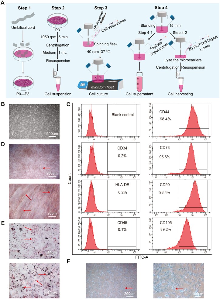
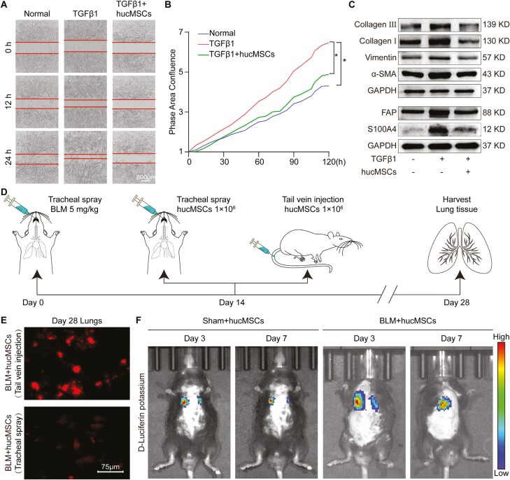
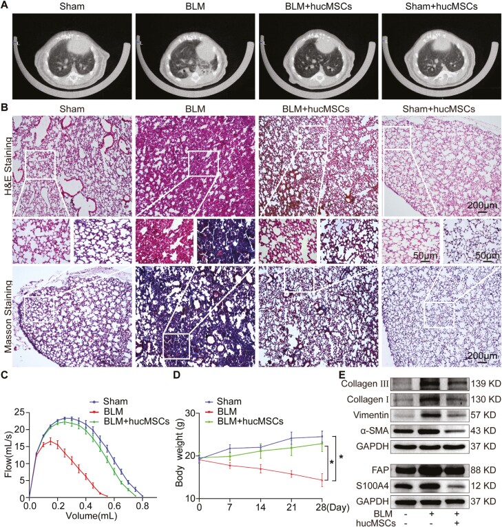
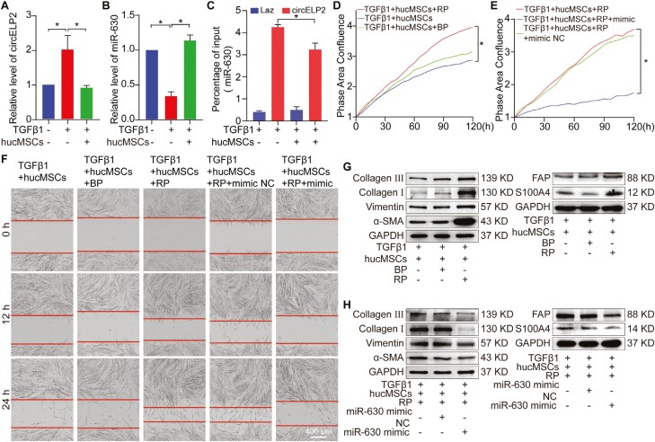
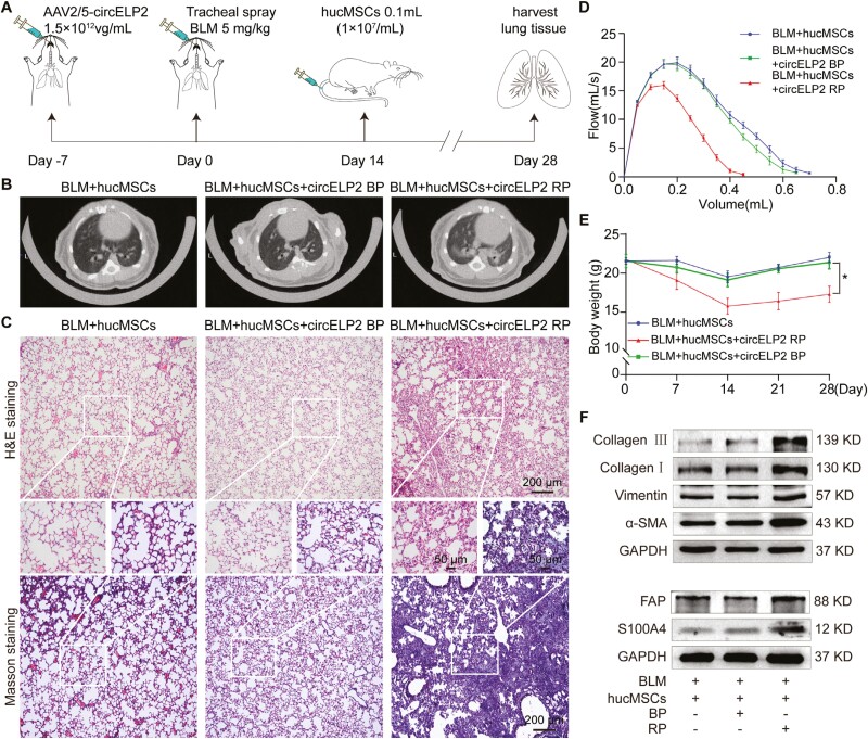
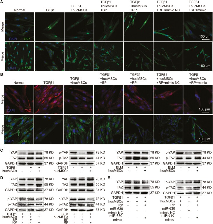
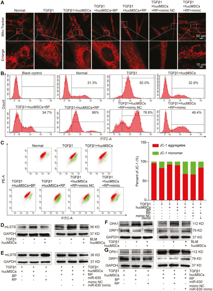
Pulmonary fibrosis is a kind of fibrotic interstitial pneumonia with poor prognosis. Aging, environmental pollution, and coronavirus disease 2019 are considered as independent risk factors for pulmonary fibrogenesis. Consequently, the morbidity and mortality striking continues to rise in recent years. However, the clinical therapeutic efficacy is very limited and unsatisfactory. So it is necessary to develop a new effective therapeutic approach for pulmonary fibrosis. Human umbilical cord mesenchymal stem cells (hucMSCs) are considered as a promising treatment for various diseases because of their multiple differentiation and immunomodulatory function. The key bottleneck in the clinical application of hucMSCs therapy is the high-quality and large-scale production. This study used FloTrix miniSpin bioreactor, a three-dimensional (3D) cell culture system, for large-scale expansion of hucMSCs in vitro, and proved 3D cultured hucMSCs inhibited the differentiation of fibroblasts into myofibroblasts and myofibroblasts proliferation and migration, leading to slow down the development of pulmonary fibrosis. Further mechanistic studies clarified that hucMSCs reduced the amount of binding between circELP2 and miR-630, resulting in blocking YAP/TAZ translocation from cytoplasm to nucleus. This condition inhibited mitochondrial fusion and promoted mitochondrial fission, and ultimately improved fusion/fission balance and cellular homeostasis. To sum up, this work clarified the anti-fibrosis and mechanism of hucMSCs cultured from the 3D FloTrix miniSpin bioreactor. We hope to provide new ideas and new methods for the clinical transformation and industrialization of hucMSCs therapy.

Keywords: YAP/TAZ; circRNA; hucMSCs; mitochondrial fission; mitochondrial fusion; pulmonary fibrosis.


Conflict of interest statement

The authors declare that they have no competing interests.

Figures

Graphical Abstract
Graphical Abstract
Figure 1.
Figure 1.
hucMSCs were identified. (A) A diagram of hucMSCs culture in 3D FloTrix miniSpin bioreactor is shown. (B) hucMSCs morphology was observed under an inverted microscope. The images showed that the cells were long spindles and arranged in parallel or whirlpool. Each group requires 1 × 106 cells. (C) Representative flow cytometry analysis of cell surface markers showed that hucMSCs highly expressed CD73, CD90, and CD44; lowly expressed CD105; and did not express HLA-DR, CD34, and CD45. Each group requires 1 × 106 cells. (D) Alizarin red staining of osteogenic differentiation of hucMSCs showed the formation of bone matrix. Each group requires 3 × 105 cells. (E) Detection of fat differentiation by oil red O staining showed a large number of fat droplets in hucMSCs. Each group requires 3 × 105 cells. (F) Identification of chondrogenic induction of hucMSCs using Alisin blue staining. Each group requires 3 × 105 cells.
Figure 2.
Figure 2.
hucMSCs demonstrated anti-pulmonary fibrotic effect in vitro and targeted mouse lungs. (A) Scratch test results showed that the migration speed of cells in the TGFβ1 group clearly accelerated compared with that in the normal group. The migration rate of cells in the hucMSCs treatment group significantly decreased compared with that in the TGFβ1 group. Each group requires 5 × 104 cells. (B) Cell proliferation curve showed that the proliferation ability of the TGFβ1 group enhanced. The cell proliferation ability of the hucMSCs treatment group decreased compared with that of the TGFβ1 group. Each group requires 1 × 105 cells. (C) Western blot detection showed that the expression levels of fibrosis-related proteins in the hucMSCs treatment group significantly decreased compared with those in the TGFβ1 group. Each group requires 1 × 106 cells. (D) BLM or hucMSCs were administered in mice by tail vein injection and tracheal spraying. (E) Cell membrane staining confirmed residual hucMSCs in the lung. The fluorescence intensity of tail vein injection was higher than that of tracheal spraying on the 28th day. (F) In-vivo imaging of small animals showed that hucMSCs could target the lungs of mice. The whole-body images of mice were observed at 3 and 7 days after hucMSCs injection via tail vein. Each bar represents mean ± SD (n = 6), *P < .05.
Figure 3.
Figure 3.
hucMSCs had anti-pulmonary fibrosis effect in vivo. (A) MicroCT imaging for small animals depicted that the BLM group had honeycomb-like changes and uneven patchy shadows compared with the sham group. These fibrotic symptoms were remarkably alleviated in the hucMSCs treatment group. There is no significant difference between the Sham + hucMSCs group and the Sham group. (B) H&E staining and Masson’s staining detected the pathological changes in lung tissue structure after hucMSCs treatment. The results showed that the BLM group had more fibrotic lesions and damaged alveolar tissue structure than the sham group. hucMSCs alleviated these morphological abnormalities and improved the lesions and symptoms. There is no significant difference between the Sham + hucMSCs group and the Sham group. (C) FVC results showed that the lung function of the BLM group decreased, and that of the hucMSCs treatment group was higher. (D) The body weight of the BLM group significantly decreased, whereas that of the hucMSCs treatment group showed a tendency to increase. (E) Western blot results showed that the expression levels of fibrosis-related proteins collagen, vimentin, α-SMA, FAP, and S100A4 in the hucMSCs treatment group significantly decreased compared with those in the BLM group. Each bar represents mean ± SD (n = 6), *P < .05.
Figure 4.
Figure 4.
circELP2-mediated hucMSCs therapy on pulmonary fibrosis depended on miR-630. (A) qRT-PCR results showed that circELP2 expression was significantly upregulated after MRC-5 cells were stimulated with TGFβ1 for 72 h compared with that in the normal group. hucMSCs treatment repressed circELP2 expression compared with TGFβ1 treatment. Each group requires 1 × 106 cells. (B) qRT-PCR results showed that miR-630 was highly expressed in the normal group. After MRC-5 cells were stimulated with TGFβ1 for 72 h, the expression of miR-630 was significantly downregulated. hucMSCs treatment promoted miR-630 expression compared with TGFβ1 treatment. Each group requires 1 × 106 cells. (C) RAP results showed that the binding of circELP2 to miR-630 reduced under the action of hucMSCs compared with TGFβ1 action. Each group requires 4 × 107 cells. (D) Compared with TGFβ1 + hucMSCs/circELP2 BP treatment, circELP2 overexpression promoted cell proliferation and reversed the effect of hucMSCs on cell proliferation. Each group requires 1 × 105 cells. (E) miR-630 mimic inhibited cell proliferation and reversed the effect of circELP2 overexpression on cell proliferation. Each group requires 1 × 105 cells. (F) Migration images were automatically monitored using the IncuCyte S3 Live-Cell Analysis System. Compared with hucMSCs treatment, circELP2 overexpression promoted cell migration and reversed the inhibitory effect of hucMSCs on cell migration. Meanwhile, miR-630 mimic inhibited cell migration and reversed the promoting effect of circELP2 overexpression on cell migration. Each group requires 5 × 104 cells. (G) Overexpressed circELP2 significantly increased the expression levels of fibrotic proteins and reversed the effect of hucMSCs. Each group requires 1 × 106 cells. (H) miR-630 mimic reduced the expression levels of fibrotic proteins and reversed the effect of overexpressed circELP2. RP represents overexpression of circELP2; BP represents overexpression of circELP2 empty plasmid; and mimic represents miR-630 analog. Each group requires 1 × 106 cells. Each bar represents mean ± SD (n = 6), *P < .05.
Figure 5.
Figure 5.
Rescue experiments were designed to prove the therapeutic effect of hucMSCs via circELP2 in mice. (A) Schematic diagram of drug administration in mice. (B) MicroCT imaging system for small animals showed that circELP2 overexpression reversed the therapeutic effect of hucMSCs and aggravated the degree of fibrosis. (C) H&E and Masson’s staining exhibited that the mice with circELP2 overexpression had thickened alveolar walls, increased collagen deposition, abnormal changes in alveolar structure, and significantly aggravated alveolar inflammation compared with those in the BLM + hucMSCs + cirELP2 BP group. (D) Compared with the hucMSCs and cirELP2 BP treatment groups, the circELP2 overexpression group demonstrated worsening of lung function. (E) The body weight of the BLM + hucMSCs + circELP2 RP group was significantly lower than that of the BLM + hucMSCs + circELP2 BP and BLM + hucMSCs groups. (F) Compared with the BLM + hucMSCs and BLM + hucMSCs + circELP2 BP groups, the BLM + hucMSCs + circELP2 RP group demonstrated a significant increase in the expression levels of pulmonary fibrosis-related proteins. Each bar represents mean ± SD (n = 6), *P < .05.
Figure 6.
Figure 6.
hucMSCs blocked nuclear translocation of YAP/TAZ by targeting circELP2-miR-630. (A) Immunofluorescence staining images showed that in the TGFβ1 group, the YAP in the nucleus increased and the p-YAP in the cytoplasm decreased. Compared with TGFβ1 treatment, hucMSCs treatment significantly decreased the expression of YAP in the nucleus and increased the cytoplasmic expression of p-YAP. Overexpressed circELP2 increased YAP in the nucleus, decreased p-YAP in the cytoplasm, and reversed the effect of hucMSCs treatment. miR-630 mimic decreased YAP in the nucleus, increased p-YAP in the cytoplasm, and reversed the effect of overexpressed circELP2 treatment. Each group requires 1 × 105 cells. (B) Immunofluorescence images showed that in the TGFβ1 group, TAZ increased in the nucleus and p-TAZ decreased in the cytoplasm compared with the normal group. hucMSCs treatment significantly decreased TAZ in the nucleus and increased the cytoplasmic expression of p-TAZ. Overexpressed circELP2 increased TAZ in the nucleus, decreased p-TAZ expression in the cytoplasm, and reversed the effect of hucMSCs treatment. miR-630 mimic decreased TAZ expression in the nucleus, increased p-TAZ expression in the cytoplasm, and reversed the effect of overexpressed circELP2 treatment. Each group requires 1 × 105 cells. (C) Western blot results showed that hucMSCs treatment significantly reduced the expression levels of YAP and TAZ and increase those of p-YAP and p-TAZ proteins increased compared TGFβ1 or BLM treatment. Each group requires 1 × 106 cells. (D) Overexpression of circELP2 increased the expression levels of YAP and TAZ proteins decreased those of p-YAP and p-TAZ proteins, and reversed the effect of hucMSCs on YAP/TAZ. Each group requires 1 × 106 cells. (E) miR-630 mimic reversed the effect of overexpressed circELP2 treatment by reducing YAP/TAZ expression and increasing p-YAP/TAZ expression. Each group requires 1 × 106 cells.
Figure 7.
Figure 7.
hucMSCs inhibited/promoted mitochondrial fusion/fission through circELP2-mediated signal pathway. (A) Mitochondrial morphology was observed using a laser scanning confocal microscope. Compared with the normal group, the TGFβ1 and hucMSCs treatment group showed an increase in mitochondrial fusion and fission, respectively. Overexpression of circELP2 promoted mitochondrial fusion, and adding miR-630 mimic increased mitochondrial fission. Each group requires 1 × 105 cells. (B) Flow cytometry was used to detect ROS. Compared with the ROS level in the normal group, those in the TGFβ1 and hucMSCs treatment groups increased and decreased, respectively. These levels were increased by overexpression of circELP2 and decreased by the addition of miR-630 mimic. Each group requires 1 × 106 cells. (C) MMP was detected by JC-1. Compared with the normal group, the TGFβ1 and hucMSCs treatment groups had decreased and increased MMP, respectively. Overexpression of circELP2 decreased MMP and reversed the effect of hucMSCs treatment. miR-630 mimic reversed the effect of overexpressed circELP2. Each group requires 1 × 106 cells. (D) Western blot experiment showed that hucMSCs reduced mLST8 expression compared with TGFβ1 or BLM. Each group requires 1 × 106 cells. (E) Rescue experiment showed that overexpression of circELP2 increased mLST8 expression and reversed the effect of hucMSCs. miR-630 mimic decreased mLST8 expression and reversed the effect of overexpressed circELP2. Each group requires 1 × 106 cells. (F) The expression of OPA1 was higher in the TGFβ1 or BLM group than in the control group, whereas that of DRP1 was lower. DRP1 was upregulated and OPA1 was downregulated by hucMSCs treatment. Each group requires 1 × 106 cells. (G) Western blot results showed that circELP2 overexpression enhanced OPA1 expression, reduced DRP1 expression, and reserved the effect of hucMSCs treatment. miR-630 mimic reversed the effect of overexpressed circELP2. Each group requires 1 × 106 cells.

Similar articles

References

    1. Podolanczuk AJ, Thomson CC, Remy-Jardin M, et al.. Idiopathic pulmonary fibrosis: state of the art for 2023. Eur Respir J. 2023;61(4):2200957. 10.1183/13993003.00957-2022 - DOI - PubMed
    1. Pugashetti JV, Adegunsoye A, Wu Z, et al.. Validation of proposed criteria for progressive pulmonary fibrosis. Am J Respir Crit Care Med. 2023;207(1):69-76. 10.1164/rccm.202201-0124OC - DOI - PMC - PubMed
    1. Flack KF, Katzen J.. Revealing the rare: pulmonary fibrosis from surfactant-related gene mutation. Chest. 2023;163(4):744-745. 10.1016/j.chest.2022.12.020 - DOI - PubMed
    1. Moss BJ, Ryter SW, Rosas IO.. Pathogenic mechanisms underlying idiopathic pulmonary fibrosis. Annu Rev Pathol. 2022;17(1553-4006):515-546. 10.1146/annurev-pathol-042320-030240 - DOI - PubMed
    1. Yang L, Liu G, Li X, et al.. Small GTPase RAB6 deficiency promotes alveolar progenitor cell renewal and attenuates PM2.5-induced lung injury and fibrosis. Cell Death Dis. 2020;11(10):827. 10.1038/s41419-020-03027-2 - DOI - PMC - PubMed