. 2024 Jul 29;21(1):165.
doi: 10.1186/s12985-024-02437-4.

Inflammatory damage caused by Echovirus 30 in the suckling mouse brain and HMC3 cells

Affiliations

Inflammatory damage caused by Echovirus 30 in the suckling mouse brain and HMC3 cells

Jichen Li et al. Virol J. .

Abstract

Echovirus 30 (E30), a member of the species B Enterovirus family, is a primary pathogen responsible for aseptic meningitis and encephalitis. E30 is associated with severe nervous system diseases and is a primary cause of child illness, disability, and even mortality. However, the mechanisms underlying E30-induced brain injury remain poorly understood. In this study, we used a neonatal mouse model of E30 to investigate the possible mechanisms of brain injury. E30 infection triggered the activation of microglia in the mouse brain and efficiently replicated within HMC3 cells. Subsequent transcriptomic analysis revealed inflammatory activation of microglia in response to E30 infection. We also detected a significant upregulation of polo-like kinase 1 (PLK1) and found that its inhibition could limit E30 infection in a sucking mouse model. Collectively, E30 infection led to brain injury in a neonatal mouse model, which may be related to excessive inflammatory responses. Our findings highlight the intricate interplay between E30 infection and neurological damage, providing crucial insights that could guide the development of interventions and strategies to address the severe clinical manifestations associated with this pathogen.

Keywords: Echovirus 30; HMC3 cells; Inflammatory response; PLK1.


Conflict of interest statement

The authors declare no competing interests.

Figures

Fig. 1
Fig. 1
E30 infection induces brain injury in a mouse model. Three-day-old IFNAR−/− mice were intracranially (i.c.) injected with TL1C strain at 105 TCID50; (n = 7 per group). Control mice received uninfected culture medium. The body weight (A), survival rate (B), and clinical scores (C) of the 3-day-old mice were monitored daily. Histopathological examination was performed on brain tissue from infected mice using H&E and Nissl’s staining. Histopathological analysis revealed cerebral edema, neuronal cell swelling, diminished or absent Nissl bodies, and nuclear vacuolation (D). Mock tissues displayed no alterations. Brain tissue sections from suckling mice were immunofluorescence stained with DAPI (blue) and VP1 (E; red)—scale bars: 200 μm, 50 μm. Data are shown as the mean ± SD. **P < 0.01; ***P < 0.001; ****P < 0.0001; ns: Not significant
Fig. 2
Fig. 2
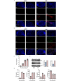
Efficient E30 replication and associated neurological pathological damage in infected mouse brains. Three-day-old IFNAR−/− mice were i.c. injected with a 105 TCID50 of E30. The spinal cords from suckling mice were immunofluorescence stained with DAPI (blue), astrocyte markers GFAP (A; red), microglia marker Iba1 (B; red), and VP1 (A, B; green). Scale bars: 1000 μm, 200 μm and 100 μm. Brain tissue virus titers were determined at 1, 3, and 5 days post-inoculation (dpi) (n = 7 at each time point), and ELISA was performed to determine the levels of IL6, IFN-g, TNF-a and IL-1b in mice (n = 7) (C, E). Western blot was applied to detect the expression of GFAP and Iba1 in mouse brains (D). The GFAP and Iba1 protein expression related to the internal control was quantified using Image J software. Data are shown as the mean ± SD. **P < 0.01; ***P < 0.001; ****P < 0.0001; ns: Not significant
Fig. 3
Fig. 3
Proficient replication of the E30 strain TL1C in HMC3 cells and dysregulation of inflammatory signaling and virus-responsive genes at the transcriptome level. Virus titer detection in E30-infected HMC3 cells (n = 7 for each time point) (A). Volcano plot analysis for differentially expressed genes in E30-infected mice relative to the control group. Red dots denote up-regulated genes, whereas blue dots represent down-regulated genes (B). Gene Ontology (GO) enrichment and Kyoto Encyclopedia of Genes and Genomes (KEGG) pathway analyses of differentially expressed genes in HMC3 cells (C, D). RNA level by RT-qPCR (n = 5) (E). Data are shown as the mean ± SD. **P < 0.01; ***P < 0.001; ****P < 0.0001; ns: Not significant
Fig. 4
Fig. 4
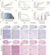
PLK1 inhibitor BI2536 alleviates pathogenic symptoms in E30-infected mice. Monitoring of body weight (A), survival rates (B), and clinical scores (C) continued until 9 dpi. Cell viability and IC50 assays of BI2536 (D, E). Brain tissue virus titers were determined at 1, 3, and 5 days post-inoculation (dpi) with the BI2536 (n = 7 for each time point) (F). Nissl’s and H&E staining of Spinal cord sections from E30-infected mice (G, H). Brain tissues collected from DMSO-treated, BI2536-treated, E30-infected, and DMSO + BI2536-treated E30 infection groups. E30-infected groups showed clear edema, bleeding, and vacuole-like changes (H). Scale bars: 1000 μm, 200 μm and 100 μm. Data represent the mean ± SD. Two-tailed Student’s t-tests were employed to determine the significance of the data. **P < 0.01; ***P < 0.001; ****P < 0.0001; ns: Not significant

Similar articles

References

    1. Lefkowitz EJ, Dempsey DM, Hendrickson RC, Orton RJ, Siddell SG, Smith DB. Virus taxonomy: the database of the International Committee on Taxonomy of Viruses (ICTV). Nucleic Acids Res. 2018;46:D708–17. 10.1093/nar/gkx932 - DOI - PMC - PubMed
    1. Benschop KSM, Broberg EK, Hodcroft E, Schmitz D, Albert J, Baicus A, Bailly JL, Baldvinsdottir G, Berginc N, Blomqvist S, et al. Molecular Epidemiology and Evolutionary Trajectory of emerging Echovirus 30, Europe. Emerg Infect Dis. 2021;27:1616–26. 10.3201/eid2706.203096 - DOI - PMC - PubMed
    1. Chen J, Han Z, Wu H, Xu W, Yu D, Zhang Y. A large-scale outbreak of Echovirus 30 in Gansu Province of China in 2015 and its phylodynamic characterization. Front Microbiol. 2020;11:1137. 10.3389/fmicb.2020.01137 - DOI - PMC - PubMed
    1. Vandesande H, Laajala M, Kantoluoto T, Ruokolainen V, Lindberg AM, Marjomaki V. Early entry events in Echovirus 30 infection. J Virol 2020, 94. - PMC - PubMed
    1. Jin Y, Li D, Sun T, Du Y, Gao Y, Ding R, Ji W, Zhang W, Yang H, Chen S, Duan G. Pathological features of Enterovirus 71-Associated brain and lung damage in mice based on quantitative proteomic analysis. Front Microbiol. 2021;12:663019. 10.3389/fmicb.2021.663019 - DOI - PMC - PubMed

Substances

LinkOut - more resources