. 2024 Oct;31(10):1465-1476.
doi: 10.1038/s41417-024-00813-4. Epub 2024 Jul 29.

Rabenosyn-5 suppresses non-small cell lung cancer metastasis via inhibiting CDC42 activity

Affiliations

Rabenosyn-5 suppresses non-small cell lung cancer metastasis via inhibiting CDC42 activity

Xiong Guo et al. Cancer Gene Ther. 2024 Oct.

Abstract

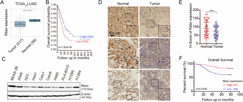
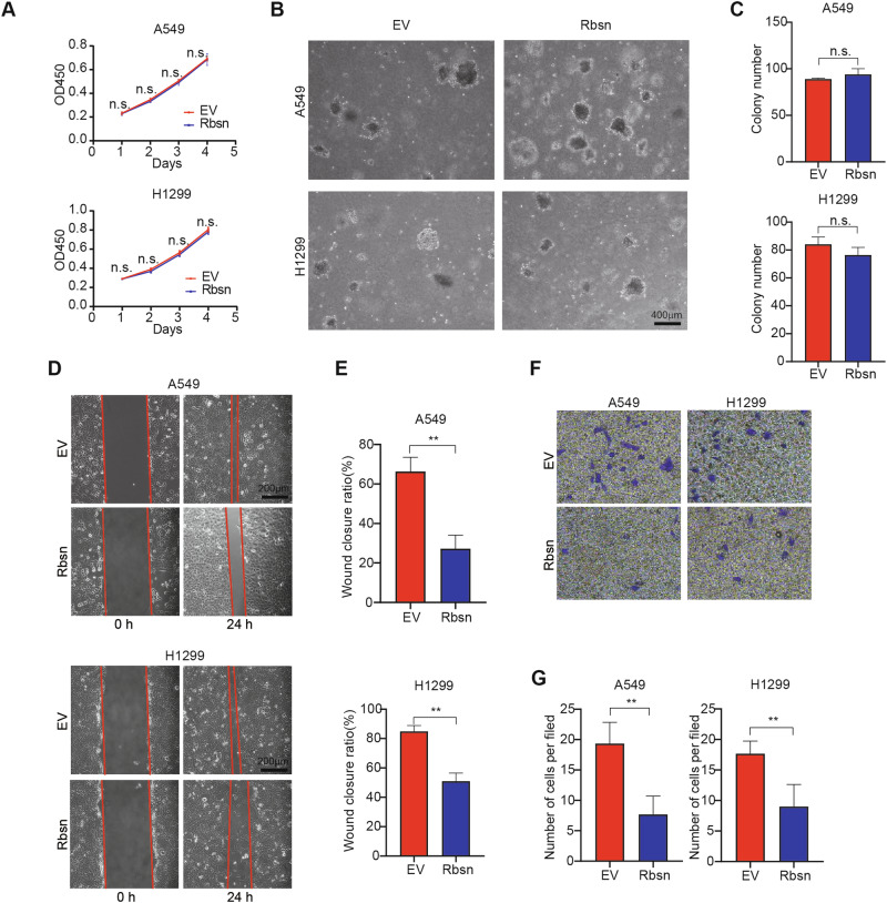
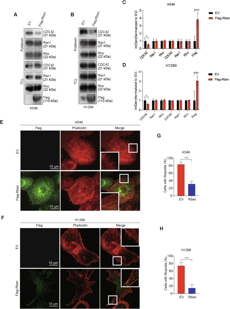
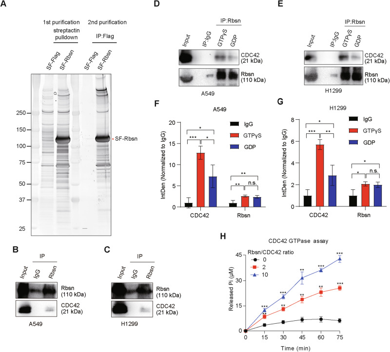
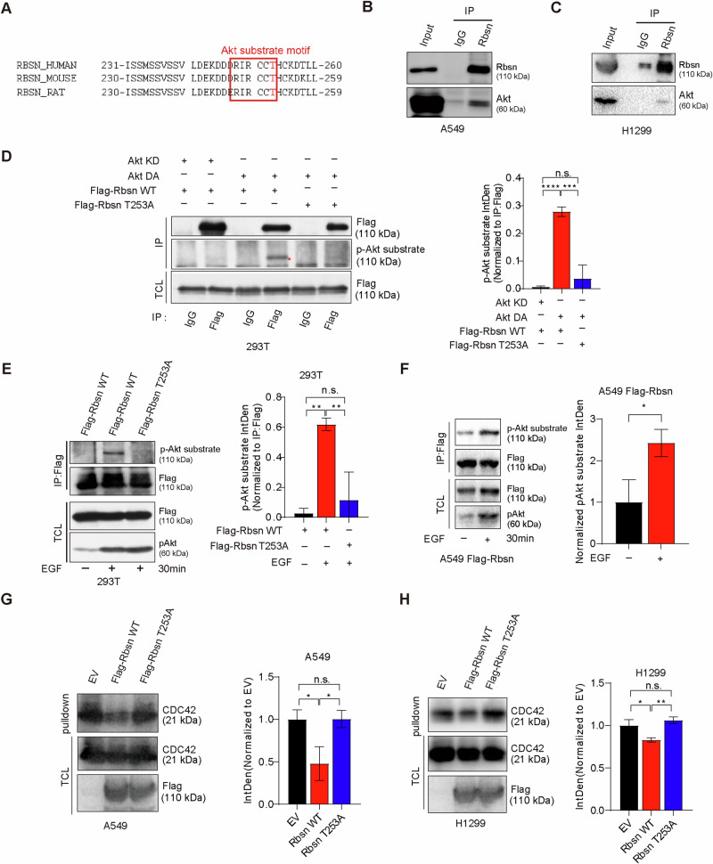
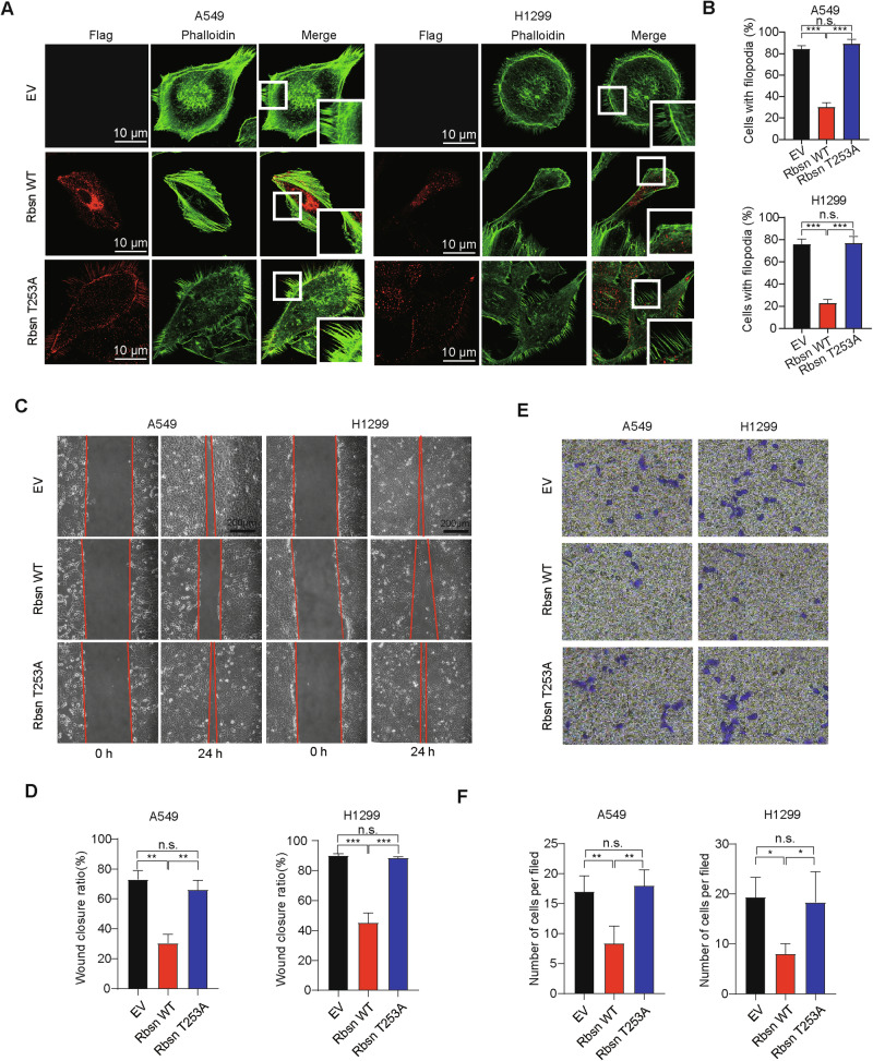
Metastasis, the primary cause of death in lung cancer patients, is facilitated by cytoskeleton remodeling, which plays a crucial role in cancer cell migration and invasion. However, the precise regulatory mechanisms of intracellular trafficking proteins involved in cytoskeleton remodeling remain unclear. In this study, we have identified Rabenosyn-5 (Rbsn) as an inhibitor of filopodia formation and lung cancer metastasis. Mechanistically, Rbsn interacts with CDC42 and functions as a GTPase activating protein (GAP), thereby inhibiting CDC42 activity and subsequent filopodia formation. Furthermore, we have discovered that Akt phosphorylates Rbsn at the Thr253 site, and this phosphorylation negates the inhibitory effect of Rbsn on CDC42 activity. Additionally, our analysis reveals that Rbsn expression is significantly downregulated in lung cancer, and this decrease is associated with a worse prognosis. These findings provide strong evidence supporting the role of Rbsn in suppressing lung cancer progression through the inhibition of metastasis.


Conflict of interest statement

The authors declare no competing interests.

Figures

Fig. 1
Fig. 1. Rbsn is lowly expressed in NSCLC.
A The expression of Rbsn in NSCLC patients was analyzed in the LUNG CANCER EXPLORER platform (TCGA_LUAD_2016 dataset). The numbers in parentheses represent the sample size. A two-tailed Student’s t-test was used for statistical analyses. B Kaplan–Meier analysis of prognosis about Rbsn expression in NSCLC patients from an R2 clinic patient database (TCGA-515-rsem-tcgars dataset). Rbsn expression levels were divided into high and low groups based on the median expression level of Rbsn. Log-rank test was used for statistical analyses. C The expression of Rbsn was assessed in different cell lines using Western blotting. D, E Immunohistochemical staining of Rbsn was performed in human NSCLC tissues. The right panels in (D) show a higher magnification of the regions within the black boxes. The quantification results of Rbsn staining are presented in (E). A two-tailed Student’s t test was used for statistical analyses. Data are presented as means ± SE (n = 95), ***p < 0.001. F Overall survival analysis was conducted to evaluate the impact of Rbsn expression on NSCLC patients. Rbsn expression levels were divided into high and low groups based on the median expression level of Rbsn. Log-rank test was used for statistical analyses.
Fig. 2
Fig. 2. Rbsn inhibits lung cancer cell migration and invasion.
A The effect of Rbsn on the proliferation of lung cancer cells was examined using CCK8 analysis (n = 3, technical replicates) and the experiments were conducted with at least three independent repetitions. B, C The colony formation of lung cancer cells in soft agar was assessed, with or without Rbsn overexpression. The quantification data are shown in (C) (n = 3, technical replicates) and the experiments were conducted with at least three independent repetitions. D, E Lung cancer cell migration was evaluated using a wound healing assay, with or without Rbsn overexpression. The quantification data are shown in (E) (n = 3, technical replicates) and the experiments were conducted with at least three independent repetitions. F, G The invasion of lung cancer cells was examined using a transwell assay, with or without Rbsn overexpression. The quantification data are shown in (G) (n = 3, technical replicates) and the experiments were conducted with at least three independent repetitions. In the results of (A), (C), (E), and (G), a two-tailed Student’s t test was used for statistical analyses. Data are presented as means ± SE, ** p < 0.01. EV indicates empty vector; n.s. indicates no significant difference.
Fig. 3
Fig. 3. Rbsn inhibits CDC42 activity.
AD The GTPase activity of CDC42, Rac1, and Rho in lung cancer cells, both with and without Rbsn overexpression, was assessed through repeated experiments using a small Rho GTPase activity assay, along with the statistical analysis related to A549 (C) and H1299 (D). EH Cells were infected with indicated plasmids, and immunofluorescence was performed 72 h later to visualize filopodia formation in lung cancer cells with or without Rbsn overexpression. Closed boxes are magnified in the insets. The percentage of cells with filopodia in each group (100 cells from three independent experiments) is quantified in (G) and (H), respectively. Two-tailed Student’s t test was used for statistical analyses. Data are presented as means ± SE, * p < 0.05, ***p < 0.001, ****p < 0.0001. EV indicates empty vector; TCL indicates total cell lysate.
Fig. 4
Fig. 4. Rbsn is a GAP for CDC42.
A Tandem affinity purification was performed to isolate the Rbsn interacting proteins in H1299 cells, followed by visualization of the protein complex using silver staining. B, C The interaction between endogenous Rbsn and CDC42 was examined by co-immunoprecipitation in A549 (B) and H1299 (C). DG The interaction between endogenous Rbsn and CDC42 was examined by co-immunoprecipitation in the presence of either GTPγS or GDP, along with the statistical analysis related to A549 (F) and H1299 (G), a two-way ANOVA was used for statistical analyses. H CDC42 GTPase activity assays were conducted to assess the impact of the Rbsn on CDC42 GTPase activity. The results demonstrated that the Rbsn increased CDC42 GTPase activity in a time- and dose-dependent manner. The numbers represent the molar ratio of the Rbsn protein to the CDC42 protein utilized in this assay. A one-way ANOVA test was used for statistical analyses. Data are presented as means ± SE (n = 6, technical replicates) and the experiments were conducted with at least three independent repetitions, *p < 0.05, **p < 0.01, ***p < 0.001. IP indicates immunoprecipitation.
Fig. 5
Fig. 5. Akt-induced Rbsn phosphorylation is essential for CDC42 activity regulation.
A Amino acid sequences of Rbsn from various species are presented, with the conserved threonine at position 253 (highlighted in red) conforming to the Akt substrate motif. B, C The endogenous interaction between Rbsn and Akt was checked by co-immunoprecipitation in lung cancer cells. D Indicated plasmids were transfected into 293T cells, and the immunoprecipitation was performed with anti-Flag antibody or control IgG, followed by p-Akt substrate to check the phosphorylation of Rbsn, along with statistical analysis of three independent experiments. A One-way ANOVA was used for statistical analyses. E, F The indicated plasmids were transfected into 293 T cells (E), or A549 Flag-Rbsn cell line (F) stimulated with or without 100 ng/ml EGF for 30 min. Subsequently, the immunoprecipitation was performed with anti-Flag antibody or control IgG. The phosphorylated Rbsn was examined with a p-Akt substrate, along with statistical analysis of three independent experiments. A One-way ANOVA and two-tailed Student’s t test was used for statistical analyses. G, H CDC42 activity assay to examine CDC42 activity in lung cancer cells stably expressing with indicated plasmids. The active CDC42 was purified by a pulldown experiment with the affinity beads, along with statistical analysis of three independent experiments. A One-way ANOVA was used for statistical analyses. WT indicates wild type; IP indicates immunoprecipitation; TCL indicates total cell lysate. Data are presented as means ± SE, *p < 0.05, **p < 0.01, ***p < 0.001, ****p < 0.0001.
Fig. 6
Fig. 6. Rbsn Thr253 phosphorylation is essential for lung cancer cell migration and invasion.
A, B Cells were infected with indicated plasmids, and immunofluorescence was performed 72 h later to visualize filopodia formation in lung cancer cells expressing the indicated plasmids. Closed boxes in (A) are magnified in the insets. The percentage of cells with filopodia in each group (100 cells from three independent experiments) is quantified in (B), and a One-way ANOVA was used for statistical analyses. C, D Wound healing assay to examine cell migration in lung cancer cells stably expressing with indicated plasmids. The quantification data from three independent experiments are shown in (D) (n = 3, technical replicates) and the experiments were conducted with at least three independent repetitions. E, F Cell invasion in lung cancer cells expressing the indicated plasmids was examined using a transwell assay. Quantification data for cell invasion are presented in (F) (n = 3, technical replicates) and the experiments were conducted with at least three independent repetitions. In the results of (B), (D), and (F), a One-way ANOVA was used for statistical analyses. Data are presented as means ± SE, *p < 0.05, **p < 0.01, ***p < 0.001. EV indicates an empty vector; WT indicates wild type.
Fig. 7
Fig. 7. Rbsn inhibits lung cancer metastasis in vivo.
A Schematic of the mice models to check lung cancer metastasis. B, C Representative images depicting the metastatic tumor foci in the lungs are presented. Quantification data for the number of metastatic tumor foci are shown in (C). A One-way ANOVA was used for statistical analyses. Data are presented as means ± SE (6 mouses per group), **p < 0.01. EV indicates an empty vector; WT indicates wild type.

Similar articles

References

    1. Vachani A, Sequist LV, Spira A. Ajrccm: 100-year anniversary. The shifting landscape for lung cancer: past, present, and future. Am J Respir Crit Care Med. 2017;195:1150–60. - PMC - PubMed
    1. Goldstraw P, Ball D, Jett JR, Le Chevalier T, Lim E, Nicholson AG, et al. Non-small-cell lung cancer. Lancet. 2011;378:1727–40. - PubMed
    1. Hirsch FR, Scagliotti GV, Mulshine JL, Kwon R, Curran WJ Jr, Wu YL, et al. Lung cancer: current therapies and new targeted treatments. Lancet. 2017;389:299–311. - PubMed
    1. Relli V, Trerotola M, Guerra E, Alberti S. Abandoning the notion of non-small cell lung cancer. Trends Mol Med. 2019;25:585–94. - PubMed
    1. Planchard D, Popat S, Kerr K, Novello S, Smit EF, Faivre-Finn C, et al. Metastatic non-small cell lung cancer: ESMO Clinical Practice Guidelines for diagnosis, treatment and follow-up. Ann Oncol. 2018;29:iv192–iv237. - PubMed