Review
. 2024 Jul 25;28(1):367-380.
doi: 10.1080/19768354.2024.2380294. eCollection 2024.

ESCRT-III: a versatile membrane remodeling machinery and its implications in cellular processes and diseases

Affiliations
Review

ESCRT-III: a versatile membrane remodeling machinery and its implications in cellular processes and diseases

Jisoo Park et al. Anim Cells Syst (Seoul). .

Abstract

The endosomal sorting complexes required for transport (ESCRT) machinery is an evolutionarily conserved cytosolic protein complex that plays a crucial role in membrane remodeling and scission events across eukaryotes. Initially discovered for its function in multivesicular body (MVB) formation, the ESCRT complex has since been implicated in a wide range of membrane-associated processes, including endocytosis, exocytosis, cytokinesis, and autophagy. Recent advances have elucidated the ESCRT assembly pathway and highlighted the distinct functions of the various ESCRT complexes and their associated partners. Among the ESCRT complexes, ESCRT-III stands out as a critical player in membrane remodeling, with its subunits assembled into higher-order multimers capable of bending and severing membranes. This review focuses on the ESCRT-III complex, exploring its diverse functions in cellular processes beyond MVB biogenesis. We delve into the molecular mechanisms underlying ESCRT-III-mediated membrane remodeling and highlight its emerging roles in processes such as viral budding, autophagosome closure, and cytokinetic abscission. We also discuss the implications of ESCRT-III dysregulation in neurodegenerative diseases. The versatile membrane remodeling capabilities of ESCRT-III across diverse cellular processes underscore its importance in maintaining proper cellular function. Furthermore, we highlight the promising potential of ESCRT-III as a therapeutic target for neurodegenerative diseases, offering insights into the treatments of the diseases and the technical applications in related research fields.

Keywords: Autophagy; ESCRT-III; MVB; membrane remodeling; neurodegenerative disease.


Conflict of interest statement

No potential conflict of interest was reported by the author(s).

Figures
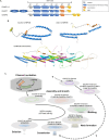
Figure 1.
Figure 1.
The architecture of ESCRT-III. (A) All CHMP proteins share a Snf7 core region consisting of six α-helices (shown as spirals). The α1-α4 helices (shown as blue spirals) facilitate polymerization and membrane binding via their positive charges, while the α5 helix (shown as orange spirals), bearing negative charges, inhibits polymerization in the cytosol. The α0 helix anchors the protein to the membrane via its hydrophobic residues called the ANCHR motif (shown as grey spirals), and the α6 helix, bearing the MIM (shown as yellow spirals), interacts with MIT domain-containing proteins like VPS4. In addition to the core Snf7 domain, CHMP7 and CHMP8 feature additional motifs and domains: CHMP7 has WH1 and WH2 domains for LEMD2 interaction, while CHMP8 has longer MIM sequences. ANCHR: amphipathic N-terminus containing hydrophobic residues motif; MIM: MIT-interacting motif; WH: winged helix domain. (B) In their cytosolic state, ESCRT-III proteins, such as CHMP2B, adopt a closed conformation mediated by electrostatic interactions between the α1-α4 helices and the negatively charged α5 helix. When the protein opens, the self-assembly region becomes exposed, allowing it to assemble into higher-order multimeric polymers. The 3D models of CHMP2B and CHMP2B assembly were generated using AlphaFold2 and visualized as cartoons using PyMOL. (C) The sequential assembly of the ESCRT-III complex. ESCRT-III polymerization generally initiates with the binding of CHMP6 to ESCRT-II, which induces CHMP4B polymerization. CHMP2A and CHMP3 then join the CHMP4B polymer, and their assembly, along with CHMP1B, drives membrane curvature formation. Subsequently, CHMP4B dissociates from the polymer, allowing for the reshaping of the membrane into a neck-like structure. The detachment of CHMP2A and the recruitment of CHMP8 induce further constriction of the membrane neck, eventually leading to scission. This sequential assembly and disassembly process is driven by the AAA-ATPase activity of the VPS4 complex. Interestingly, CHMP5 can initiate ESCRT-III assembly independently of CHMP4B, but its precise physiological function remains unclear.
Figure 2.
Figure 2.
The involvement of ESCRT-III proteins in various membrane dynamics. (A) The pathogenic virus utilizes the host ESCRT-III complex for its budding and propagation from infected cells at the plasma membrane. Similar to virus budding, the addition of VPS4 into the ESCRT-III polymer induces membrane bending, leading to the formation of buds and tubules protruding from the cell surface for exocytosis. (B) Along with the ESCRT-II complex and CHMP6 on endosomal membranes, the ESCRT-III complex regulates the biogenesis of ILVs within MVBs derived from early endosomes containing EGF-R trafficked through endocytosis. ESCRT-III proteins mediate the repair of damaged lysosomal membranes upon the recruitment of TSG101 and ALIX. (C) The ESCRT-III complex plays crucial roles in phagophore sealing during degradative and secretory autophagosome formation. For termination of the STING signaling, the degradation of STING is regulated by TSG101 and ESCRT-III-mediated microautophagy. (D) At the end of cytokinesis, ESCRT complex and adaptor proteins are sequentially recruited to the constriction zones in intercellular bridges, resulting in the formation of spiral-like ESCRT-III polymers at the abscission site. Then, VPS4, spastin, and MITD1 induce constriction of the abscission site, which eventually drives a membrane cut. On the other hand, the ESCRT-III complex is transiently recruited to the reassembling site of the nuclear envelope during the anaphase. The inner nuclear membrane protein LEMD2 recruits CHMP7, which promotes nuclear membrane sealing.

Similar articles

References

    1. Adell MAY, Teis D.. 2011. Assembly and disassembly of the ESCRT-III membrane scission complex. FEBS Lett. 585:3191–3196. doi:10.1016/j.febslet.2011.09.001. - DOI - PMC - PubMed
    1. Adell MAY, Vogel GF, Pakdel M, Müller M, Lindner H, Hess MW, Teis D.. 2014. Coordinated binding of Vps4 to ESCRT-III drives membrane neck constriction during MVB vesicle formation. J Cell Biol. 205:33–49. doi:10.1083/jcb.201310114. - DOI - PMC - PubMed
    1. Agromayor M, Carlton JG, Phelan JP, Matthews DR, Carlin LM, Ameer-Beg S, Bowers K, Martin-Serrano J.. 2009. Essential role of hIST1 in cytokinesis. Mol Biol Cell. 20:1374–1387. doi:10.1091/mbc.e08-05-0474. - DOI - PMC - PubMed
    1. Allison R, Edgar JR, Pearson G, Rizo T, Newton T, Günther S, Berner F, Hague J, Connell JW, Winkler J.. 2017. Defects in ER–endosome contacts impact lysosome function in hereditary spastic paraplegia. J Cell Biol. 216:1337–1355. doi:10.1083/jcb.201609033. - DOI - PMC - PubMed
    1. Antonioli M, Rienzo D, Piacentini M, Fimia M, M G.. 2017. Emerging mechanisms in initiating and terminating autophagy. Trends Biochem Sci. 42:28–41. doi:10.1016/j.tibs.2016.09.008. - DOI - PubMed

Grants and funding

This work was supported by grants from the National Research Foundation of Korea (NRF) funded by the Ministry of Science and ICT (NRF-2021R1A5A2031612).

LinkOut - more resources