Review
. 2024 Jul 17;29(14):3356.
doi: 10.3390/molecules29143356.

Primed for Interactions: Investigating the Primed Substrate Channel of the Proteasome for Improved Molecular Engagement

Affiliations
Review

Primed for Interactions: Investigating the Primed Substrate Channel of the Proteasome for Improved Molecular Engagement

Cody A Loy et al. Molecules. .

Abstract

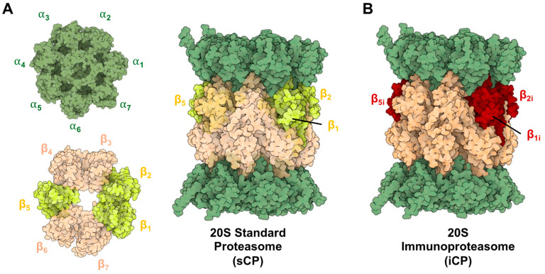
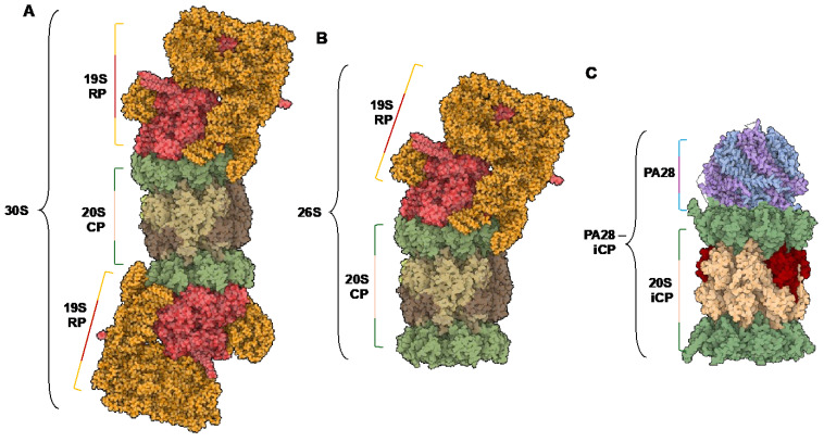
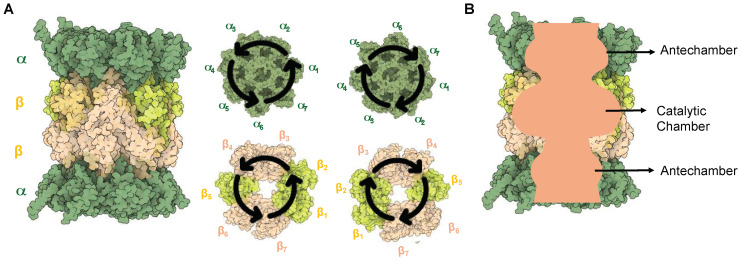
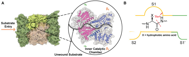
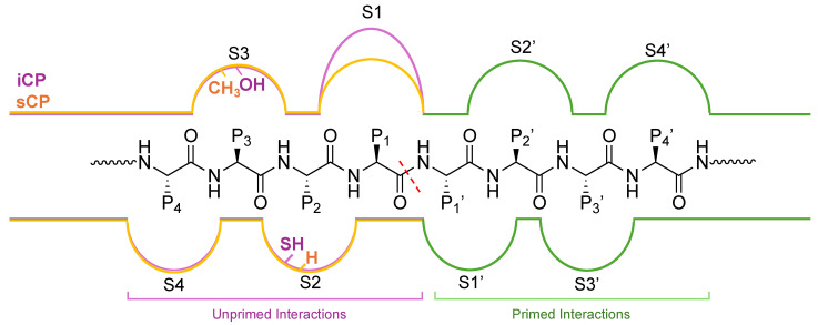
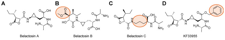
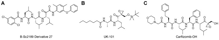
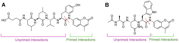
Protein homeostasis is a tightly conserved process that is regulated through the ubiquitin proteasome system (UPS) in a ubiquitin-independent or ubiquitin-dependent manner. Over the past two decades, the proteasome has become an excellent therapeutic target through inhibition of the catalytic core particle, inhibition of subunits responsible for recognizing and binding ubiquitinated proteins, and more recently, through targeted protein degradation using proteolysis targeting chimeras (PROTACs). The majority of the developed inhibitors of the proteasome's core particle rely on gaining selectivity through binding interactions within the unprimed substrate channel. Although this has allowed for selective inhibitors and chemical probes to be generated for the different proteasome isoforms, much remains unknown about the interactions that could be harnessed within the primed substrate channel to increase potency or selectivity. Herein, we discuss small molecules that interact with the primed substrate pocket and how their differences may give rise to altered activity. Taking advantage of additional interactions with the primed substrate pocket of the proteasome could allow for the generation of improved chemical tools for perturbing or monitoring proteasome activity.

Keywords: inhibitor; proteasome; substrate channel.


Conflict of interest statement

Prof. Trader is a shareholder and consultant for Booster Therapeutics, GmbH. Other authors declares no conflicts of interest.

Figures

Figure 1
Figure 1
(A) Structure of the 20S Standard Core Particle (sCP), containing 14 distinct subunits repeated twice (28 total proteins), forming heptoheteromic rings that assemble into the active barrel-like structure. Catalytically active subunits are highlighted (yellow). (B) Structure of the 20S immunoproteasome (iCP) containing the same barrel-like assembly of subunits but with altered catalytic subunits (red). These isoforms are capable of degrading proteins that have been oxidatively damaged or unstructured, but their cleavage products will differ due to their altered substate specificities. PDB ID: 4R3O and 5LE5.
Figure 2
Figure 2
(A) Structure of the 30S isoform of the proteasome, containing a 20S catalytic core particle and two 19S regulatory particles. (B) Structure of the 26S isoform of the proteasome, containing a 20S catalytic core particle and one 19S regulatory particle. (C) Structure of the PA28—iCP complex. PDB IDs: 5GJR, 7DR6.
Figure 3
Figure 3
(A) Structure of the 20S sCP. The symmetry of the proteasome allows one set of α and β subunits to be clockwise, while the other two are counterclockwise in orientation. (B) A sliced view of the proteasome to show the two antechambers and the catalytic chamber.
Figure 4
Figure 4
(A) Substrate entry into the 20S proteasome through the α-subunit portal into the inner catalytic chamber. (B) Active site hydrolysis of an unwound substrate by the β5 subunit. All subunits utilize an active site Thr (red) for hydrolysis of substrates but have altered substrate specificities due to differences in S1 and S2 substrate binding pockets.
Figure 5
Figure 5
Substrate channel for the β5i (purple) and β5 (orange) subunits of the iCP and sCP, respectively. The unprimed channel has been thoroughly explored for differences in the substrate binding pockets S1–S4. The differences in substrate binding pockets S1′–S4′ have remained relatively understudied; however, several inhibitors and chemical probes have been found to take advantage of interactions in this channel to gain selectivity and potency. These indications hint that the primed substrate channel has interactions that can be harnessed when developing proteasome inhibitors or probes, and work is needed to further identify the crucial interactors.
Figure 6
Figure 6
Structures of Belactosin A and its derivatives that engage with the primed substrate channel to inhibit proteasome activity. (A) Belactosin A contains a lactone ring to react with the 20S CP catalytic Thr. (B) Belactosin B does not have a lactone ring (orange circle) and is no longer effective at inhibiting 20S CP. (C) Belactosin C no longer has the trans-cycloproane ring but can still engage and inhibit 20S CP because of the lactone. (D) Optimization of Belactosin A led to improved inhibition with the introduction of the phenyl ring at the carboxylic acid. This demonstrates that there are moieties that can be explored to better engage with the primed substrate channel that lead to improved selectivity or potency.
Figure 7
Figure 7
(A) SAR of Belactosin A from its trans-isomer to cis- was found to increase its IC50 value. Further optimization of the scaffold led to the development of compound 3e, which has toxicity similar to that of Bortezomib, all through interactions, highlighted in orange, in the primed substrate channel. (B) Structure of Marizomib that interacts with all catalytic subunits of the 20S CP and is orally available.
Figure 8
Figure 8
(A) α-ketoamide was developed to engage the primed substrate channel to increase selectivity. SAR derivative 27 was the most potent and selective of the inhibitors developed, with IC50 values in low nM concentrations as well as over 4-fold selectivity for the sCP over iCP. (B) UK-101 was developed to gain selectivity for the LMP2 subunit of the iCP by engaging with the primed substrate pocket. (C) Derivatives of the FDA-approved proteasome inhibitor Carfilzomib have been developed to overcome resistance seen with current proteasome inhibitors. By adding interactions to the primed substrate channel, this derivative increased its potency and was able to be effective against proteasome inhibitor resistance cells.
Figure 9
Figure 9
Fluorescent activity-based AMC probes for 20S CP. Intact probes are non-fluorescent until liberated by cleavage from the proteasome. Specificity has generally been achieved through unprimed substrate interactions (purple). As substrate (green) is cleaved (dashed red line), AMC is released, and fluorescence increases are monitored over time. (A) Structure of Suc-LLVY-AMC, which is selective for the chymotrypsin-like activity of the β5 subunit. (B) Structure of Ac-ANW-AMC, which is selective for the chymotrypsin-like activity of the β5i subunit.
Figure 10
Figure 10
(A) TED FRET reporter to monitor sCP cleavage activity. When intact, FRET reporter signal for EDANS is quenched by the acceptor, DABCYL. Upon interaction with the 20S sCP, the bond between phenylalanine and alanine is cleaved (dashed red line), leading to an increase in fluorescent signal from liberated EDANS. (B) Rhodamine-based probes to monitor iCP activity biochemically or in cells (TBZ-1). iCP recognition sequence ATMW conjugates a rhodamine-peptoid that is non-fluorescent until interaction with the β5i subunit to cleave the bond between Trp and rhodamine (red-dashed line) that allows for increase in fluorescent signal to be monitored over time. (C) Rhodamine-based probes to monitor sCP activity biochemically or in cells (TAS-1). The sCP recognition sequence LLVY conjugates a rhodamine-peptoid that is non-fluorescent until interaction with the β5 subunit to cleave the bond between Tyr and rhodamine (red-dashed line), which allows for increase in fluorescent signal to be monitored over time. (D) FRET probe generated through combinatorial library for primed substrate interactors. Primed interactions led to probes being selective for iCP, demonstrating that engagements in this channel can lead to more selective probes/inhibitors.

Similar articles

References

    1. Groll M., Ditzel L., Löwe J., Stock D., Bochtler M., Bartunik H.D., Huber R. Structure of 20S Proteasome from Yeast at 2.4Å Resolution. Nature. 1997;386:463–471. doi: 10.1038/386463a0. - DOI - PubMed
    1. Coux O., Tanaka K., Goldberg A.L. Structure and functions of the 20S and 26S proteasomes. Annu. Rev. Biochem. 1996;65:801–847. doi: 10.1146/annurev.bi.65.070196.004101. - DOI - PubMed
    1. Boes B., Hengel H., Ruppert T., Multhaup G., Koszinowski U.H., Kloetzel P.M. Interferon Gamma Stimulation Modulates the Proteolytic Activity and Cleavage Site Preference of 20S Mouse Proteasomes. J. Exp. Med. 1994;179:901–909. doi: 10.1084/jem.179.3.901. - DOI - PMC - PubMed
    1. Myers N., Olender T., Savidor A., Levin Y., Reuven N., Shaul Y. The Disordered Landscape of the 20S Proteasome Substrates Reveals Tight Association with Phase Separated Granules. Proteomics. 2018;18:1800076. doi: 10.1002/pmic.201800076. - DOI - PubMed
    1. Chondrogianni N., Georgila K., Kourtis N., Tavernarakis N., Gonos E.S. 20S Proteasome Activation Promotes Life Span Extension and Resistance to Proteotoxicity in Caenorhabditis Elegans. FASEB J. 2015;29:611–622. doi: 10.1096/fj.14-252189. - DOI - PMC - PubMed