Review
. 2024 Jul 16;13(14):4145.
doi: 10.3390/jcm13144145.

Crohn's Disease: Radiological Answers to Clinical Questions and Review of the Literature

Affiliations
Review

Crohn's Disease: Radiological Answers to Clinical Questions and Review of the Literature

Laura Maria Minordi et al. J Clin Med. .

Abstract

Background: Crohn's disease (CD) is a chronic, progressive inflammatory condition, involving primarily the bowel, characterized by a typical remitting-relapsing pattern. Despite endoscopy representing the reference standard for the diagnosis and assessment of disease activity, radiological imaging has a key role, providing information about mural and extra-visceral involvement. Methods: Computed Tomography and Magnetic Resonance Imaging are the most frequently used radiological techniques in clinical practice for both the diagnosis and staging of CD involving the small bowel in non-urgent settings. The contribution of imaging in the management of CD is reported on by answering the following practical questions: (1) What is the best technique for the assessment of small bowel CD? (2) Is imaging a good option to assess colonic disease? (3) Which disease pattern is present: inflammatory, fibrotic or fistulizing? (4) Is it possible to identify the presence of strictures and to discriminate inflammatory from fibrotic ones? (5) How does imaging help in defining disease extension and localization? (6) Can imaging assess disease activity? (7) Is it possible to evaluate post-operative recurrence? Results: Imaging is suitable for assessing disease activity, extension and characterizing disease patterns. CT and MRI can both answer the abovementioned questions, but MRI has a greater sensitivity and specificity for assessing disease activity and does not use ionizing radiation. Conclusions: Radiologists are essential healthcare professionals to be involved in multidisciplinary teams for the management of CD patients to obtain the necessary answers for clinically relevant questions.

Keywords: Computed Tomography; Crohn’s disease; Magnetic Resonance Imaging; inflammatory bowel disease.


Conflict of interest statement

The authors declare no conflict of interest.

Figures
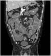
Figure 1
Figure 1
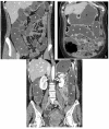
Coronal CT-E image after iodinated contrast medium injection: example of optimal distention of the ileal loops obtained by oral administration contrast medium (arrowheads); jejunal loops are on the top left (white asterisk). Original figures by LMM and LL.
Figure 2
Figure 2
Axial CT-E image after iodinated contrast medium injection: example of intestinal loops’ distension by a hypodense contrast medium (polyethylene glycol solution), showing the hypodensity of the jejunal lumen (asterisks) and the hyperdensity of the intestinal wall (arrowheads). Original figures by LMM and LL.
Figure 3
Figure 3
MR-E: example of intestinal loops’ distention by biphasic contrast medium (polyethylene glycol solution). Intestinal lumen (asterisks) is hyperintense in T2-weighted axial image (a) and hypointense in T1-weighted coronal image after gadolinium injection (b). Original figures by LMM and LL.
Figure 4
Figure 4
CT-E axial image: example of intestinal distension by oral administration of hydrosoluble iodine contrast medium in patient who has undergone intestinal resection with suspected post-surgical fistula. The image shows the hyperdense appearance of the ileal lumen (asterisks). Original figures by LMM and LL.
Figure 5
Figure 5
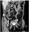
CT after iodinated contrast injection: example of colon distension using air by endorectal insufflation in the coronal (a) and axial (b) planes (asterisks). (c) Another example of colonic distension using air by endorectal insufflation in the axial plane shows a polypoid thickening of the sigmoid colon (arrow); hyperdense contrast agent for faecal tagging is also evident in the lumen (black asterisks). Original figures by LMM, LL and BB.
Figure 6
Figure 6
Hydro-CT. Coronal (ac) images show good distension of the colon obtained by both endorectal (white asterisks) and oral (black asterisks) administration of water and hypodense contrast medium (polyethylene glycol solution). Original figures by LMM and LL.
Figure 7
Figure 7
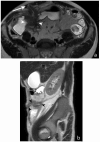
Assessment of disease activity. MR-E after gadolinium injection and administration of polyethylene glycol solution: axial (a) and coronal (b) images show pathological thickening of the distal ileum (arrow) with hyperintensity of the inner layer (mucosa) referred to hyperaemia, hypointensity of the intermediate layer (submucosa) referred to oedema, and hyperintensity of outer layer (serosa) referred to hyperaemia. Original figures by LMM and LL.
Figure 8
Figure 8
Assessment of disease activity. Axial CT-E after iodinated contrast injection and administration of polyethylene glycol solution (a,b) shows thickening of the descending colon with perivisceral fat stranding surrounding the pathological segment (white arrowheads in (a)). A bowel wall ulcer is also evident in another plane (white arrowheads in (b)). Original figures by LMM and LL.
Figure 9
Figure 9
Assessment of disease activity. Axial MR-E after gadolinium injection image and administration of polyethylene glycol solution shows ileal thickening with stratified contrast enhancement (white arrows); a «minus» spot is present in the intestinal wall, indicative of deep parietal ulcer (black arrow). Original figures by LMM and LL.
Figure 10
Figure 10
Fibro-stenotic subtype. MR-E after gadolinium injection and administration of polyethylene glycol solution, axial image shows fibrotic ileal loop with a stretched appearance (white arrowheads). Original figures by LMM and LL.
Figure 11
Figure 11
Fistulizing disease. MR-E after administration of polyethylene glycol solution; the T2-weighted axial image (a), T2-weighted coronal image (b) and contrast-enhanced fat-sat T1-weighted coronal image (c) show an ileo-ileal fistula in the distal ileum (arrows); focal fatty depositions in the submucosal layer are evident in a (asterisks). Original figures by LMM and LL.
Figure 12
Figure 12
Fistulizing disease. MR-E after gadolinium injection and administration of polyethylene glycol solution, coronal image shows pathological intestinal loops in hypogastrium-right iliac fossa, with entero-enteric fistulas (white arrowheads). Original figures by LMM and LL.
Figure 13
Figure 13
Entero-enteric fistula: CT-E coronal image after iodinated contrast injection and administration of polyethylene glycol solution shows entero-enteric fistula between the descending colon and an adjacent ileal loop (arrows). Letter “A” indicates the coronal view of the CT-E image. Original figures by LMM and LL.
Figure 14
Figure 14
Intraparietal abscess and mesenteric inflammation. MR-E after gadolinium injection and administration of polyethylene glycol solution: axial image shows nodular formation with hypointense central core and hyperintense peripheral rim in the wall of a pathological loop (intraparietal abscess, arrowhead). In the adjacent mesentery, hyperintensity of the mesenteric fat is observed with local hypervascularization (hypertrophy of vasa recta, white arrow). Original figures by LMM and LL.
Figure 15
Figure 15
Entero-enteric fistula and entero-cutaneous fistula: MR-E after administration of polyethylene glycol solution. T2-weighted axial image (a) shows pathological loops with entero-enteric fistulas (white arrowheads). Further fistula with hyperintense T2 signal can be observed between the pathological loop and the anterior abdominal wall (arrow). T2-weighted sagittal image (b) shows the fistula between the pathological loop and the anterior abdominal wall; hyperintensity of signal in the lumen of fistula is present (black arrowheads). Original figures by LMM and LL.
Figure 16
Figure 16
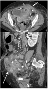
Abscess/phlegmon: CT-E after iodinated contrast injection and administration of polyethylene glycol solution. Axial image (a) shows an abscess/phlegmon (arrowheads) with extension to the wall of the left anterior rectus muscle (white arrow). Sagittal image (b) shows the extension of the abscess/phlegmon (arrowheads) to the wall of the left anterior rectus muscle (white arrow); perienteric fat stranding is present in pelvis (asterisk). Inhomogeneous hypodense tissue portion is present with anti-slope air components. Original figures by LMM and LL.
Figure 17
Figure 17
Fibro-stenotic subtype. MR-E after gadolinium injection and administration of polyethylene glycol solution; axial (a) and coronal (b) images show a homogeneous enhanced intestinal wall with narrowing of the terminal ileum (white arrow) and with dilation of the upstream small bowel loop (black arrow). Original figures by LMM and LL.
Figure 18
Figure 18
Intestinal stenosis: CT-E after iodinated contrast injection and administration of polyethylene glycol solution; coronal image (a) and sagittal image (b) show tight stenosis of the descendent colon (arrowheads), with stratified contrast enhancement; mild overdistention of the upstream descending colon (asterisk) is evident. Original figures by LMM and LL.
Figure 19
Figure 19
Calculation of the length of unaffected intestine using postprocessing software; a tubular view of the small bowel loops from MR-E is shown. The green line indicates the center of the bowel lumen. Original figures by LMM and LL.
Figure 20
Figure 20
Calculation of the length of affected intestine using postprocessing software; a tubular view of the small bowel loops from CT-E is shown. There is evidence of two areas of pathological wall thickening on the right of the picture (arrowheads). The green line indicates the center of the bowel lumen. Original figures by LMM and LL.

Similar articles

References

    1. Chang J.T. Pathophysiology of Inflammatory Bowel Diseases. N. Engl. J. Med. 2020;383:2652–2664. doi: 10.1056/NEJMra2002697. - DOI - PubMed
    1. Kaplan G.G., Windsor J.W. The Four Epidemiological Stages in the Global Evolution of Inflammatory Bowel Disease. Nat. Rev. Gastroenterol. Hepatol. 2020;18:56–66. doi: 10.1038/s41575-020-00360-x. - DOI - PMC - PubMed
    1. Le Berre C., Honap S., Peyrin-Biroulet L. Ulcerative Colitis. Lancet. 2023;402:571–584. doi: 10.1016/S0140-6736(23)00966-2. - DOI - PubMed
    1. Dolinger M., Torres J., Vermeire S. Crohn’s Disease. Lancet. 2024;403:1177–1191. doi: 10.1016/S0140-6736(23)02586-2. - DOI - PubMed
    1. Maaser C., Sturm A., Vavricka S.R., Kucharzik T., Fiorino G., Annese V., Calabrese E., Baumgart D.C., Bettenworth D., Borralho Nunes P., et al. ECCO-ESGAR Guideline for Diagnostic Assessment in IBD Part 1: Initial Diagnosis, Monitoring of Known IBD, Detection of Complications. J. Crohns Colitis. 2019;13:144–164. doi: 10.1093/ecco-jcc/jjy113. - DOI - PubMed

Grants and funding

This research received no external funding.

LinkOut - more resources