. 2024 Jul 21;25(14):7972.
doi: 10.3390/ijms25147972.

The Mechanism of Action of L-Tyrosine Derivatives against Chikungunya Virus Infection In Vitro Depends on Structural Changes

Affiliations

The Mechanism of Action of L-Tyrosine Derivatives against Chikungunya Virus Infection In Vitro Depends on Structural Changes

Vanessa Loaiza-Cano et al. Int J Mol Sci. .

Abstract

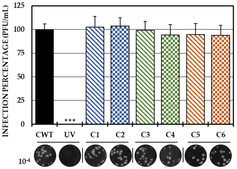
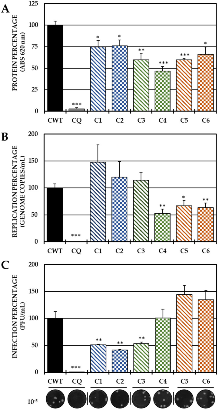
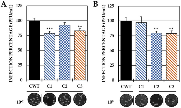
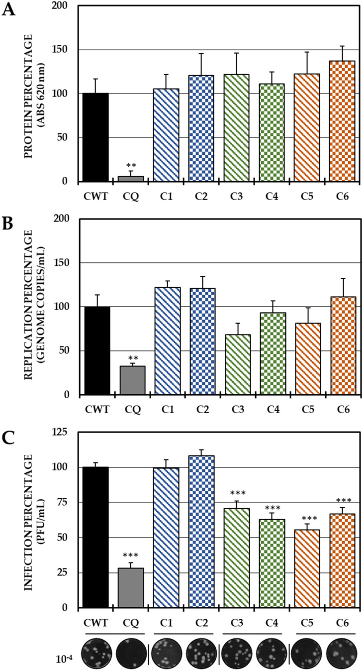
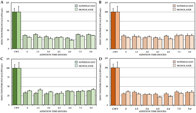
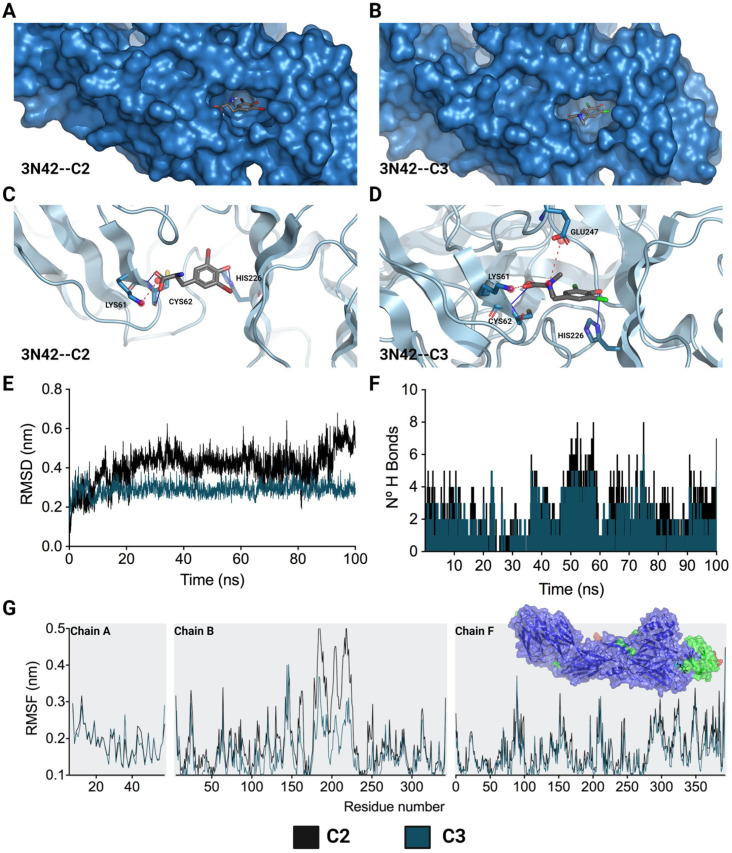
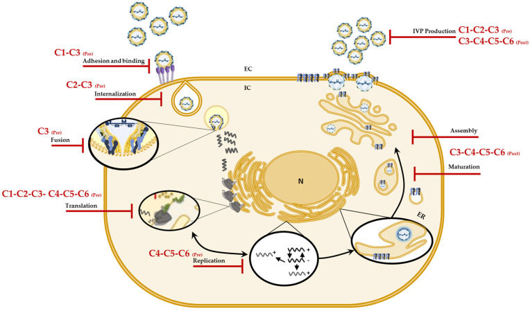
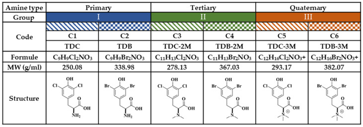
Although the disease caused by chikungunya virus (CHIKV) is of great interest to public health organizations around the world, there are still no authorized antivirals for its treatment. Previously, dihalogenated anti-CHIKV compounds derived from L-tyrosine (dH-Y) were identified as being effective against in vitro infection by this virus, so the objective of this study was to determine the mechanisms of its antiviral action. Six dH-Y compounds (C1 to C6) dihalogenated with bromine or chlorine and modified in their amino groups were evaluated by different in vitro antiviral strategies and in silico tools. When the cells were exposed before infection, all compounds decreased the expression of viral proteins; only C4, C5 and C6 inhibited the genome; and C1, C2 and C3 inhibited infectious viral particles (IVPs). Furthermore, C1 and C3 reduce adhesion, while C2 and C3 reduce internalization, which could be related to the in silico interaction with the fusion peptide of the E1 viral protein. Only C3, C4, C5 and C6 inhibited IVPs when the cells were exposed after infection, and their effect occurred in late stages after viral translation and replication, such as assembly, and not during budding. In summary, the structural changes of these compounds determine their mechanism of action. Additionally, C3 was the only compound that inhibited CHIKV infection at different stages of the replicative cycle, making it a compound of interest for conversion as a potential drug.

Keywords: antiviral; chikungunya virus; computational biology; in vitro; mechanism of action; tyrosine.


Conflict of interest statement

The authors declare no conflict of interest.

Figures

Figure 1
Figure 1
Direct effect of compounds on IVP. Percentages of infection calculated according to the results obtained by the plaque assay (plaque forming units per milliliter, PFU/mL) of the mixtures between the virus and each of the compounds. The inhibition control of the technique was UV radiation (0.0%, <1.0 × 10² PFU/mL). At the bottom is shown the representative plaques of titration on VERO cells corresponding to each experimental condition and the log title of the images. In all cases, control cultures without treatment (CWTs) were assumed as 100.0% infection. The asterisks indicate statistically significant differences with respect to the control without treatment (*** p < 0.001; Student’s t test), and the error bars indicate the standard error of the mean (n = 6).
Figure 2
Figure 2
Effect of tyrosine-derived compounds on pre-treatment CHIKV infection. The relative percentages of infection were calculated according to the results obtained using the cell-ELISA (A) and qPCR (B) assays in monolayers and through the plaque assay (PFU/mL) of the supernatants (C) of the pre-treatment antiviral assay (treatment prior to CHIKV/Col virus inoculation at a multiplicity of infection (MOI) of 5). The inhibition control technique was 50 µM CQ (2.9% infection in the case of cell-ELISA (A); 0.5% infection and 8.6 × 10² genomic copies/mL in the case of qPCR (B) and 0.2% infection and 1.9 × 10⁴ PFU/mL in the case of the plaque assay (C)). At the bottom is shown representative plaques of titration on VERO cells corresponding to each experimental condition and the log title of the images. In all cases, the CWT was assumed to be 100.0% infection (* p < 0.05, ** p < 0.01 and *** p < 0.001; Student’s t test), and the error bars indicate the standard error of the mean (n = 4).
Figure 3
Figure 3
Effect of compounds in the early stages of infection. Percentages of infection calculated according to the results obtained by the plaque assay (PFU/mL) of the supernatants collected from the adhesion inhibition assay (A) and from the internalization inhibition assay monolayers (B). In all cases, the CWT was assumed to be 100.0% infection. At the bottom is shown representative plaques of titration on VERO cells corresponding to each experimental condition and the log title of the images. In all the experimental conditions, the cells were infected with CHIKV/Col at an MOI of 5. The asterisks indicate that the results were significantly different from those in the CWT group (** p < 0.01 and *** p < 0.001; t test). Two independent experiments were carried out with three replicates each (n = 6). The error bars indicate the standard error of the mean.
Figure 4
Figure 4
Effect of dH-Y on post-treatment CHIKV infection. The relative percentages of infection were calculated according to the results obtained by the cell-ELISA (A) and qPCR (B) assays in monolayers and by the plaque assay (PFU/mL) of the supernatants of the post-treatment antiviral assay (treatment after CHIKV/Col virus inoculation at an MOI of 5). The inhibition control technique was 50 µM chloroquine (2.9% infection in the case of cell-ELISA (A), 0.5% infection, 8.6 × 10² genomic copies/mL in the case of qPCR (B) and 28.3% infection, 8.1 × 10⁵ PFU/mL (C)). At the bottom is shown representative plaques of titration on VERO cells corresponding to each experimental condition and the log title of the images. In all cases, the CWT was assumed as 100.0% infection (** p < 0.01 and *** p < 0.001; Student’s t test), and the error bars indicate the standard error of the mean (n = 4).
Figure 5
Figure 5
Effect of compounds on the production of IVP in the CHIKV/Col replication cycle. Percentages of infection calculated according to the results obtained by plaque assay (PFU/mL) in both the supernatant and monolayer of VERO cells infected with the CHIKV/Col virus at an MOI of 5 from the addition time assay for the compounds of Group II (A,C) and Group III (B,D). All the experimental samples were collected at 10.5 h after the removal of the inoculum. In all cases, the CWT was assumed to be 100.0% infection. All experimental conditions were significantly different from those of the control without treatment (p < 0.05; Student’s t test), and the error bars indicate the standard error of the mean (n = 4).
Figure 6
Figure 6
Lowest-energy docked pose and stability of complexes formed by L-tyrosine derivatives with 3N42. C2-3N42 binding pocket and interaction modes (A,C). C3-3N42 binding pocket and interaction modes (B,D). Root mean square deviation (RMSD) (E). Hydrogen bonds (F). Root mean square fluctuation (RMSF) (G). In (C,D), the interactions are represented as hydrogen bonds (blue lines) and ionic bonds (red dotted lines). In (EG), the black and dark green lines correspond to C2 and C3, respectively.
Figure 7
Figure 7
Possible mechanisms of action of the compounds against CHIKV. Graphic representation of the sites on the CHIKV replicative cycle on which, according to the results, the compounds (C1 to C6) could have an effect and jointly explain the in vitro antiviral activity.
Figure 8
Figure 8
Study compounds. The six dH-Y were classified into three groups according to the substitution of the amino group. Each group has a chlorinated compound (Group I, 2-amino-3-(3,5-dichloro-4-hydroxyphenyl) propanoic acid; Group II, 3-(3,5-dichloro-4-hydroxyphenyl)-2-(dimethylamino) propanoic acid; and Group III, 1-carboxy-2-(3,5-dichloro-4-hydroxyphenyl)-N,N-trimethylethan-1-aminium; corresponding to C1, C3 and C5, respectively); and a brominated compound (Group I, 2-amino-3-(3,5-dibromo-4-hydroxyphenyl) propanoic acid; Group II, 3-(5-dibromo-4-hydroxyphenyl)-2- (dimethylamino) propanoic acid; and Group III, 1-carboxy-2-(3,5-dibromo-4-hydroxyphenyl)-N, N,N-trimethylethan-1-aminium; corresponding to C2, C4 and C6, respectively).

Similar articles

References

    1. Morrison T.E. Reemergence of chikungunya virus. J. Virol. 2014;88:11644–11647. doi: 10.1128/JVI.01432-14. - DOI - PMC - PubMed
    1. Ramirez R.M.G., Bohers C., Mousson L., Madec Y., Vazeille M., Piorkowski G., Moutailler S., Diaz F.J., Rua-Uribe G., Villar L.A., et al. Increased threat of urban arboviral diseases from Aedes aegypti mosquitoes in Colombia. IJID Reg. 2024;11:100360. doi: 10.1016/j.ijregi.2024.100360. - DOI - PMC - PubMed
    1. de Souza W.M., Fumagalli M.J., de Lima S.T.S., Parise P.L., Carvalho D.C.M., Hernandez C., de Jesus R., Delafiori J., Candido D.S., Carregari V.C., et al. Pathophysiology of chikungunya virus infection associated with fatal outcomes. Cell Host Microbe. 2024;32:606–622.e8. doi: 10.1016/j.chom.2024.02.011. - DOI - PMC - PubMed
    1. Jones R., Kulkarni M.A., Davidson T.M., Team R.-L.R., Talbot B. Arbovirus vectors of epidemiological concern in the Americas: A scoping review of entomological studies on Zika, dengue and chikungunya virus vectors. PLoS ONE. 2020;15:e0220753. doi: 10.1371/journal.pone.0220753. - DOI - PMC - PubMed
    1. Ruiz-López F., González-Mazo A., Vélez-Mira A., Gómez G.F., Zuleta L., Uribe S., Vélez-Bernal I.D. Presencia de Aedes (Stegomyia) aegypti (Linnaeus, 1762) y su infección natural con el virus del dengue en alturas no registradas para Colombia. Biomédica. 2016;36:303–308. doi: 10.7705/biomedica.v36i2.3301. - DOI - PubMed

LinkOut - more resources