Review
. 2024 Jul 8;12(7):1518.
doi: 10.3390/biomedicines12071518.

Interactions Involving Glycine and Other Amino Acid Neurotransmitters: Focus on Transporter-Mediated Regulation of Release and Glycine-Glutamate Crosstalk

Affiliations
Review

Interactions Involving Glycine and Other Amino Acid Neurotransmitters: Focus on Transporter-Mediated Regulation of Release and Glycine-Glutamate Crosstalk

Luca Raiteri. Biomedicines. .

Abstract

Glycine plays a pivotal role in the Central Nervous System (CNS), being a major inhibitory neurotransmitter as well as a co-agonist of Glutamate at excitatory NMDA receptors. Interactions involving Glycine and other neurotransmitters are the subject of different studies. Functional interactions among neurotransmitters include the modulation of release through release-regulating receptors but also through transporter-mediated mechanisms. Many transporter-mediated interactions involve the amino acid transmitters Glycine, Glutamate, and GABA. Different studies published during the last two decades investigated a number of transporter-mediated interactions in depth involving amino acid transmitters at the nerve terminal level in different CNS areas, providing details of mechanisms involved and suggesting pathophysiological significances. Here, this evidence is reviewed also considering additional recent information available in the literature, with a special (but not exclusive) focus on glycinergic neurotransmission and Glycine-Glutamate interactions. Some possible pharmacological implications, although partly speculative, are also discussed. Dysregulations in glycinergic and glutamatergic transmission are involved in relevant CNS pathologies. Pharmacological interventions on glycinergic targets (including receptors and transporters) are under study to develop novel therapies against serious CNS pathological states including pain, schizophrenia, epilepsy, and neurodegenerative diseases. Although with limitations, it is hoped to possibly contribute to a better understanding of the complex interactions between glycine-mediated neurotransmission and other major amino acid transmitters, also in view of the current interest in potential drugs acting on "glycinergic" targets.

Keywords: GABA; NMDA receptors; glutamate; glycine; glycine transporter 1 (GlyT1); glycine transporter 2 (GlyT2); neurotransmitter release.


Conflict of interest statement

The author declares no conflicts of interest.

Figures

Figure 1
Figure 1
Representative scheme of the stimulation of release of neurotransmitter “B” following activation of the “heterotransporter A” by “neurotransmitter A”. This release can occur through different mechanisms, mostly non-exocytotic in the case of amino acid NTs, as indicated by the evidence discussed in the present work. The whole process is proposed to occur mainly in basal conditions as a mode of neuromodulation that is additional but not alternative to the depolarization-induced “classical exocytosis” of “neurotransmitter B” that depends on action potentials [31].
Figure 2
Figure 2
Representation of the Glu release evoked by Gly through activation of heterotransporters (either of GlyT1 or GlyT2 type) from spinal cord glutamatergic, vGLUT-1 positive nerve terminals, according to the evidence described in Section 3.1. Red curved arrow highlights the reversal of transporters for Glu, likely facilitated by Na+ entry in nerve terminals due to Gly/Na+ cotransport (red straight arrows: Na+ ions entry; light blue arrows: Cl ions entry; black arrows: Glu efflux).
Figure 3
Figure 3
Left: Release of Glu induced by Gly through activation of GlyT1/GlyT2 heterotransporters from spinal cord glutamatergic nerve terminals in control mice (left) and FALS mice (right). Left panel is representative of the situation already depicted in Figure 2 (see above) and reported here for comparison. Right: the constitutively excessive exocytotic activity in spinal glutamatergic nerve terminals of FALS mice leads to increased trafficking of GlyT1 and/or GlyT2 heterotransporters to the glutamatergic nerve terminal plasma membrane (black dotted arrows) in FALS mice, thus explaining the excessive effect of Gly transporter activation on Glu release (Section 3.2). Arrow with black outline: excessive release of Glu.
Figure 4
Figure 4
Gly evokes the release of Glu (thick grey arrow) in the cerebellum following activation of GlyT1 transporters likely located on glutamatergic parallel fiber nerve terminals (Section 3.3). Detailed mechanism of Glu release in this CNS area was not investigated (“question mark”), although the similarity with the Gly-evoked Glu release in the spinal cord (Figure 2) suggests that the mechanism involved could be similar and partially involve reversal of the Glu transporter GLT-1/EAAT2 (black thin arrow), found to be colocalized with GlyT1 [28]. Straight red arrows: Na+ ions entry; blue arrows: Cl ions entry; curve red arrow: possible transporter reversal facilitated by Na+ ions.
Figure 5
Figure 5
Representation of the transporter-mediated Gly–Glu interactions in a subset of hippocampal nerve terminals in which we proposed that Gly and Glu could be co-stored, on the basis of reported evidence suggesting Gly–Glu cotransmission and of our recent results [64], as described in Section 3.4. The parts circled with a green dashed line are still partly speculative (presence of a question mark); in the presence of cotransmission, Gly and Glu could be released together by exocytosis following depolarization (grey dotted arrow), in agreement with the study by Muller et al. [57], and could also participate in regulating each other’s release (⊕) through activation of their transporters (pink and grey outside-inside arrows). In the absence of cotransmission, the two reciprocal interactions between Gly and Glu, depicted here in the same nerve terminal, would occur in separate (Gly-releasing and Glu-releasing) terminals. The two amino acids can reciprocally regulate their release onto postsynaptic NMDARs (see [64]).
Figure 6
Figure 6
Left: Glu release evoked by activation of GAT-1 heterotransporters (blue arrows) from spinal cord glutamatergic, vGLUT-1-positive nerve terminals, according to the evidence proposed (Section 4.1). Curve arrows: Cl and Na+ ions entry. Straight red arrows: efflux of Glu. Right: Excessive Glu release (inside-out red straight arrows) evoked by activation of GAT-1 heterotransporters whose “trafficking” to the plasma membranes of glutamatergic nerve terminals of FALS mice (black dotted arrows) is augmented [42] (Section 4.1).
Figure 7
Figure 7
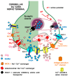
Representation of the functional reciprocal interactions between Gly and GABA in the cerebellum, according to the evidence from functional and immunocytochemical data, in nerve terminals where the two amino acids have been proposed to possibly behave as cotransmitters. The left part of the Figure mainly depicts the Gly-evoked GABA release following activation of GlyT2 (pink arrow), where it is proposed that Na+ cotransported with Gly (red curve arrows) contributes to GAT-1 reversal and the increase in local internal Ca2+ availability through plasmalemmal Na+/Ca2+ exchangers (NCX) and mitochondrial Na+/Ca2+ exchangers, finally facilitating the activation of “Ca2+-sensitive anion channels”. GABA is released both by GAT-1 reversal and through anion channels [41] (blue arrows). On the other hand (right part of the figure), GABA can induce Gly release following cotransport of Na+ through GAT-1 (red curve arrow); it is proposed that Na+ leads to increased availability of internal Ca2+ through exchange via mitochondrial Na+/Ca2+ exchangers (red long-dashed arrows) and, in part, by inducing mild depolarization, sufficient to cause the opening of voltage-sensitive Ca2+ channels (orange arrows). This leads to entry of Ca2+ ions that do not trigger exocytosis but contribute to the activation of “Ca2+-sensitive anion channels” (red thin dotted arrow) through which Gly is released (for details, see [69]). Due to the complexity of the scheme, synaptic versus extrasynaptic locations of transporters, channels, and other structures are not reported.

Similar articles

Cited by

  • Glycine Transporter 1 Inhibitors Minimize the Analgesic Tolerance to Morphine.
    Galambos AR, Essmat N, Lakatos PP, Szücs E, Boldizsár I Jr, Abbood SK, Karádi DÁ, Kirchlechner-Farkas JM, Király K, Benyhe S, Riba P, Tábi T, Harsing LG Jr, Zádor F, Al-Khrasani M. Galambos AR, et al. Int J Mol Sci. 2024 Oct 17;25(20):11136. doi: 10.3390/ijms252011136. Int J Mol Sci. 2024. PMID: 39456918 Free PMC article.

References

    1. Baer K., Waldvogel H.J., Faull R.L.M., Rees M.I. Localization of Glycine Receptors in the Human Forebrain, Brainstem, and Cervical Spinal Cord: An Immunohistochemical Review. Front. Mol. Neurosci. 2009;2:25. doi: 10.3389/neuro.02.025.2009. - DOI - PMC - PubMed
    1. Lynch J.W. Native Glycine Receptor Subtypes and Their Physiological Roles. Neuropharmacology. 2009;56:303–309. doi: 10.1016/j.neuropharm.2008.07.034. - DOI - PubMed
    1. Johnson J.W., Ascher P. Glycine Potentiates the NMDA Response in Cultured Mouse Brain Neurons. Nature. 1987;325:529–531. doi: 10.1038/325529a0. - DOI - PubMed
    1. Chatterton J.E., Awobuluyi M., Premkumar L.S., Takahashi H., Talantova M., Shin Y., Cui J., Tu S., Sevarino K.A., Nakanishi N., et al. Excitatory Glycine Receptors Containing the NR3 Family of NMDA Receptor Subunits. Nature. 2002;415:793–798. doi: 10.1038/nature715. - DOI - PubMed
    1. Grand T., Abi Gerges S., David M., Diana M.A., Paoletti P. Unmasking GluN1/GluN3A Excitatory Glycine NMDA Receptors. Nat. Commun. 2018;9:4769. doi: 10.1038/s41467-018-07236-4. - DOI - PMC - PubMed

Grants and funding

This research received no external funding.

LinkOut - more resources