. 2024 Jul 3;13(7):560.
doi: 10.3390/pathogens13070560.

Recombinant Ixodes scapularis Calreticulin Binds Complement Proteins but Does Not Protect Borrelia burgdorferi from Complement Killing

Affiliations

Recombinant Ixodes scapularis Calreticulin Binds Complement Proteins but Does Not Protect Borrelia burgdorferi from Complement Killing

Moiz Ashraf Ansari et al. Pathogens. .

Abstract

Ixodes scapularis is a blood-feeding obligate ectoparasite responsible for transmitting the Lyme disease (LD) agent, Borrelia burgdorferi. During the feeding process, I. scapularis injects B. burgdorferi into the host along with its saliva, facilitating the transmission and colonization of the LD agent. Tick calreticulin (CRT) is one of the earliest tick saliva proteins identified and is currently utilized as a biomarker for tick bites. Our recent findings revealed elevated levels of CRT in the saliva proteome of B. burgdorferi-infected I. scapularis nymphs compared to uninfected ticks. Differential precipitation of proteins (DiffPOP) and LC-MS/MS analyses were used to identify the interactions between Ixs (I. scapularis) CRT and human plasma proteins and further explore its potential role in shielding B. burgdorferi from complement killing. We observed that although yeast-expressed recombinant (r) IxsCRT binds to the C1 complex (C1q, C1r, and C1s), the activator of complement via the classical cascade, it did not inhibit the deposition of the membrane attack complex (MAC) via the classical pathway. Intriguingly, rIxsCRT binds intermediate complement proteins (C3, C5, and C9) and reduces MAC deposition through the lectin pathway. Despite the inhibition of MAC deposition in the lectin pathway, rIxsCRT did not protect a serum-sensitive B. burgdorferi strain (B314/pBBE22Luc) from complement-induced killing. As B. burgdorferi establishes a local dermal infection before disseminating to secondary organs, it is noteworthy that rIxsCRT promotes the replication of B. burgdorferi in culture. We hypothesize that rIxsCRT may contribute to the transmission and/or host colonization of B. burgdorferi by acting as a decoy activator of complement and by fostering B. burgdorferi replication at the transmission site.

Keywords: Borrelia burgdorferi; Ixodes scapularis; complement cascade; tick calreticulin.


Conflict of interest statement

The authors declare no conflicts of interest.

Figures

Figure 1
Figure 1
Expression and affinity purification of recombinant I. scapularis calreticulin. Recombinant I. scapularis calreticulin (rIxsCRT, 47.61 kDa) with His-tag was expressed in Pichia pastoris over 3 days and affinity-purified under native conditions. Affinity purification of rIxsCRT was validated by standard SDS-PAGE followed by silver staining (A) and Western blotting using the antibody for the Histidine fusion tag (B). Lanes 1–4 represent the 50, 75, 100, and 200 imidazole concentrations (mM) at which the rIxsCRT was eluted, dialyzed in PBS, concentrated, and used for the assays.
Figure 2
Figure 2
Differential Precipitation of Proteins (DiffPOP) reveals interactions between rIxsCRT and complement proteins. Affinity-purified rIxsCRT (10 µg) was pre-incubated with 10% normal human serum (NHS) for 90 min at 37 °C, while NHS and rIxsCRT served as controls. The reactions were then stabilized and subjected to differential precipitation using escalating amounts of precipitating buffer, as described in the materials and methods section (see Supplementary Table S1). Different fractions obtained from the precipitation process were analyzed by standard SDS-PAGE and silver staining. (A) shows the protein profile of NHS only, (B) illustrates the profile of the NHS + rIxsCRT mixture, and (C) displays the profile of rIxsCRT only. Additionally, the NHS + rIxsCRT mixture was subjected to Western blotting analysis using an antibody against the histidine fusion tag to track the precipitation of rIxsCRT (D). The distinct protein profile between NHS and NHS + rIxsCRT, highlighted by the red arrowhead in panels (A,B), indicated potential interactions between rIxsCRT and specific proteins. The precipitation profile depicted in (D) guided the selection of fractions for subsequent LCMS/MS analysis, shedding light on the specific complement proteins interacting with rIxsCRT. L: ladder depicting molecular weight (kDa), Lanes 1–10 = DiffPOP fractions 1–10.
Figure 3
Figure 3
Volcano plot analyses revealed human serum proteins that differentially co-precipitated with rIxsCRT. Normalized abundance values of three biological replicates were used in Proteome Discoverer™ 2.4 software (Thermo Fisher Scientific, Dallas, TX, USA) to generate the volcano plot. In the volcano plot, the Y-axis shows the −log10 p-value and the X-axis shows the magnitude of change (log2 fold change). Red dots represent proteins that co-precipitated in high amounts with rIxsCRT, while green dots represent those that were absent or co-precipitated in low amounts in the presence of rIxsCRT. An adjusted p-value ≤ 0.05 and log2 fold change of more than 2 were used as cut-offs to select proteins that co-precipitated in high amounts with rIxsCRT.
Figure 4
Figure 4
Differential precipitation of complement proteins in the presence of rIxsCRT. Fractions obtained from the differential precipitation (DiffPOP) were pooled into group A (fractions 1–5), group B (fraction 6), and group C (fractions 7 and 8). These fractions were subjected to Liquid Chromatography-Mass Spectrometry (LCMS/MS) analysis to investigate the abundance of complement proteins interacting with rIxsCRT. (A) illustrates the abundance of complement proteins interacting with rIxsCRT in group A, while (B) depicts the corresponding interactions in group B. The Y-axis in (A,B) illustrates the protein abundance, indicating the levels of proteins co-precipitated with rIxsCRT (depicted by the red graph) in comparison to NHS only (represented by the blue graph).
Figure 5
Figure 5
Validation of complement protein and rIxsCRT interactions via Western blotting analysis. To confirm the interactions observed in LCMS/MS analysis (referenced in Figure 3), DiffPOP fractions were subjected to standard Western blotting using antibodies specific for complement proteins: C1q, C1r, C1s, C3, C5, and C9, as indicated. Distinctive binding patterns, highlighting differences between NHS + rIxsCRT and the NHS control, are denoted by red arrowheads. Panels (A,C,E,G,I,K) depict immunoblots of NHS-only samples, while panels (B,D,F,H,J,L) represent immunoblots of NHS + rIxsCRT samples.
Figure 6
Figure 6
Pull-down assay validated the rIxsCRT binding of complement proteins. Affinity-purified rIxsCRT bound to His specific magnetic beads (Dynabeads™, Thermo Fisher Scientific, Waltham, MA, USA) was used to pull down complement proteins from human complement serum (HCS). The beads were washed and the eluted protein complexes were subjected to Western blotting analysis using antibodies for the histidine tag (A) and complement proteins C1q (B), C1r (C), C1s (D), C3 (E), C5 (F), and C9 (G). In all the panels (AG), HCS is the human complement serum that was eluted from the empty beads, i.e., without the bait protein (rIxsCRT), and HCS + rIxsCRT denotes the human complement proteins that were pulled down and eluted from the beads loaded with rIxsCRT. Red arrowheads denote the detected complement proteins at their expected molecular weight size (kDa).
Figure 7
Figure 7
ELISA analysis demonstrates the rIxsCRT binding of complement proteins. High binding ELISA plates coated with affinity-purified rIxsCRT (250 ng) were incubated with normal human serum (NHS) followed by antibodies for C1q (A), C1r (B), C1s (C), C3 (D), activated C3 (E), C5 (F), C9 (G), and C5b-9 or MAC (H) antibodies. Y-axis denotes the absorbance measured at A450nm, which reflected the intensity of the specific antibody binding to rIxsCRT. Non-coated wells blocked with 1% BSA were incubated with NHS and used as a negative control (Neg. Cont.). Data represent mean ± SEM of 3 biological replicates. For statistical analysis, t-test was performed on GraphPad Prism 9 and ** represents p < 0.01, *** represents p < 0.001, **** represents p < 0.0001, and ns represents not significant.
Figure 8
Figure 8
rIxsCRT apparently enhances membrane attack complex (MAC) deposition in the classical pathway and alternate pathway but inhibits MAC deposition in the lectin pathway. The Wieslab® Complement System Kit (Svar Life Science AB, Malmo, Sweden) was used to detect the effects of rIxsCRT on MAC deposition via the classical pathway, alternative pathway, and lectin pathway, as described in the materials and methods section. In brief, rIxsCRT (4 μM) was incubated with NHS (provided with the kit) at 37 °C for 30 min and then added to wells pre-coated with the antibody for MAC. Diluent and kit-provided reagent served as negative and positive controls, respectively. After washing, the conjugate and substrate were added according to the manufacturer’s instructions for each kit. NC denotes negative control and NHS denotes normal human serum, used as the positive control. % MAC deposition (Y-axis) was calculated as mentioned in the methodology section and the MAC deposition in the positive control was represented as 100% (denoted by the black dotted line). Data are presented as deposited MAC ± SEM calculated from 3 biological replicates. Statistical significance was determined by Student’s t-test in GraphPad Prism 9. * Represents p ≤ 0.05, ** represents p ≤ 0.01, and ns represents not significant.
Figure 9
Figure 9
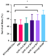
rIxsCRT does not protect B. burgdorferi from complement killing. Normal human serum (NHS) was pre-incubated with serial dilutions of rIxsCRT (1, 2, and 4 µM) or phosphate-buffered saline (PBS) at 37 °C for 30 min prior to the addition of 85 µL of 106 cells/mL of B. burgdorferi B314/pBBE22luc (complement-sensitive strain) and incubated in a bio-shaker at 32 °C and 100 rpm. NHS incubated with B. burgdorferi B314/pPCD100 (complement-resistant strain) was used as a positive control. Survival rates of B. burgdorferi were assessed at 3 h post-incubation. Data represent mean ± SEM of 3 biological replicates. Statistical significance was evaluated using t-test in GraphPad Prism 9 (ns: no significance, * represents p-value ≤ 0.05).
Figure 10
Figure 10
rIxsCRT promotes the growth of B. burgdorferi in culture. B. burgdorferi (strain MSK5) cultured in the presence of rIxsCRT at 2.6 mM (rIxsCRT 125) and 5.2 mM (rIxsCRT 250) were sampled at days 2, 5, and 7. (A) In triplicate, cells were quantified by manual counts on a Petroff-Hausser chamber and quantified using the following formula: number of cells/mL = Average of cells counted in all chambers × Dilution factor x 50,000. (B) B. burgdorferi was quantified by qPCR using the genomic DNA of the B. burgdorferi and fLaB primers. For ELISA, Student’s t-test was used for statistical analysis on GraphPad Prism 9 and p-value ≤ 0.05 (denoted by *) was considered significant for 3 biological replicates.
Figure 11
Figure 11
One-time exposure of rabbits to B. burgdorferi-infected I. scapularis nymphs triggers high IgG antibody levels for rIxsCRT. (A) Affinity-purified rIxsCRT (250 ng) was subjected to standard ELISA using serially diluted purified IgG of rabbits that were fed upon for a single time by uninfected (blue line graph) and B. burgdorferi-infected (red line graph) I. scapularis nymph ticks. The black line graph represents pre-immune IgG binding, which was used for a negative control. (BD) Various quantities of affinity-purified rIxsCRT (100, 300, and 500 ng) were subjected to Western blotting analysis using purified IgG from rabbits that were fed upon for a single time by uninfected (B) and B. burgdorferi-infected ticks (C) as well as the antibody for the histidine tag, which served as a positive control (D).

Similar articles

References

    1. Sosa J.P., Ferreira Caceres M.M., Agadi K., Pandav K., Mehendale M., Mehta J.M., Go C.C., Matos W.F., Guntipalli P., Belizaire M.E. Diseases Transmitted by the Black-Legged Ticks in the United States: A Comprehensive Review of the Literature. Cureus. 2021;13:e17526. doi: 10.7759/cureus.17526. - DOI - PMC - PubMed
    1. Wolf M.J., Watkins H.R., Schwan W.R. Ixodes scapularis: Vector to an Increasing Diversity of Human Pathogens in the Upper Midwest. WMJ. 2020;119:16–21. - PMC - PubMed
    1. Guzman N., Yarrarapu S.N.S., Beidas S.O. StatPearls. StatPearls Publishing; Treasure Island, FL, USA: 2023. Anaplasma Phagocytophilum. - PubMed
    1. Krause P.J. Human babesiosis. Int. J. Parasitol. 2019;49:165–174. doi: 10.1016/j.ijpara.2018.11.007. - DOI - PubMed
    1. Piantadosi A., Solomon I.H. Powassan Virus Encephalitis. Infect. Dis. Clin. N. Am. 2022;36:671–688. doi: 10.1016/j.idc.2022.03.003. - DOI - PMC - PubMed

LinkOut - more resources